


Abstract
Unstable fracture-dislocation of the ankle is a common lower-extremity injury. Treatment is challenging when the fracture-dislocation is open and cannot be treated with conventional open reduction and internal fixation (ORIF). Immediate ORIF may not be possible for severe, unstable ankle injuries, such as those with ischemic foot because of a poor blood supply caused by soft-tissue injury, or open fracture-dislocation of the ankle with a deltoid ligament rupture. We describe a staged treatment for unstable open fracture-dislocation of the ankle with a deltoid ligament rupture. The first stage involves temporary vertical transarticular pinning combined with external fixation. The second stage involves delayed definitive plating with autogenous bone graft for the bone defect of the distal fibula. This staged management is useful in select emergency cases of unstable open fracture-dislocations of the ankle combined with deltoid ligament rupture for which conventional ORIF cannot be performed.
Ankle fractures are common injuries of the lower extremities. The goal of treatment is to obtain anatomical restoration, and open reduction and internal fixation (ORIF) is the standard treatment to achieve this goal [1,2]. However, some situations are not amenable to ORIF. Immediate ORIF is not the best approach in some cases, such as open fracture-dislocations of the ankle with deltoid ligament rupture, and serious medical conditions resulting from multiple injuries [3,4].
In these complex situations, a staged approach will lead to better results [5,6]. We describe a staged treatment for an open fracture-dislocation of the ankle with deltoid ligament rupture. For the first stage of treatment, we used vertical transarticular percutaneous pinning [4,7–10]. in combination with external fixation. For the second stage of treatment, we performed delayed plating with bone grafting into the bone defect of the distal fibula after wound care and soft-tissue healing.
Case Report
A 75-year-old woman sustained an unstable open fracture-dislocation of the ankle with complete rupture of the deltoid ligament. The patient had no comorbidities.
The foot seemed ischemic because of a poor blood supply caused by extensive soft-tissue injury and ankle dislocation. The dorsalis pedis artery pulse was not palpable. All toes on the injured side were cold and purple (Fig. 1). However, no vascular injuries were present on computed tomographic angiography.
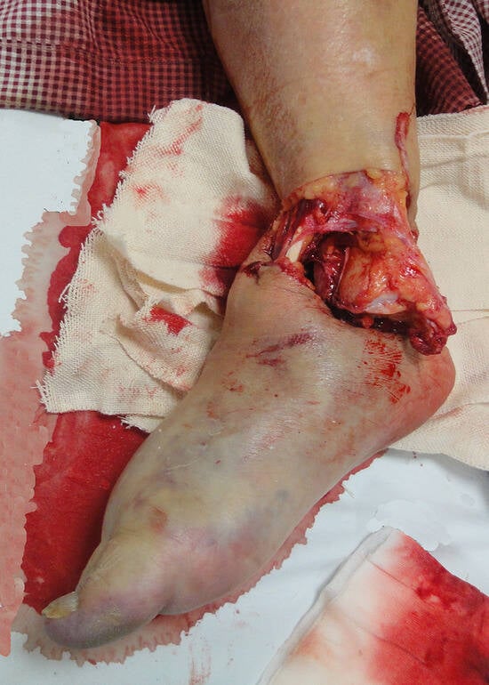
Figure 1.
Photograph showing unstable open fracture-dislocation of the ankle: the toes on the affected side were cold and purple because of a poor blood supply resulting from extensive soft-tissue injury.
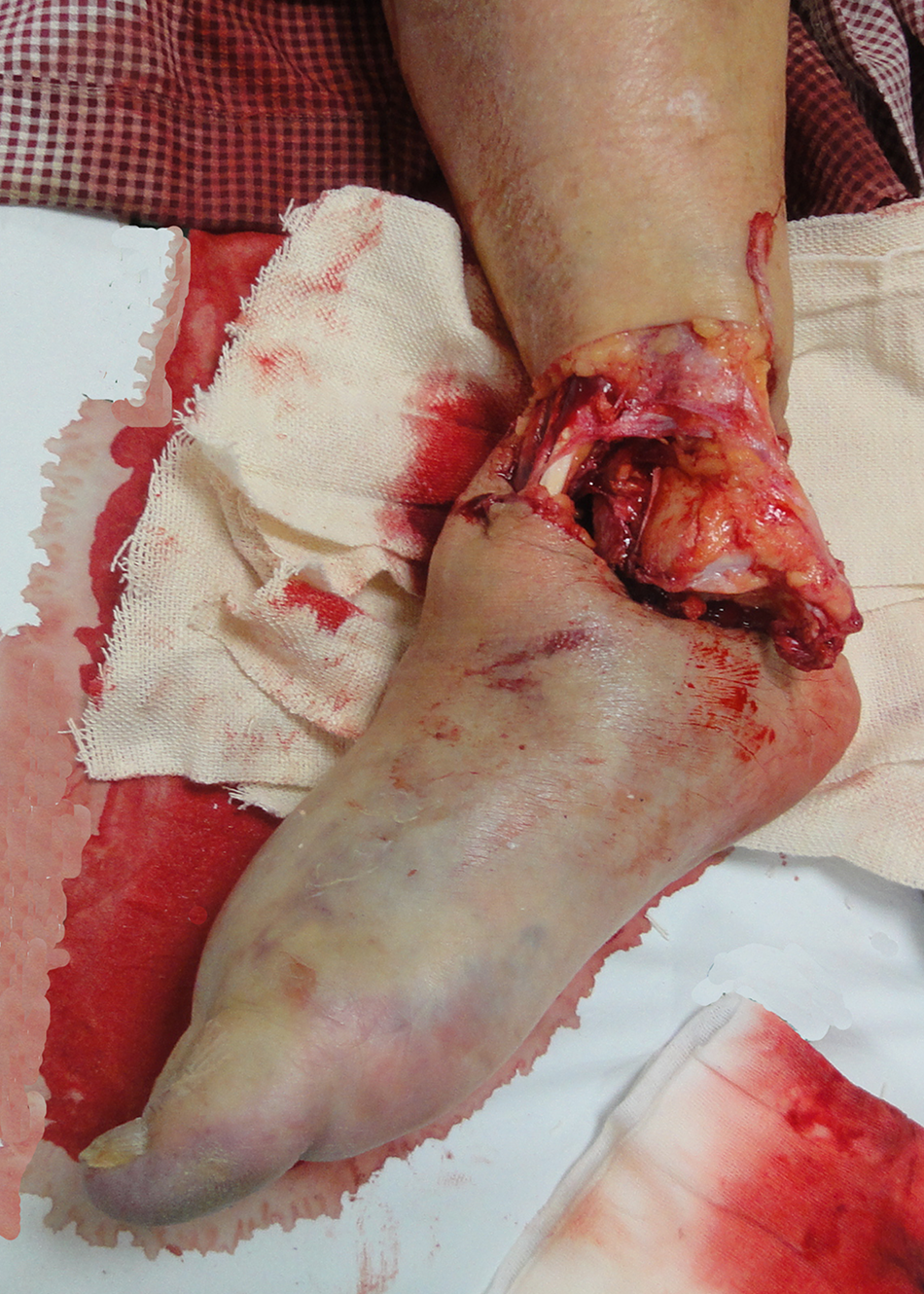
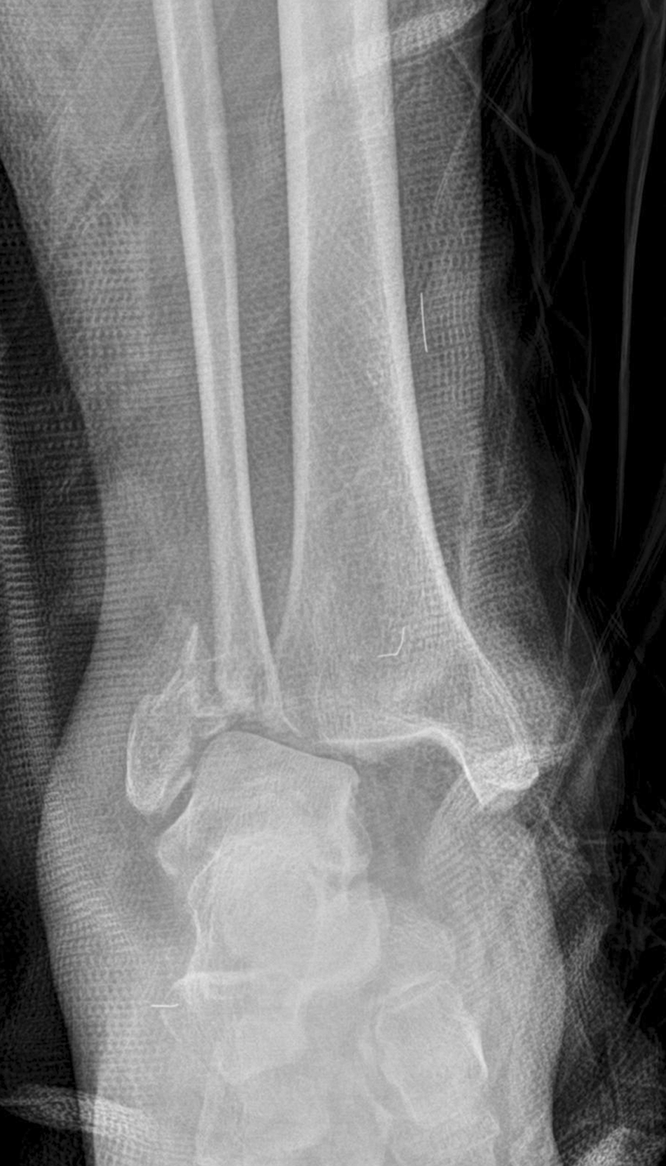
The fracture pattern of the distal fibula was 44-B2 according to the Orthopedic Trauma Association classification (Fig. 2). The open fracture classification was type IIIA according to the Gustilo open fracture classification (Fig. 1) [11]. The fibular fracture was at the level of the syndesmotic ligaments with a deltoid ligament rupture. Soft-tissue coverage was possible even with extensive soft-tissue damage. Closed reduction was attempted in the emergency room but failed because of ankle instability.
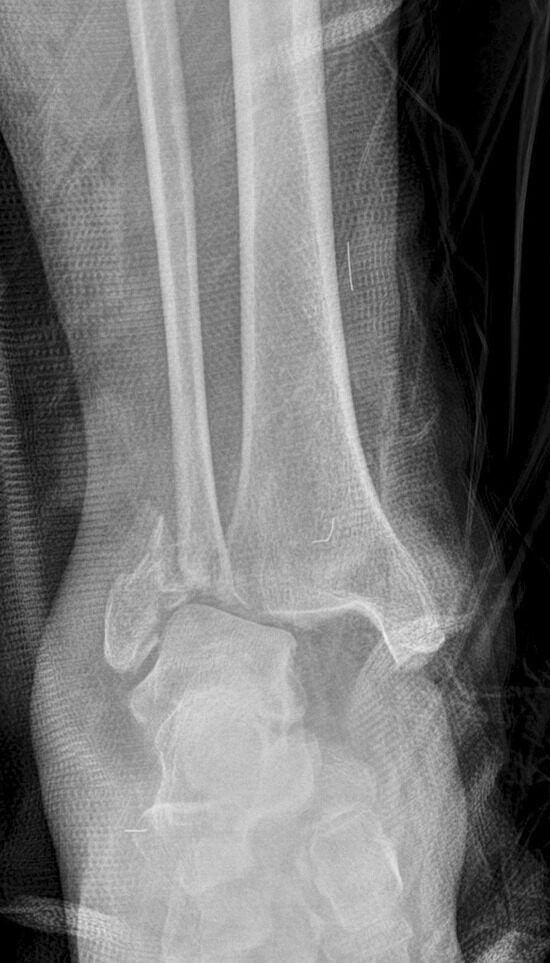
Figure 2.
Radiograph of the fracture-dislocation of the ankle with a comminuted distal fibula fracture but no medial malleolar fracture, suggesting complete deltoid ligament rupture.
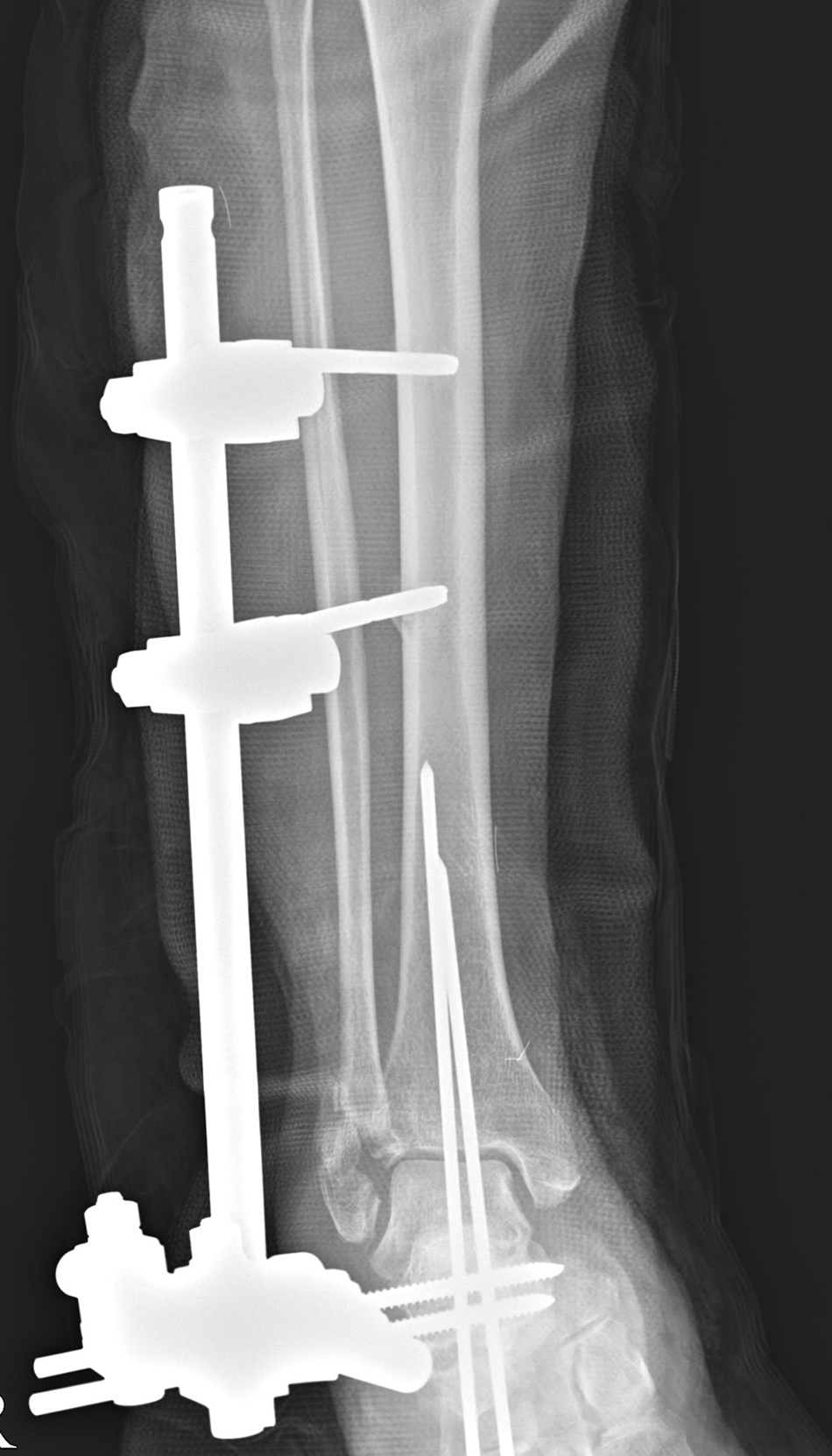
The patient was taken to the operating room 8 hours after the injury. The distal fibula fracture was comminuted with bone defect. The fracture could not be treated with conventional ORIF because of severe soft-tissue damage and the osteoporotic small fragment of the distal fibula (Figs. 1 and 3).
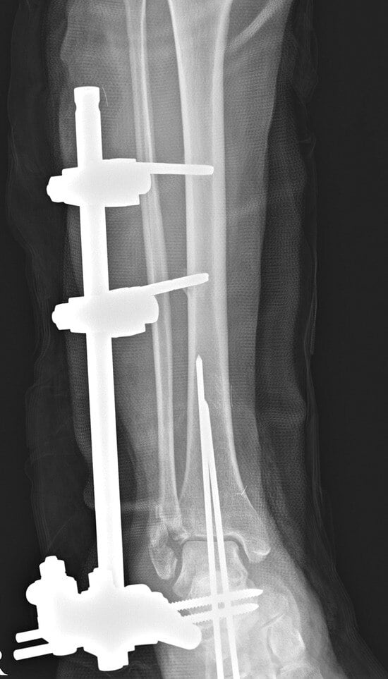
Figure 3.
Immediate postoperative radiograph showing vertical transarticular pinning and supplemental external fixation. Conventional open reduction and internal fixation was not feasible because of severe soft-tissue damage and the osteoporotic small fragment of the distal fibula.
Staged operative treatment was planned for soft-tissue care and to wait until soft-tissue consolidation could make internal fixation feasible. The first stage of treatment consisted of temporary vertical transarticular pinning combined with external fixation. The tibiotalar instability was too severe to directly repair the deltoid ligament rupture without provisional fixation because of an associated distal fibular fracture. Therefore, percutaneous pinning across the subtalar and tibiotalar joints was performed using two 2.6-mm Steinmann pins with the ankle in slight plantarflexion [7]. After pinning, the tibiotalar joint became stable enough for us to repair the ruptured deltoid ligament. A complete intrasubstance tear of the deltoid ligament was found. Simple reapproximation was possible and repaired with nonabsorbable sutures.
Vertical pinning alone was insufficient to stabilize the ankle until soft-tissue consolidation because a combined distal fibular fracture could not be treated with conventional ORIF. Splint or cast immobilization would not allow for proper wound care and soft-tissue monitoring. A unilateral external fixator was applied to the proximal tibia and calcaneus while avoiding the severely damaged soft tissue after repair of the deltoid ligament. The wound cultures obtained after debridement during surgery were sent to the laboratory [12]. The open wound was closed after thorough debridement and irrigation.
The postoperative antibiotic regimen was a combination of aminoglycoside and second-generation cephalosporin for broad-spectrum coverage. The initial wound culture was negative, and the open wound around the ankle was not infected. Laboratory assessment of infection, including erythrocyte sedimentation rate, C-reactive protein, and white blood cell count, indicated no evidence of infection. The Steinmann pins were removed at 6 weeks after the index operation to prevent further risk of infection. The external fixator was removed at 8 weeks.
The second stage of treatment consisted of both external fixator removal and definitive plating. After external fixator removal, definitive plating was performed simultaneously with a distal fibular locking plate (Zimmer, Warsaw, Indiana) and autogenous cancellous bone graft at the bone defect of the distal fibula. The medial fibrous wall (Figs. 4 and 5) was spared during curettage of the bone defect site to prevent bone graft from entering the tibiotalar joint. Autogenous cancellous bone graft from the iliac crest could be applied to the bone defect site without the involvement of the tibiotalar joint.
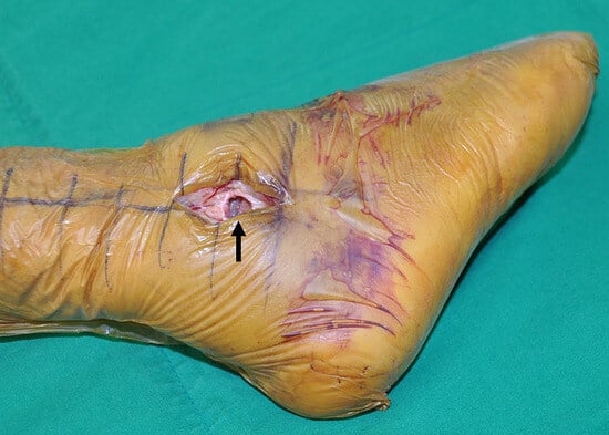
Figure 4.
Autogenous cancellous bone graft from the iliac crest was safely impacted to the bone defect (arrow) of the distal fibula because the medial fibrous wall prevented bone graft from entering the tibiotalar joint.
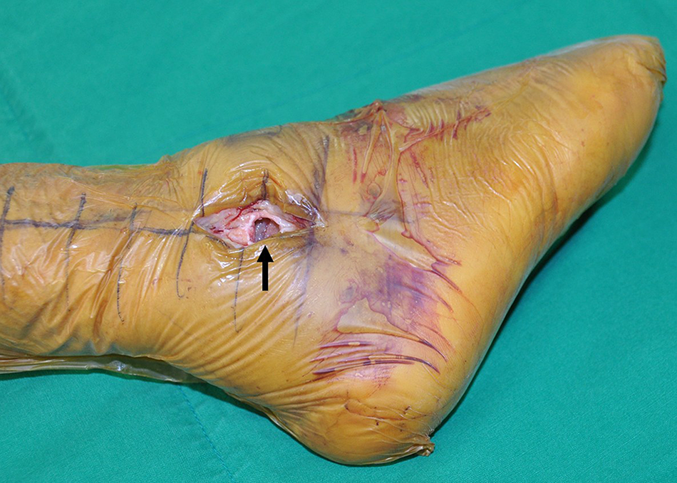
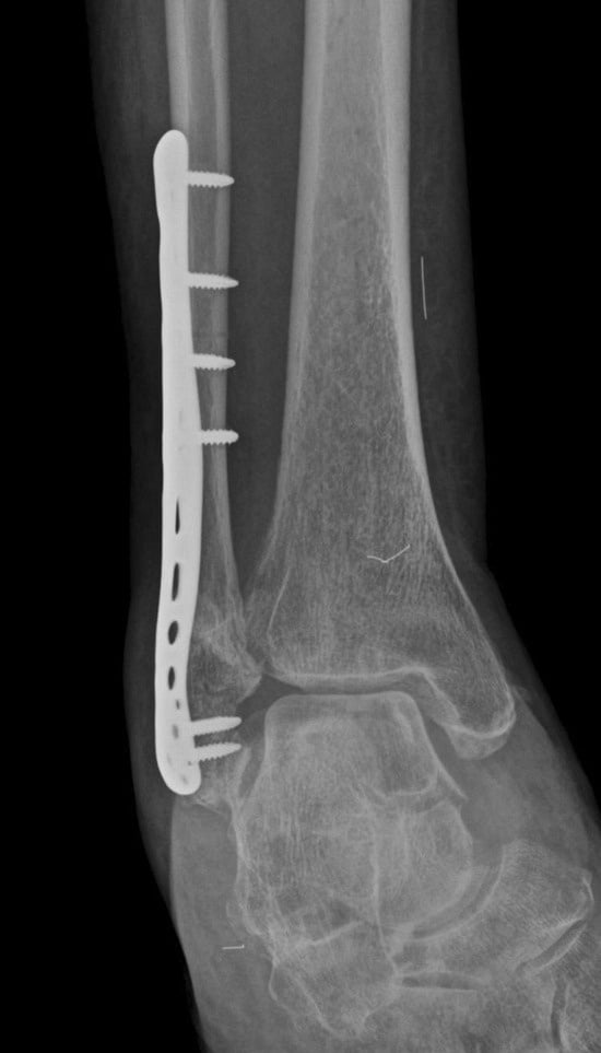
Figure 5.
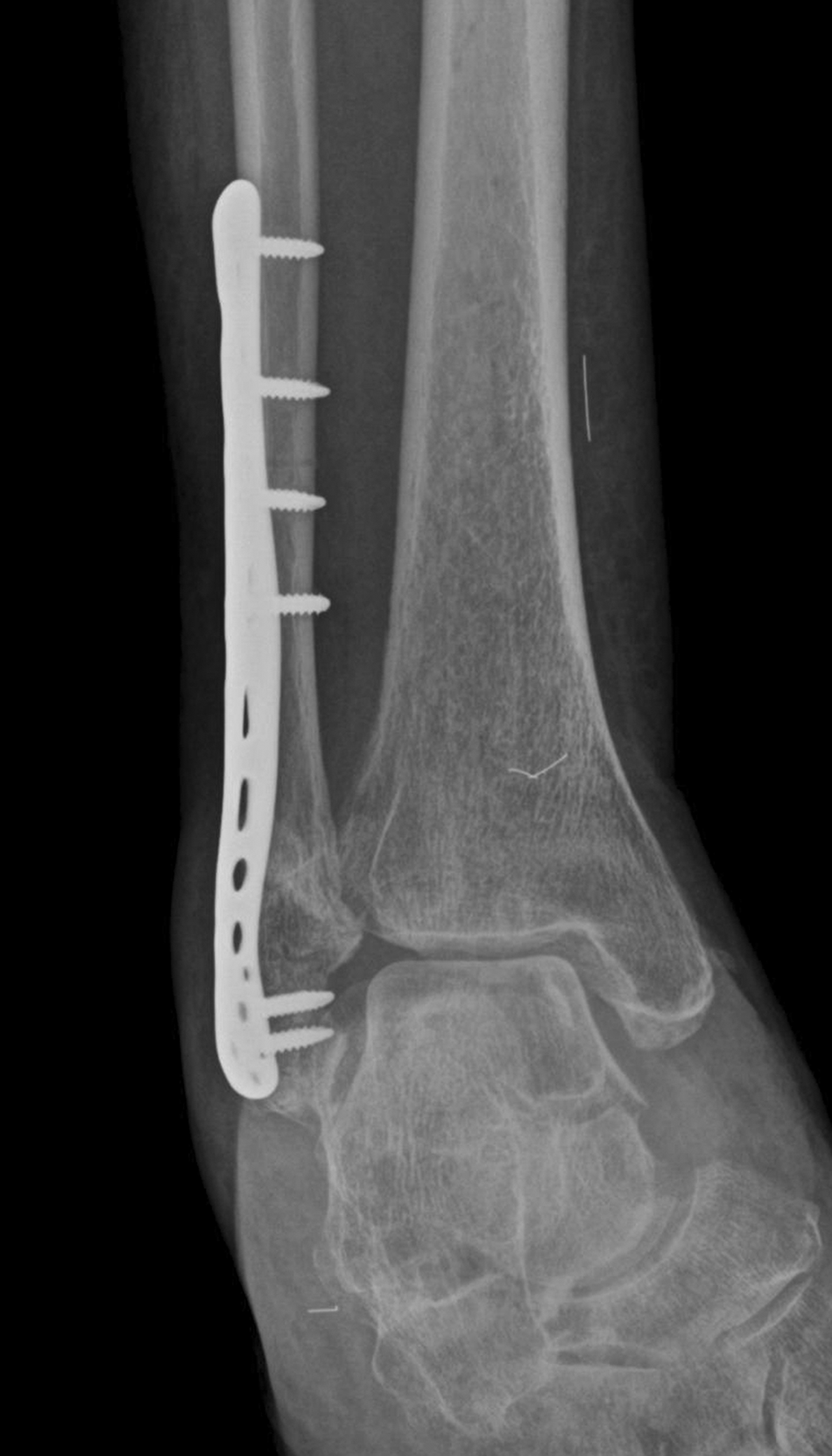
Radiographic union was obtained through bridging callus formation in at least three cortices at 3 months after definitive plating with bone graft.
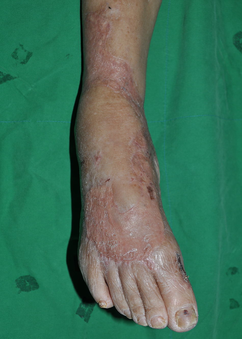
Although superficial skin necrosis occurred, the affected area was debrided and split-thickness skin graft from the lateral thigh was used at 10 weeks after the index operation. All wounds were healed at 3 months after plating with simple wound dressing (Fig. 6).
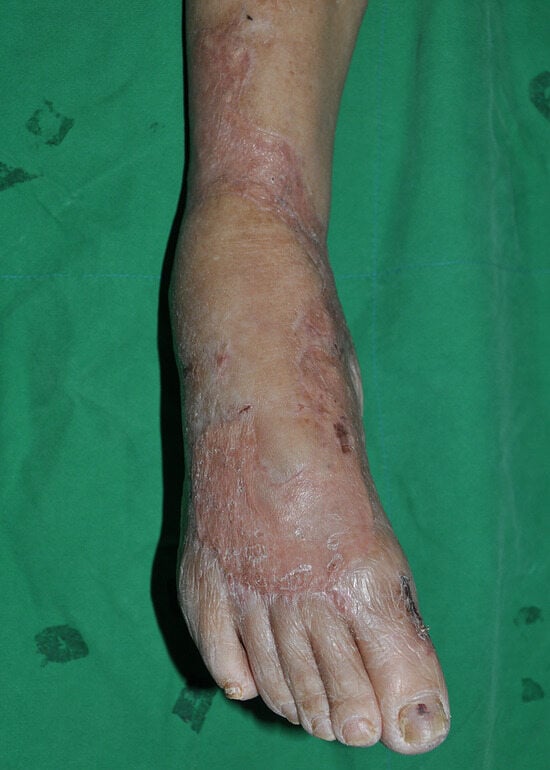
Figure 6.
The injured ankle showing that all superficial necrotic wounds were healed at 3 months after plating.
The patient was allowed to perform range-of-motion exercises immediately after plating. Partial weightbearing was begun at 2 weeks after definitive plating and progressed to full weightbearing at 2 months after plating. At 3 months after plating, radiographic union (Fig. 5) was achieved through bridging callus formation in at least three cortices. The range of motion of the ankle joint was between 0° (neutral) and 30° of plantar flexion at 3 months after plating, and remained unchanged at 1 year after plating. The American Orthopaedic Foot and Ankle Society Ankle-Hindfoot score at 3-month and 1-year follow-up was 76 and 72 points, respectively.
Discussion
Treatment of an open fracture-dislocation of the ankle is difficult because immediate conventional ORIF may not be possible, and staged management should be considered [8,10]. As the first stage of treatment, many studies have reported treatment options such as vertical transarticular pinning and external fixation [7,8,13. Each procedure has been reported separately [4,7,8,13].
Although external fixation is the standard of care for unstable ankle fractures that are not ready for internal fixation because it allows easier access for wound care [13], external fixation cannot replace vertical pinning in all cases of unstable ankles. The use of transarticular pinning is a quick and effective way to provide additional fixation for these difficult fractures.
The combination of both treatment options is necessary in select cases of unstable ankles. However, to our knowledge, there has been no report of a combined vertical pinning and external fixation as the first stage of treatment.
It can be difficult to achieve ankle stability with external fixation alone for cases in which the open fracture-dislocation of the ankle cannot be treated with conventional ORIF, and the fracture-dislocation occurs in combination with a ligament injury, such as a deltoid ligament rupture [7,8]. Ankle fracture-dislocations with a deltoid ligament injury can be more difficult to manage than those with a medial malleolar fracture because surgical repair of a torn deltoid ligament is not possible until provisional ankle stability is achieved [7,8].
In these situations, temporary vertical transarticular percutaneous pinning [4,7–9]. can quickly be used to achieve the stability necessary to repair a deltoid ligament rupture instead of using external fixation. Then, external fixation can be supplemented for an associated distal fibular fracture that cannot be treated with conventional ORIF and for wound care instead of a splint or cast application.
External fixation may seem redundant. However, in cases of unstable ankles, external fixation is needed to prevent a gradual lateral translation of the talus or pin breakage until soft-tissue consolidation and to take care of the wound. Vertical pinning alone is vulnerable to pin loosening for the time needed until soft-tissue consolidation in a splint or cast. Delta external fixation is better than unilateral fixation in terms of stability. However, the delta external fixator may be unavailable in some institutions.
For the second stage of treatment, delayed ORIF (definitive plating) with bone graft was performed. This procedure was simple because a satisfactory primary reduction (anatomical reduction of the ankle mortise) was maintained with Steinmann pins. Therefore, plating in situ was easily performed without the need for intraoperative reduction.
Cancellous bone graft was planned instead of block bone graft, curettage was performed with caution, and the medial fibrous wall (Fig. 4) of the bone defect site of the distal fibula was left intact to prevent intraarticular involvement of the bone graft. Although vertical transarticular pinning is a necessary procedure for unstable ankles combined with a deltoid ligament rupture, this pinning penetrates articular surfaces. Therefore, procedures including vertical pinning should be limited to exceptional cases in which tibiotalar stability cannot be obtained with conventional closed or open methods.
Conclusions
In conclusion, we suggest that a staged approach, in which vertical transarticular pinning with external fixation is used for the first stage and delayed definitive plating with bone graft is used for the second stage, is an effective way to treat select cases of unstable open fracture-dislocation of the ankle combined with a deltoid ligament rupture for which conventional ORIF cannot be performed.
Funding
None reported.
Conflicts of Interest
None reported.
References
- Mitchell WG, Shaftan GW, Sclafani SJ: Mandatory open reduction: its role in displaced ankle fractures. J Trauma 19: 602, 1979.
- Makwana NK, Bhowal B, Harper WM, : Conservative versus operative treatment for displaced ankle fractures in patients over 55 years of age. A prospective, randomised study. J Bone Joint Surg Br 83: 525, 2001.
- Hoiness P, Stromsoe K: Early complications of surgically managed ankle fractures related to the AO classification. A review of 118 ankle fractures treated with open reduction and internal fixation. Arch Orthop Trauma Surg 119: 276, 1999.
- Przkora R, Kayser R, Ertel W, : Temporary vertical transarticular-pin fixation of unstable ankle fractures with critical soft tissue conditions. Injury 37: 905, 2006.
- Seibert F, Schatz B, Bratschitsch G, : Temporary Kirschner wire ankle transfixation in the treatment of unstable ankle fractures. Foot Ankle Surg 7: 85, 2001.
- Park J, Lee HB, Kim GL, : Surgical treatment approach for pilon and talar dome fracture. JAPMA 110: Article_14, 2020.
- Childress HM: Vertical transarticular-pin fixation for unstable ankle fractures. J Bone Joint Surg Am 47: 1323, 1965.
- Childress HM: Vertical transarticular pin fixation for unstable ankle fractures: impressions after 16 years of experience. Clin Orthop Relat Res 120: 164, 1976.
- Scioscia TN, Ziran BH: Use of a vertical transarticular pin for stabilization of severe ankle fractures. Am J Orthop (Belle Mead NJ) 32: 46, 2003.
- Meier C, Schefold JC, Hug U, : Temporary Kirschner wire ankle transfixation and delayed ORIF. Eur J Trauma 30: 371, 2004.
- Gustilo RB, Anderson JT: JSBS classics. Prevention of infection in the treatment of one thousand and twenty-five open fractures of long bones. Retrospective and prospective analyses. J Bone Joint Surg Am 84: 682, 2002.
- Copeland-Halperin LR, Kaminsky AJ, Bluefeld N, : Sample procurement for cultures of infected wounds: a systematic review. J Wound Care 25: S4, 2016.
- Seibert FJ, Fankhauser F, Elliott B, : External fixation in trauma of the foot and ankle. Clin Podiatr Med Surg 20: 159, 2003.
© 2022 American Podiatric Medical Association