


Abstract
Objective: To investigate the predictive value of erythrocyte sedimentation rate (ESR) and C-reactive protein (CRP) in persons with and without diabetes with osteomyelitis (OM). Methods: We evaluated 455 patients in a retrospective cohort study of patients admitted to the hospital with diabetic foot OM (n = 177), diabetic foot soft-tissue infections (STIs) (n = 176), nondiabetic OM (n = 51), and nondiabetic STIs (n = 51). Infection diagnosis was determined through bone culture, histopathologic examination for OM, and/or imaging (magnetic resonance imaging/single-photon emission computed tomography) for STI. The optimal cutoff values of ESR and CRP in predicting OM were determined by receiver operating characteristic curve analysis. Sensitivity, specificity, positive and negative predictive values, and positive and negative likelihood ratios were determined through contingency tables. Results: In persons without diabetes with STI or OM, the mean ESR and CRP differences were 10.0 mm/h and 2.6 mg/dL, respectively. In contrast, persons with diabetes had higher levels of each: 24.8 mm/h and 6.8 mg/dL, respectively. As a result, ESR and CRP predicted OM better in patients with diabetes. However, when patients were stratified by neuropathy status, ESR remained predictive of OM in diabetic patients with neuropathy (75% sensitivity, 58% specificity) but not in diabetic patients without neuropathy (50% sensitivity, 44% specificity). Also, CRP remained predictive irrespective of neuropathy status. A similar trend was observed in patients without diabetes. Conclusions: Previous studies have reported that ESR and CRP are predictive of OM. However, this study suggests that neuropathy influences the predictive value of inflammatory biomarkers. The underlying mechanisms require further study.
Osteomyelitis (OM) of the foot is challenging and expensive for clinicians to diagnose and treat. The diagnosis of OM incorporates various diagnostic tests, including physical examination [1,2], radiography [3], magnetic resonance imaging (MRI) [4], bone scanning, single-photon emission computed tomography/computed tomography (SPECT/CT) after injection of radiolabeled white blood cells [5,6], histopathologic examination, and bone culture [7,8]. Erythrocyte sedimentation rate (ESR) and C-reactive protein (CRP) are perhaps the most commonly used inflammatory biomarkers in diagnostic algorithms for OM [9]. These simple laboratory tests are readily available, relatively inexpensive, and have well-established diagnostic utility in diagnosing vertebral OM, long bone infections, and periprosthetic joint infections. However, the literature reports variability in the threshold values based on the anatomical site of infection and the mechanism of infection [10,11,12,13]. The cutoff values for CRP and ESR to diagnose diabetic foot OM are two to five times higher than reports of OM at other anatomical site [9].
The existing literature for OM of the foot is limited because of small sample sizes, inconsistent reference standards to diagnose OM, and the exclusive evaluation of people with diabetes. In addition, ESR is usually the only biomarker evaluated, so there is little evidence to support the use of CRP to diagnose OM [9]. Most infected foot ulcers occur in people with diabetes. In fact, there are few published reports of foot infections in people without diabetes [14,15,16,17] Therefore, there is little evidence concerning the diagnostic value of commonly used tests to evaluate OM in people without diabetes [14]. Health-care providers tend to extrapolate the medical evidence for diabetic foot infections to people without diabetes. Conversely, it is not uncommon for providers to apply evidence from other sites (spine, pelvis, and long bones) to the foot. The goal of this study was to evaluate the sensitivity, specificity, positive and negative predictive values, and positive and negative likelihood ratios of ESR and CRP in patients with and without diabetes to predict pedal OM.
Methods
After the study received institutional review board approval by the University of Texas Southwestern Medical Center IRB, inpatient medical records were reviewed, identifying 455 adult patients who had been admitted to the hospital with a foot infection. The records reflected patients admitted between June 12, 2009, and February 21, 2017. We separated the patients into four groups: diabetic patients with OM, diabetic patients with STIs, nondiabetic patients with OM, and nondiabetic patients with STIs. The diagnosis of OM was based on positive bacterial cultures from bone specimens and/or histologic changes consistent with OM. Patients with STIs by definition had negative bone culture and histologic findings or an MRI or SPECT study that was negative for OM. Other inclusion criteria were age 18 to 89 years and baseline ESR and CRP measurements within 72 hours of admission. We excluded patients with comorbidities that could affect baseline ESR and CRP levels (ie, autoimmune disease and other sites of infection, such as endocarditis, were excluded).
A detailed medical review also collected data regarding demographics, medical history, and results of other serologic tests, imaging, and cultures. Diagnosis of diabetes mellitus was based on criteria from the American Diabetes Association. We defined sensory neuropathy as abnormal vibration sensation, abnormal sensation with a 10-g Semmes-Weinstein monofilament, and/or absent Achilles deep tendon reflexes [18,19]. We evaluated ESR and CRP levels from the medical records which were originally analyzed by the hospital laboratory.
All of the continuous variables were tested for normality using Shapiro-Wilk analysis. Descriptive statistical analyses were used to determine frequencies of categorical variables and median values of continuous variables with 25th to 75th interquartile ranges (Table 1). Continuous variables between the STI and diabetic OM study groups were analyzed using either the Student t test (if normally distributed) or the Mann-Whitney U test (for nonparametric data), and categorical variables were analyzed using the Pearson χ2 test or the Fisher exact test when appropriate. Contingency tables were used to calculate descriptive epidemiologic measures, and area under the receiver operating characteristic curve values for OM were calculated for ESR and CRP. Various cutoff levels of ESR and CRP were tested to maximize sensitivity and specificity. Optimal threshold values of ESR and CRP were identified by the highest combined rate of sensitivity and specificity results for each cutoff point (the Youden J statistic) [12]. Potential interaction effects between patient factors such as comorbidities were determined through interaction plots to evaluate for effect modification when correlating factors with ESR and CRP. An α = .05 was used in all of the statistical analyses to denote significance. All of the statistical analyses were performed using R version 3.3.1 (Vienna, Austria).

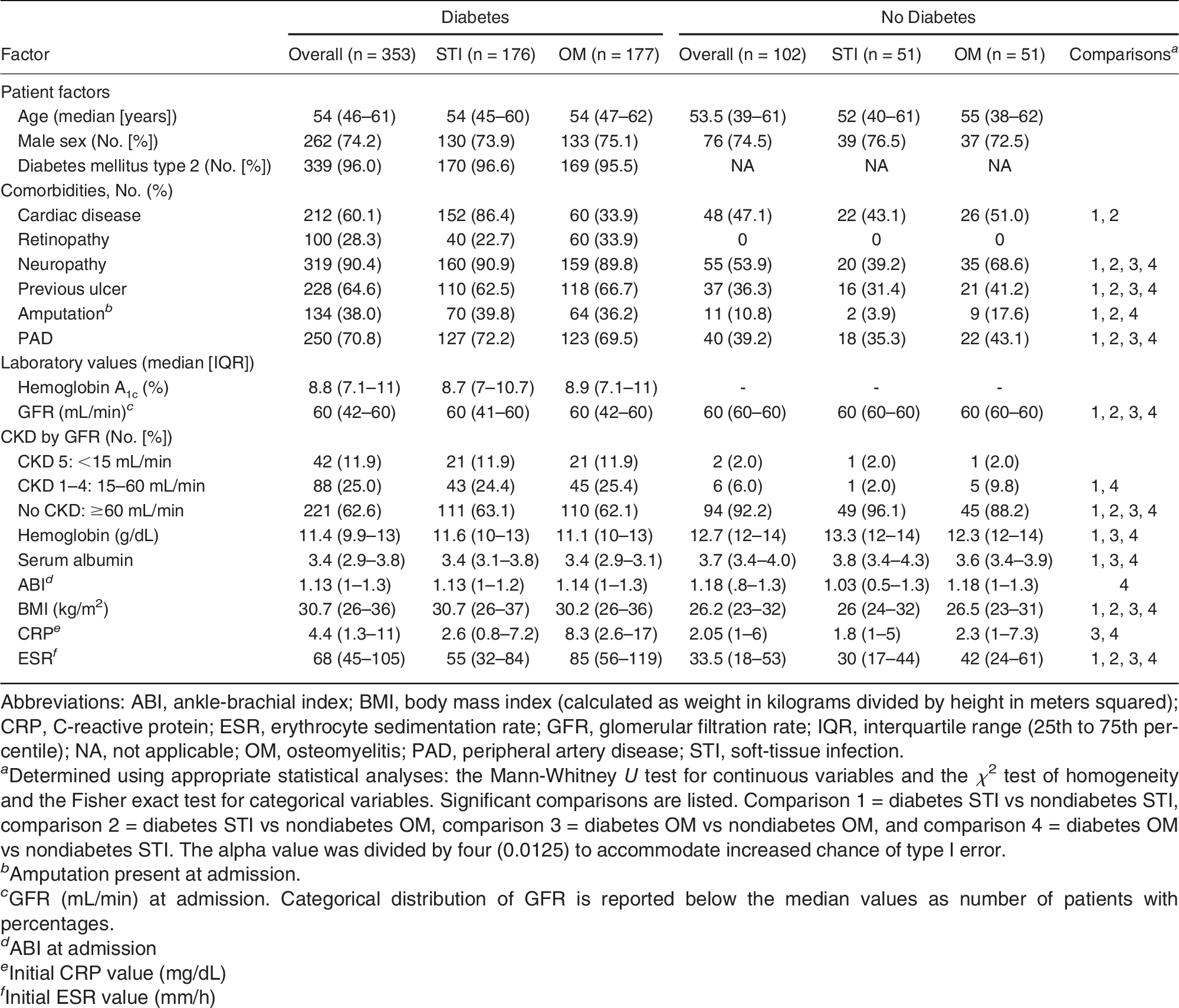
Table 1.
Patient Characteristics and Laboratory Values Grouped by Presence or Absence of Diabetes and Presence of STI or OM
Results
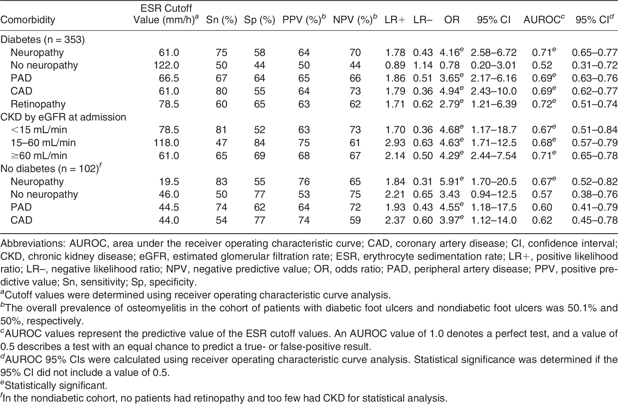
The median age of patients was 54 years (interquartile range, 46–61 years) (Table 1). Seventy-five percent of patients were male. No differences in age or sex were observed between patients with and without diabetes. However, patients with diabetes had a significantly greater body mass index, history of foot ulceration, ankle-brachial index, white blood cell count, ESR, CRP level, and presence of peripheral artery disease (PAD), neuropathy, and retinopathy. Furthermore, patients with diabetes had lower glomerular filtration rates, serum albumin levels, and hemoglobin levels. Next, we compared the mean differences of ESR and CRP. The mean difference was the difference between the averages. We subtracted the ESR and CRP averages of the diabetes OM group from those of the diabetes STI group, which resulted in differences of 10.0 mm/h and 2.6 mg/dL respectively. However, persons with diabetes with STIs or OM had greater mean differences in ESR and CRP of 24.8 mm/h and 6.8 mg/dL, respectively. In addition, ESR and CRP tended to predict OM versus STI with greater sensitivity and specificity in patients with diabetes than in those without diabetes. However, when patients were stratified by presence or absence of neuropathy, ESR remained predictive of OM in diabetic patients with neuropathy (odds ratio [OR], 4.16; 95% confidence interval [CI], 2.58–6.72), whereas ESR was not significantly predictive in diabetic patients without neuropathy (OR, 0.78; 95% CI, 0.20–3.01) (Table 2). This relationship was not observed with CRP values, as CRP remained predictive regardless of neuropathic status. When patients without diabetes were stratified by presence or absence of neuropathy, a similar change was seen, with ESR becoming more predictive of OM in neuropathic patients (OR, 5.91; 95% CI, 1.70–20.5) and not significantly predictive in nonneuropathic patients (OR, 3.43; 95% CI, 0.94–12.5). None of the patient factors demonstrated statistically significant interaction. As such, it is unlikely that any factors modified the effect of comorbidities on ESR and CRP.
Table 2.
Predictive Value of ESR for Osteomyelitis with Presence or Absence of Comorbidities in Patients with or Without Diabetes
Discussion
ESR and CRP are inflammatory biomarkers that are inexpensive and readily available in clinical practice. The cutoff values used to define OM are much higher in people with diabetes than are those used to predict OM at other anatomical locations [10,11,12,13,20,21,22,23]. This is the first study to evaluate the differences in ESR and CRP and evaluate the role of diabetes, neuropathy, PAD, retinopathy, and chronic kidney disease. In contrast, these comorbidities are often not documented in other studies of STIs and bone infections at other anatomical sites and from other mechanisms of infection.
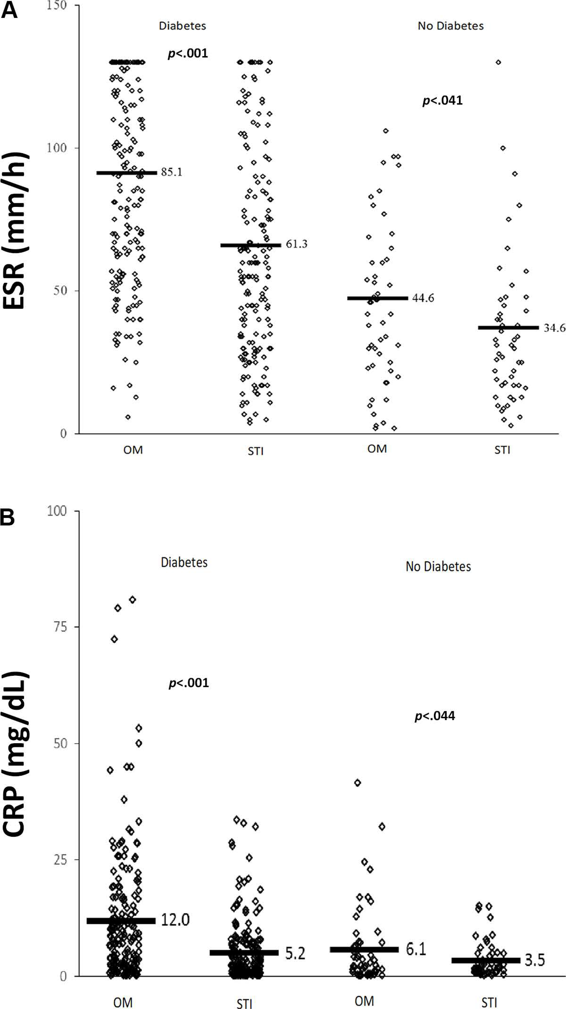
Diabetic foot infections were associated with significantly higher inflammatory biomarker levels than were nondiabetic foot infections. In patients with foot infections, both those with and without diabetes had significantly higher ESRs with OM (Fig. 1A); however, CRP was significantly different only in patients with diabetes (Fig. 1B). To determine which factors were responsible for the apparent effect in diabetic patients, the common diabetes-associated comorbidities of PAD, retinopathy, and neuropathy were independently investigated. We found that in patients without neuropathy, ESR could not predict OM in patients with and without diabetes. However, in diabetic patients without neuropathy, CRP remained a significant predictor of OM, with sensitivity of 56% and specificity of 88%.
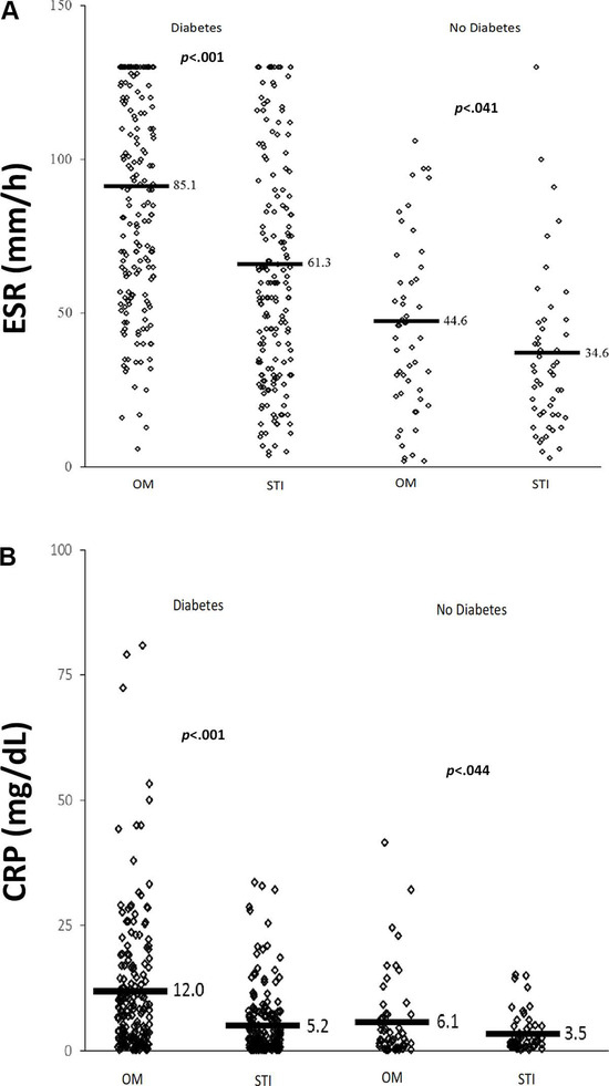
Figure 1.
Scatterplots of erythrocyte sedimentation rate (ESR) (A) and C-reactive protein (CRP) (B) values in diabetic and nondiabetic patients with soft-tissue infection (STI) or osteomyelitis (OM).
Elevations in CRP and ESR correlate with several clinical conditions (ie, diabetes, neuropathy, vascular complication, obesity, and inflammatory arthritis) and have been used as markers of morbidity and mortality [24,25,26,27,28]. C-reactive protein has been used as a diagnostic biomarker for sensory neuropathy in people without diabetes [29]. The patient profile for many of the patients who are admitted to the hospital with foot infections include many of the comorbidities associated with an elevation in ESR and CRP level. For example, in this study, patients with diabetes had a higher body mass index and a higher prevalence of sensory neuropathy, PAD, retinopathy, and chronic kidney disease (Table 1). Baseline CRP and ESR values may be elevated before STI or bone infection develops.
Although peripheral sensory neuropathy significantly altered the predictive value of ESR for OM of the foot, the presence or absence of neuropathy did not sufficiently explain the difference in ESR and CRP between patients with and without diabetes. As a result, further study is needed to explain the difference in inflammatory biomarkers in patients with diabetes. Furthermore, the presence of multiple comorbidities may affect ESR and CRP, but we did not observe any interaction effects using linear modeling between neuropathy and the other comorbidities based on diabetes status. Therefore, the effect of neuropathy on ESR was not modified by other patient comorbidities.
We acknowledge the limitations of this study. This is a retrospective study, and, therefore, some of the disease processes can have variable operational definitions and inconsistent testing and documentation. One of the strengths of the present data set is that it was uniformly obtained by faculty and fellows working on the University of Texas Southwestern Medical Center Diabetic Limb Salvage Service (Dallas, TX, USA), who follow an algorithmic approach to perform and document clinical examination in patients with foot infections. Examination for sensory neuropathy is one of the standard aspects of this evaluation, and this is rarely reported by advanced practice providers, primary care physicians, and surgeons not specialized in diabetic foot disease. Another limitation of this study is that we did not evaluate the severity of peripheral sensory neuropathy; however, the present finding should stimulate further study.
This study has several advantages compared with other studies that evaluate biomarkers to predict OM. This is a large study compared with previous reports in diabetic foot infections [9]. We compared ESR and CRP in patients with and without diabetes and evaluated the effect of common comorbidities. We used bone biopsy as the reference standard to define OM. Most other studies use a combination of clinical examination, radiographic changes, and probe to bone, MRI, or SPECT. We used imaging that has low rates of false-negatives and low negative likelihood ratios to identify patients who did not have OM when bone cultures were not indicated or available [4]. In addition, we evaluated both ESR and CRP. Most studies that evaluate foot infections use only ESR [9].
In conclusion, these data support assessing diabetes status and peripheral sensory neuropathy in patients with infected foot ulcers as an important initial step when evaluating OM. Further study is needed to elucidate the association between neuropathy and inflammatory biomarkers and to evaluate the potential impact of neuropathy on the utility of ESR and CRP in the diagnostic algorithm of foot OM.
Financial Disclosure:
Dr. La Fontaine received research funding from Cardinal Health, Avazzia, KCI, EO2 Concepts, and Osiris Therapeutics. Dr. Crisologo received research funding from the American Diabetes Association, Cardinal Health, EO2 Concepts, Osiris Therapeutics, Avazzia, and Pluristem Therapeutics. Dr. Malone received research funding from Medline Industries. Dr. Oz received research funding from the American Diabetes Association, Osiris Therapeutics, MedImmune, and Pluristem Therapeutics. Dr. Lavery received research funding from Cardinal Health, KCI, EO2 Concepts, Osiris Therapeutics, Avazzia, and Pluristem Therapeutics.
Conflicts of Interest:
Dr. Wukich is in a consulting and advisory role for Orthofix and Arthrex. Dr. Lavery has consulting agreements with EO2 Concepts, Cardinal Health, Bayer, Medline Industries, Boehringer Ingelheim, and MedImmune.
References
- Lam K, van Asten SA, Nguyen T, : Diagnostic accuracy of probe to bone to detect osteomyelitis in the diabetic foot: a systematic review. Clin Infect Dis 63: 944, 2016.
- Aragon-Sanchez J, Lipsky BA, Lazaro-Martinez JL: Diagnosing diabetic foot osteomyelitis: is the combination of probe-to-bone test and plain radiography sufficient for high-risk inpatients? Diabet Med 28: 191, 2011.
- Alvaro-Afonso FJ, Lazaro-Martinez JL, Garcia-Morales E, : Cortical disruption is the most reliable and accurate plain radiographic sign in the diagnosis of diabetic foot osteomyelitis. Diabet Med 36: 258, 2019.
- Butalia S, Palda VA, Sargeant RJ, : Does this patient with diabetes have osteomyelitis of the lower extremity? JAMA 299: 806, 2008.
- La Fontaine J, Bhavan K, Lam K, : Comparison between Tc-99m WBC SPECT/CT and MRI for the diagnosis of biopsy-proven diabetic foot osteomyelitis. Wounds 28: 271, 2016.
- Lipsky BA, Senneville E, Abbas ZG, : Guidelines on the diagnosis and treatment of foot infection in persons with diabetes (IWGDF 2019 update). Diabetes Metab Res Rev 36(suppl 1): e3280, 2020.
- Senneville E, Lipsky BA, Abbas ZG, : Diagnosis of infection in the foot in diabetes: a systematic review. Diabetes Metab Res Rev 36(suppl 1): e3281, 2020.
- Sybenga AB, Jupiter DC, Speights VO, : Diagnosing osteomyelitis: a histology guide for pathologists. J Foot Ankle Surg 59: 75, 2020.
- Victoria van Asten SA, Geradus Peters EJ, Xi Y, : The role of biomarkers to diagnose diabetic foot osteomyelitis: a meta-analysis. Curr Diabetes Rev 12: 396, 2016.
- Jiang N, Ma YF, Jiang Y, : Clinical characteristics and treatment of extremity chronic osteomyelitis in Southern China: a retrospective analysis of 394 consecutive patients. Medicine 94: e1874, 2015.
- Khan MH, Smith PN, Rao N, : Serum C-reactive protein levels correlate with clinical response in patients treated with antibiotics for wound infections after spinal surgery. Spine J 6: 311, 2006.
- Radtke K, Tetzlaff T, Vaske B, : Arthroplasty-center related retrospective analysis of risk factors for periprosthetic joint infection after primary and after revision total hip arthroplasty. Technol Health Care 24: 721, 2016.
- Yoon SH, Chung SK, Kim KJ, : Pyogenic vertebral osteomyelitis: identification of microorganism and laboratory markers used to predict clinical outcome. Eur Spine J 19: 575, 2010.
- Ryan EC, Ahn J, Wukich DK, : Diagnostic utility of erythrocyte sedimentation rate and C-reactive protein in osteomyelitis of the foot in persons without diabetes. J Foot Ankle Surg 58: 484, 2019.
- Bristow I: Foot ulceration in a non-diabetic population: a cross-sectional audit of staff in one health district. J Wound Care 17: 445, 2008.
- Haji Zaine N, Hitos K, Vicaretti M, : Characteristics of non-diabetic foot ulcers in Western Sydney, Australia. J Foot Ankle Res 9: 6, 2016.
- Lavery LA, Walker SC, Harkless LB, : Infected puncture wounds in diabetic and nondiabetic adults. Diabetes Care 18: 1588, 1995.
- Wunderlich RP, Armstrong DG, Husain K, : Defining loss of protective sensation in the diabetic foot. Adv Wound Care 11: 123, 1998.
- Lavery LA, Armstrong DG, Vela SA, : Practical criteria for screening patients at high risk for diabetic foot ulceration. Arch Intern Med 158: 157, 1998.
- Ghanem E, Antoci V Jr, Pulido L, : The use of receiver operating characteristics analysis in determining erythrocyte sedimentation rate and C-reactive protein levels in diagnosing periprosthetic infection prior to revision total hip arthroplasty. Int J Infect Dis 13: e444, 2009.
- Greidanus NV, Masri BA, Garbuz DS, : Use of erythrocyte sedimentation rate and C-reactive protein level to diagnose infection before revision total knee arthroplasty: a prospective evaluation. J Bone Joint Surg Am 89: 1409, 2007.
- Tsai CE, Lee FT, Chang MC, : Primary cervical osteomyelitis. J Chin Med Assoc 76: 640, 2013.
- Wang C, Ao Y, Fan X, : C-reactive protein and erythrocyte sedimentation rate changes after arthroscopic anterior cruciate ligament reconstruction: guideline to diagnose and monitor postoperative infection. Arthroscopy 30: 1110, 2014.
- Aulich J, Cho YH, Januszewski AS, : Associations between circulating inflammatory markers, diabetes type and complications in youth. Pediatr Diabetes 20: 1118, 2019.
- Chuengsamarn S, Rattanamongkolgul S, Sittithumcharee G, : Association of serum high-sensitivity C-reactive protein with metabolic control and diabetic chronic vascular complications in patients with type 2 diabetes. Diabetes Metab Syndr 11: 103, 2017.
- Aryan Z, Ghajar A, Faghihi-Kashani S, : Baseline high-sensitivity C-reactive protein predicts macrovascular and microvascular complications of type 2 diabetes: a population-based study. Ann Nutr Metab 72: 287, 2018.
- George MD, Giles JT, Katz PP, : Impact of obesity and adiposity on inflammatory markers in patients with rheumatoid arthritis. Arthritis Care Res (Hoboken) 69: 1789, 2017.
- Sudhakar M, Silambanan S, Chandran AS, : C-reactive protein (CRP) and leptin receptor in obesity: binding of monomeric CRP to leptin receptor. Front Immunol 9: 1167, 2018.
- Rashad NM, El-Shabrawy RM, Sabry HM, : Interleukin-6 and hs-CRP as early diagnostic biomarkers for obesity-related peripheral polyneuropathy in non-diabetic patients. Egypt J Immunol 25: 153, 2018.
© 2025 American Podiatric Medical Association