


Abstract
Background: The aim of this study is to compare clinical and radiologic outcomes of self-adhesive taping (SAT) or a short- leg cast (SLC) groups with base of fifth metatarsi. Methods: Functional outcome was assessed by the Visual-Analogue-Scale Foot and Ankle (VAS-FA) at the Emergency and at 2, 4, 6, and 12 weeks. Labour loss, bone union and The American Orthopedic Foot and Ankle Score (AOFAS) at 12 weeks were also assessed. Results: There was no difference between the SAT group and SLC group in VAS-FA scores at time of injury, 6 and 12 weeks. The SAT group had a significantly higher mean VAS-FA score at the second and fourth weeks of follow-up compared with the SLC group (P = .001 and P = .039, respectively). No correlation was observed between the fracture gap and functional scores for both groups. There was no difference in AOFAS between two groups at 12 weeks. Twenty one patients were unable to work for a mean of 38.2 days during the treatment. 10 patients with the SAT missed 37.5 days and eleven patients with the SLC g missed 40.2 (p: 0.41). The bone union was also achieved for all patients within 12 weeks. Conclusion: Treatment with SAT in these fractures had satisfactory functional results compared with traditional SLC. Although there were no significant differences in labor loss and use of assistive devices, The VAS-FA score was significantly higher in SAT group than the SLC group at the second and fourth weeks of treatment.
Fracture of the base of the fifth metatarsal (zone 1) is one of the most common injuries [1] in adult ankle trauma, and it has been classified into three zones by Lawrence and Botte [2]. Zone 1 is the most proximal zone and the most prevalent aspect. The zone 1 fracture is a typical avulsion fractures seen after the pull of peroneus brevis muscle tendon where the tendon insert to this region [3,4].
There are many conservative treatment protocols for zone 1 fractures, which are clinically successful as much as 99% of the time [5]. Short-leg casting (SLC) and a walking boot are conservative treatment methods that aim to protect weightbearing. Also, methods such as an elastic bandage allow partial weightbearing with crutches [6]. In addition to these treatment methods, we applied self-adhesive taping (SAT) for treatment of fracture of zone 1, which allowed weightbearing.
In this study we aimed to compare the clinical results, including loss of labor and using assistive devices, functional results, and radiologic results, including bone union, between patients with fracture of zone 1 who treated with SLC and those with SAT. We hypothesized that SAT methods have similar clinical, functional and radiologic results when comparing SLC methods in management of patients with fracture of zone 1.
Materials and Methods
After obtaining institutional review board of Umraniye Training and Research Hospital approval, we prospectively treated 105 patients who had a zone 1 fracture between February 2016 and October 2017. All of the patients signed an informed consent form before participating in the study for expected complications and results of the treatment procedure. The patients were selected as two groups in a nonrandomized manner. Patients older than 18 years with an acute zone 1 fracture were considered eligible. Patients who had a zone 2 or 3 fracture, concomitant ligament injuries (diagnosed by magnetic resonance imaging), diabetes mellitus, osteoporotic bone (T score less than –2.5), a history of osteoporotic drug therapy, previous ipsilateral foot surgery or fracture, chronic fracture, and open fracture were excluded from this study.
Both types of treatment were applied in the emergency department by the same physician (S.G.B.)F. A double-layered SAT was applied to patients in the SAT group. Patients wear their footwear with the SAT. The SAT group was advised to use the SAT continuously for the first week and were allowed to remove it for 1 hour a day after the first week. The bandage was removed at the third week. A standard plaster below-the-knee cast was applied for the SLC group [7].
Both groups were provided crutches to walk with until they had no pain. Patients in the SAT group were encouraged to bear weight in the shortest time they could. Although patients in the SLC group were advised not to bear weight until the cast was removed, we allowed them to bear weight as soon as tolerated. Both groups received standard oral analgesics, including acetaminophen 500 mg twice a day. In the SLC group, patients were assessed for the development of lower-extremity deep venous thromboembolism and managed with 100 mg of acetylsalicylic acid once a day as prophylaxis until the cast was removed.
Follow-up Management
The anteroposterior, lateral, and oblique views of both feet were performed using a standard radiographic device for initial evaluation of patients in the emergency department. Magnetic resonance imaging was used for excluding the simultaneous ligament injuries in suspected patients. All of the patients were followed up 2, 4, 6, and 12 weeks after the injury for clinical and radiologic evaluation. Radiographs were taken until union was diagnosed radiographically. The Visual Analogue Scale Foot and Ankle (VAS-FA) score was used to evaluate the clinical outcomes [8]. The score was obtained at the initial consult and 2, 4, 6, and 12 weeks after treatment for each group.
Bone union was determined according to radiologic and clinical evaluation. Clinical evaluation determined union of the fracture as the time when the patient reported no pain over the fracture site or only mild activity-related pain. Radiologic evaluation was based on the presence of bridging callus at three of four cortices on anteroposterior and lateral views.
Secondary outcome measures included labor loss, use of assistive devices, bone healing, complications, and American Orthopaedic Foot & Ankle Society (AOFAS) score at 12-week follow-up.
Statistical Analysis
The Student t test was used to compare quantitative, normally distributed variables of descriptive statistics (mean, standard deviation, range), whereas the Mann-Whitney U test was used to compare nonnormally distributed variables. The relationships between amount of fracture displacement and VAS-FA scores were investigated using the Pearson correlation. A P < .05 was considered statistically significant for all of the measures. Statistical analysis was performed using IBM SPSS Statistics for Windows, Version 22.0 (IBM Corp, Armonk, New York).
Results
One hundred five patients (58 in the SAT group, 47 in the SLC group) were evaluated. During follow-up, ten patients (six in the SAT group, four in the SLC group) were excluded because of loss of follow-up. Ninety-five patients (52 in the SAT group, 43 in the SLC group) were included the study. There were no statistically significant differences between the groups in terms of age and sex. The demographic data of all patients are presented in Table 1.

Table 1.
Baseline Demographic Data for the Study Groups

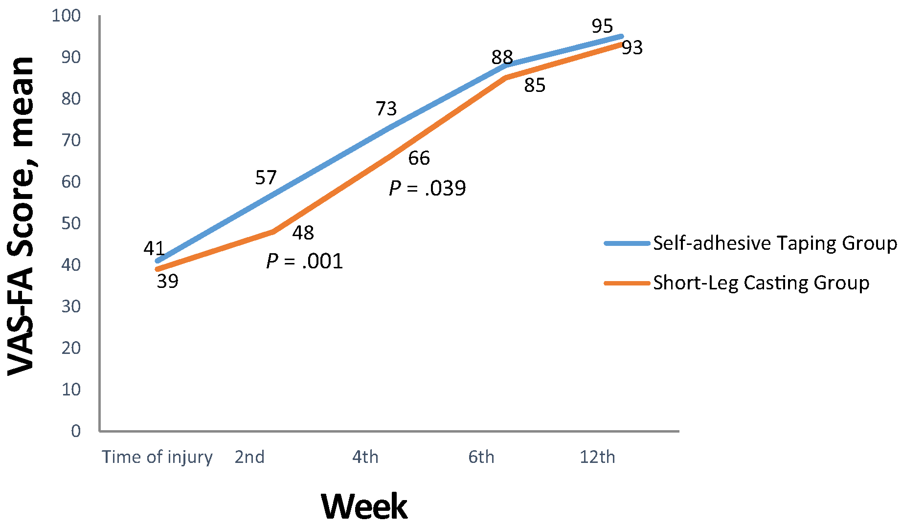
The mean (range) VAS-FA scores were 41 (24–72) and 95 (85–100) in the SAT group and 39 (15–55) and 93 (85–100) in the SLC group at the time of injury and at the 12th-week follow-up visit, respectively (Fig. 1). The SAT group had a significantly higher mean VAS-FA score at the second and fourth weeks of follow-up compared with the SLC group (P = .001 and P = .039, respectively).
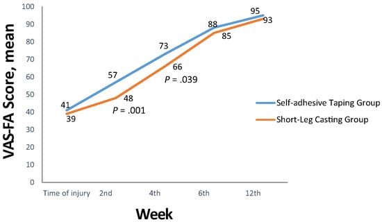
Figure 1.
Comparison of Visual Analogue Scale Foot and Ankle (VAS-FA) scores.
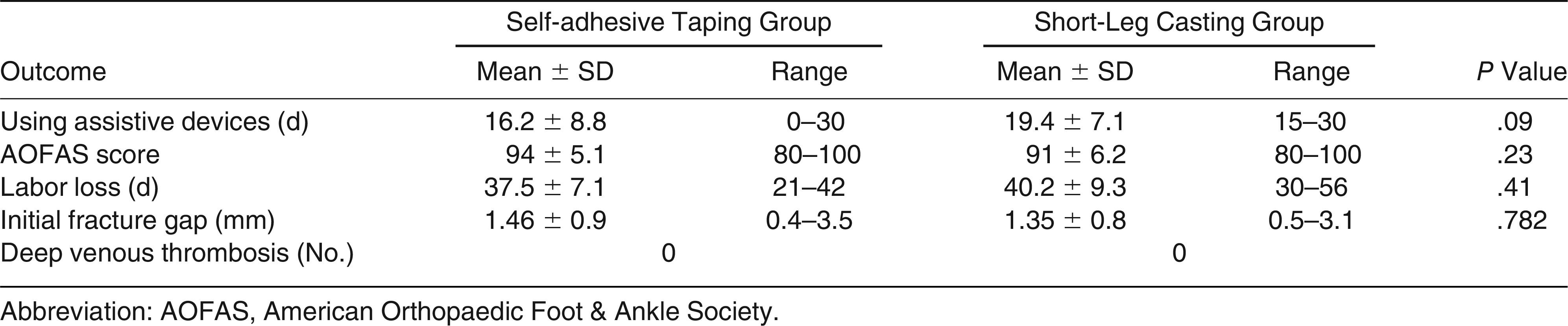
Twenty-one patients were unable to work a mean of 38.2 days (range, 21–56 days) during follow-up. Ten patients in the SAT group missed a mean of 37.5 days of work and 11 patients in the SLC group missed a mean of 40.2 days (P = .41). There was no difference between the two groups in the amount of time taken to walk independently without crutches from the time of injury (Table 2).
Table 2.
Results of Clinical Outcomes
All of the patients in both groups showed complete union at 12 weeks. There was no significant difference in bone healing between the two groups (Fig. 2).
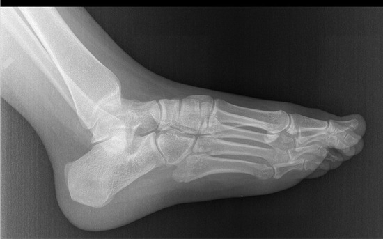
Figure 2.
Radiographic fracture healing observed in a 12th-week control.
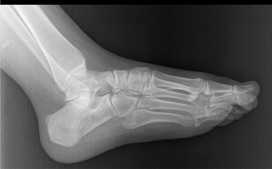
The mean (range) AOFAS score at 12 weeks was 94 (80–100) in patients with SAT and 91 (80–100) in patients with SLC, with no significant differences between the groups (P = .23).
The mean (range) initial amount of fracture displacement was 1.46 mm (0.4–3.5mm) in patients with SAT and 1.35 mm (0.5–3.1 mm) in patients with SLC. Eleven patients in the SAT group had more than 2 mm of fracture gap at the time of injury compared with ten patients in the SLC group. There was no deep venous thrombosis in either group.
Discussion
Zone 1 fracture of the fifth metatarsal is usually treated with conservative methods. Quill [9] recommended that weightbearing be protected in a cast for 3 to 6 weeks. However, Wiener et al [10] recommended less rigid immobilization, which resulted in better pain and functional outcomes in their prospective study. In the present study, we suggest that zone 1 fractures can be treated adequately with SAT. There were no differences in pain, function, and radiologic union between the SAT and SLC except functional results in the second and fourth weeks.
There were two comparative studies about pain and functional outcomes for use of the validated foot-specific outcome measure (VAS-FA score) for fifth metatarsal bone fracture. One of these studies compared the Aircast walking boot (Aircast Inc, Summit, New Jersey) and SLC. In that study, the walking boot had better VAS-FA scores at the third-, sixth-, and ninth-week visits after injury [11]. Akimau et al [12] compared an elastic bandage and SLC using VAS-FA scoring and found no significant differences between the two methods. In the present study, we also did not observe any significant difference between the SAT and SLC groups. At the second- and fourth-week visits, the SAT group had significantly higher VAS-FA scores than the SLC group. Gray et al [6] evaluated their patients with a modified AOFOS midfoot clinical rating system. They found that 93 patients had better outcomes for pain and function after 2 weeks with a plaster slipper than with an elasticated tubular bandage (Tubigrip; Mölnlycke Health Care, Peachtree Corners, Georgia), which is in contrast to the present result [6]. Zenios et al [13] compared an elastic bandage and SLC and found better AOFAS scores with the elastic bandage at 3-month follow-up. In the present study, AOFAS scores between the two groups had no statistically significant differences at 3-month follow-up [13].
Similar to the literature, we found that the zone 1 fracture is a cause of labor loss. Gosele et al [14] found in their series that 52 patients with zone 1 fractures treated with an orthopedic shoe had an average of 19 days of labor loss. Egol et al [3] reported on 52 similar patients treated with hard-soled shoe who lost an average of 22 days of work; 47% took up to 10 days off and 37% more than 10 days. Shahid et al [11] found 31.5 missed workdays in a walking boot group and 39.2 days in an SLC group. In the present study, the mean number of missed workdays for patients treated with SAT is 37.5 days and for the SLC group is 40.2 days. Although we found a much higher duration of labor loss compared with the cited studies, labor loss was seen in only 21 of 95 patients. Some of these patients have physical jobs, such as those requiring a long time standing. Although they walked without an assistive device, they were not able to work.
Zwitser et al [15] recommend that fractures with more than 2 mm of displacement or significant comminution at the metatarsocuboid joint should be treated operatively. The lack of consensus regarding the maximum size of the fracture gap that can be treated nonoperatively and the theoretical difficulty of the healing of comminuted fractures warrants further studies. In their study, Lee et al [16] compared operative and conservative treatment methods in patients with a fracture gap of more than 2 mm. They found that there were no significant differences between the groups in VAS and AOFAS scores at the week 4 visit, but union time was significantly less in the conservative group. In the present study, we did not find any relation between the fracture gap and the treatment methods. The SAT and SLC groups were treated successfully, and no differences between the two groups were observed.
It is reported in the literature that prolonged immobilization can cause deep venous thrombosis in patients [17]. In the present study, no patient had deep venous thrombosis during treatment. This result might be due to the fact that we did not use Doppler ultrasonography or the relatively small number of patients included in this study.
The main limitation of this study was the relatively short patient follow-up. The VAS-FA is a validated score but might not be representative of the overall outcome of the patients. We did not evaluate time to bone union. Cost-effectiveness was not compared in this study. The length of fifth metatarsi was not assessed for these study results. Last, we did not evaluate the duration of analgesic drug use.
Conclusions
There are many treatment options for fifth metatarsal zone 1 fractures. Treatment with SAT in these fractures had satisfactory functional results compared with traditional SLC. Although there were no significant differences in labor loss and use of assistive devices, The VAS-FA score was significantly higher in the SAT group than the SLC group at the second and fourth weeks of treatment.
Financial Disclosure
None reported.
Conflict of Interest
None reported.
References
- Kane JM, Sandrowski K, Saffel H, : The epidemiology of fifth metatarsal fracture. Foot Ankle Spec 8: 354, 2015.
- Lawrence SJ, Botte MJ: Jones’ fractures and related fractures of the proximal fifth metatarsal. Foot Ankle 14: 358, 1993.
- Egol K, Walsh M, Rosenblatt K, : Avulsion fractures of the fifth metatarsal base: a prospective outcome study. Foot Ankle Int 28: 581, 2007.
- Morris PM, Francois AG, Marcus RE, : The effect of peroneus brevis tendon anatomy on the stability of fractures at the fifth metatarsal base. Foot Ankle Int 36: 579, 2015.
- Zwitser EW, Breederveld RS: Fractures of the fifth metatarsal; diagnosis and treatment. Injury 41: 555, 2010.
- Gray AC, Rooney BP, Ingram R: A prospective comparison of two treatment options for tuberosity fractures of the proximal fifth metatarsal. Foot (Edinb) 18: 156, 2008.
- Sarmiento A: A functional below-the-knee cast for tibial fractures. J Bone Joint Surg Am 49: 855, 1967.
- Richter M, Zech S, Gearing J, : A new foot and ankle outcome score: questionnaire based, subjective, visual analogue scale, validated and computerised. Foot Ankle Surg 12: 191, 1996.
- Quill GE Jr: Fractures of the proximal fifth metatarsal. Orthop Clin North Am 26: 353, 1995.
- Wiener BD, Linder JF, Giattini JF: Treatment of fractures of the fifth metatarsal: a prospective study. Foot Ankle Int 18: 267, 1997.
- Shahid MK, Punwar S, Boulind C, : Aircast walking boot and below-knee walking cast for avulsion fractures of the base of the fifth metatarsal: a comparative cohort study. Foot Ankle Int 34: 75, 2013.
- Akimau PI, Cawthron KL, Dakin WM, : Symptomatic treatment or cast immobilisation for avulsion fractures of the base of the fifth metatarsal: a prospective, randomised, single-blinded non-inferiority controlled trial. Bone Joint J 98-B: 806, 2016.
- Zenios M, Kim WY, Sampath J, : Functional treatment of acute metatarsal fractures: a prospective randomised comparison of management in a cast versus elasticated support bandage. Injury 36: 832, 2005.
- Gosele A, Schulenburg J, Oschsner PE: Early functional treatment of a fifth metatarsal fracture using an orthopaedic boot. Swiss Surg 3: 81, 1997.
- Zwitser EW, Breederveld RS: Fractures of the fifth metatarsal; diagnosis and treatment. Injury 41: 555, 2010.
- Lee TH, Lee JH, Chay SW, : Comparison of clinical and radiologic outcomes between non-operative and operative treatment in 5th metatarsal base fractures (Zone 1). Injury 47: 1789, 2016.
- Lapidus LJ, Rosfors S, Ponzer S, : Prolonged thromboprophylaxis with dalteparin after surgical treatment of Achilles tendon rupture: a randomized, placebo-controlled study. J Orthop Trauma 21: 52, 2007.
© 2022 American Podiatric Medical Association