


Abstract
There are many theoretical models that attempt to accurately and consistently link kinematic and kinetic information to musculoskeletal pain and deformity of the foot. Biomechanical theory of the foot lacks a consensual model: clinicians are enticed to draw from numerous paradigms, each having different levels of supportive evidence and contrasting methods of evaluation, in order to engage in clinical deduction and treatment planning. Contriving to find a link between form and function lies at the heart of most of these competing theories and the physical nature of the discipline has prompted an engineering approach. Physics is of great importance in biology and helps us to model the forces that the foot has to deal with in order for it to work effectively. However, the tissues of the body have complex processes that are in place to protect them and they are variable between individuals. Research is uncovering why these differences exist and how these processes are governed. The emerging explanations for adaptability of foot structure and musculoskeletal homeostasis offer new insights into how clinical variation in outcomes and treatment effects might arise. These biological processes underlie how variation in the performance and use of common traits, even within apparently similar subgroups, make anatomical distinction less meaningful and are likely to undermine the justification of a “foot type.” Furthermore, mechanobiology introduces a probabilistic element to morphology based on genetic and epigenetic factors.
In recent years, the principles of physics have usefully enhanced the modeling of foot behavior. In reality, this is an integration of several different fields, including applied mathematics, anatomy, and physiology. Within this biomechanical model, the morphology of the foot has been used persistently to correlate symptoms or predict specific behavioral patterns within numerous manifestations of “foot typing.” Such models (even if underpinned with some engineering concepts) represent a heuristic strategy to find a solution to a complex problem; however; morphologic categorization is an artificial construct and (unless acknowledged) concomitantly fails to capture the natural spread of overlapping variation between groups and within them. If a foot typing model implies, in a post hoc fashion, that configuration of anatomy is nominally in control of limb activity, the proposition is that from such a mechanistic viewpoint physical laws and computerized analysis of data can be clinically correlated and used to understand the basis for what we regard as foot dysfunction. This approach to diagnosis and treatment planning needs to be scrutinized within the context of a valid biological explanation. Unlike physical laws, biological processes are variable, and this is significant when clinical reasoning and treatment planning incorporate kinetic and kinematic data or include assumptions about the relationship between foot shape and function.
The laws of physics that underpin biomechanics cannot be violated; they act as a first principle. As they apply to constituent parts of the foot; however, their biological impact is variable within component tissues. Consequently, the assumption that physical laws can always be predictably applied to the foot (or foot type) as a whole is flawed. Physiologic and pathologic responses are likely to demonstrate variability—and possibly be contradictory—as biological responses to forces emerge over time. Therefore, to understand its behavior, the laws of physics must be applied to the constituent parts of the foot and ankle complex. The behavior and proficiency of the foot to deliver function or undergo adaptive or pathologic change will emerge over time as a consequence of these interactions, which involve concepts in biological adaptability that this article will explore.
The biological responses of tissues and their susceptibility to variance—influenced by genetic and nongenetic factors—need to be better understood by advocates of foot typing systems (and by researchers when designing cohort studies of foot function). In particular, the influence of aging demands special attention, as biological adaptability diminishes over time, potentially reducing the sensitivity of older patients to adapt to physical and orthotic intervention for biomechanical dysfunction.
Form-Function Complex
The initial use of the term “form-function complex” as we understand it was offered by Walter Bock and Gerd von Wahlert [1] as an alternative to contrasting between the two terms. This shift recognized the concept that anatomical form was intrinsically linked to the overall biological purpose (or purposes) that it served and that both are observable and measurable. However, despite its influence, this work predates detailed explanation of biological adaptability of a part or feature through interaction with the environment.
Although similarity of form might serve as a useful principle for categorization, the form-function complexes it helps constitute must also be catalogued in terms of associated behavior when compared to contrasting categories; however, the statistical spread of responses across any population will produce overlap across any arbitrarily defined boundary of classification. A historical example is the simplistic association of a low-arch foot to subtalar joint pronation and high-arch feet to supination, which remains a mainstay of popular description. Although some supportive evidence for significant functional differences might exist for many [2], some high-arch feet demonstrate significant subtalar joint pronation kinematics, and so the initial hypothetical contention that there is a direct causative association between arch height and subtalar pronation is easily disproved and this comparative form-function model is scientifically undermined. Many other modeling systems for the foot’s form-function complexes have emerged and become fixed through commercial exploitation or maintained by homophilous bias, but clinicians still lack a consistent model that accommodates physical and functional variance and is supported by empirical evidence.
In isolation, the term “function” is more problematic, as it implies performance, the multiple attributes of a phenotype or those things which it does. Functional attributes convey a suitability to deliver a beneficial task and often present an opportunity for a measurable trait (eg, contractility of cardiac muscle [function] creates blood pressure [measurable trait]); thus, the form-function complex of heart muscle cells is well adapted to deliver a beneficial trait. We also know that blood vessels remodel themselves in response to altered blood pressure and shear stress [3,4] and this is phenotypic adaptability of another form-function complex to an underlying change in tissue stress.
The foot also exhibits normal characteristic functional traits that are the result of interactions between its component parts and other form-function complexes of the lower limb. As with all form-function complexes, it is not unreasonable to seek a correlation between the two elements and understand the possible cause-and-effect relationship for any differences between the two, when this is pathoetiologic and how treatment can be delivered and scaled to be effective.
The embryologic and juvenile foot structure emerges during development to achieve early stability during stance. As it does so, the growth and development of children is enhanced by their interactions with the environment to experiment with and calibrate postural torque reactions within the lower limb to offset balance perturbations [5]. The general trend to reproduce muscle righting reactions that gradually approach those of adults is an example of broad canalization (a drift toward robust biological similarity) with a tolerance for variability, as individuals with a given morphology fine-tune the fit between form and behavior of the body to achieve bipedal stance and movement.
Mechanobiology
Mechanobiology is an interdisciplinary study that is concerned with the cells’ biological responses to mechanical loads and the mechanotransduction mechanisms by which these loads are transduced into a cascade of cellular and molecular events [6]. During embryonic development, each new generation of cells becomes gradually more specialized than its parent cell; thus, there is a gradual transition from stem cells to specific differentiated specialist cells (eg, osteocytes within bone). This differentiation of tissues is governed by the selective regulatory expression of genes with the result that specialized cells are consequently limited in their repertoire of functions. To influence this, stem cells have been shown to specialize according to the level of local elasticity of the tissue matrix that they are within [7]. Once specialized, each group of differentiated cells has a form and function available to it that is defined by the range of genes still available for expression. For each cell, the integration of physical and biochemical factors within the cellular environment can control gene transcription, alter ion flow across cell membranes, and transmit force through cell adhesions [8]. In fact, many fundamental aspects of cell behavior are mechanosensitive, including adhesion, spreading, migration, gene expression, and cell-cell interactions [9-15]. It has been known for more than a century that bone tissue adapts to stress by altering its endochondral and appositional growth. This is encapsulated within the “Heuter-Volkmann law” that proposes growth retardation or acceleration depending on whether local mechanical forces exceed or fall below a physiologic threshold. Longitudinal bone growth is governed by the rate of proliferation and hyperplasia in the proliferative zone that is modulated through sustained or cyclic loading [16]. The concept of bone remodelling throughout life to adapt to the physical environment is known as “Wolff’s law.” This law actually comprises a number of different processes that have been proposed to be best considered separately [17]:
- The mechanical problems that confront bones (axial compression, bending, twisting and shear), and the stress and strain characteristics.
- The sensing of loading through mechanotransduction whereby an external mechanical stimulus is translated by fluid and ionic flow and into a cellular signal to potentially evoke a response.
- Induced osteogenic responses that may be quiescent, bone forming (osteoblastic), resorptive (osteoclastic), or remodelling (osteoblastic and osteoclastic). Responses are relative to the size, duration, and direction of the stimulus in relation to a threshold and decrease with aging.
- Cellular coordination of bone cells through mechanotransduction feedback; both genes and environment including health and nutrition combine over a long period to influence bone morphology.
The mechanotransduction processes in bone demonstrate the complexity of genetic and environmental factors interacting to produce variable phenotypes over long periods, although an investigation into the mechanoregulation of tissue differentiation in the healing of osteotomies encountered a greater likelihood of encountering cartilage rather than woven bone in the presence of higher shear strain, indicating shorter term relevance [18].
As demonstrated by Mueller and Maluf [19], tissue stress responses are intrinsic to all musculoskeletal tissues as a homeostatic mechanism that is sensitive to the environment and facilitates adaptability to physical forces. Treatment can therefore be directed to normalize forces or guide activity level to ameliorate their effect. So as a set of adult foot form-function complexes emerge, inherent adaptability of the developing lower limb responds to environmental forces to control, exploit, or protect it as function emerges. The shape of bones is influenced by the mechanical forces they experience during development [20], and this process is exemplified in the juvenile adaptive change in talar articular surfaces as a response to increasing body weight [21]. In a similar manner, associated soft tissues (eg, tendon) have the capacity to adapt to their mechanical environment [22-25]. The heterogeneous nature of such responses is highlighted by a craniomandibular study that concludes that the component of the adaptive change in mammalian joint proportions and biomineralization represents a compensatory mechanism to cartilage degradation that maintains the overall functional integrity of such composite tissue systems [26]. Therefore, the structure of the anatomical form is modified and adapted (within canalized limits) in response to remodeling forces to retain and preserve function that minimizes the effect of difference. Furthermore, such remodelling of form or adaptive processes must also be subject to regulation to preserve the function for which it has been retained over generations [27].
The adaptability of tissues to physical stress including the existence of thresholds to alter physiologic responses also supports no one ideal foot posture for individuals, as tissues themselves adapt to the physical stressors (with magnitude, duration, and direction of force) imposed on them [19]. Such insights promote the use of a tissue stress approach to treatment [28]. and, although a pragmatic approach as to how this is achieved with therapies such as foot orthoses has remained unclear for the majority of practitioners [29], useful engineering concepts have been applied to foot biomechanics and pathology [30]. Although rigorous theory is applied in this and similar important studies, there is an unintended proposition that predictable or consistent responses will follow. Thereby, the more we focus on methodologic details, the less we notice the variable homeostatic connection to cell and tissue biology, the response of which may be analyzed statistically but may not be predicted precisely because of difference in adaptability and random variation in cell mechanobiology [31]. It is conceivable that this phenomenon, other than the potential for technical failure to offset deforming forces, offers some explanation for the unpredictable response of the first metatarsophalangeal joint to interventions directed at prevention of juvenile hallux valgus [32,33].
Adaptive Plasticity and Epigenetics
During growth, the foot emerges as an integral part of the lower limb during development and retains common though variable features because of varied genetic regulation in the timing and extent of local development [34,35]. The progressive modification of the foot’s quantitative (polygenic) structure is an example of how phenotypes are commonly modified by environmental forces. The spectrum of morphology produced by any species genotype across different environments is referred to as a “norm of reaction,” [36]. and the extent to which the phenotype is modified by the environment is described as “adaptive plasticity.” [37]. As an outcome of developing form-function complexes, variations in common traits will inevitably arise. Simply put, not all feet are the same before being exposed to altering forces from the environment, including direct force transmission and moments resulting from internal and external forces. Genetic influence can thereafter act to disrupt the most advantageously developed foot by delivering variation in plasticity.
The adaptive plasticity of a character trait is most likely attributable to an epistasis (genetically interactive) effect directed by remote DNA loci from those determining the mean of the characteristic (eg, proportions, alignment) in question [38]. In the event that such processes lend themselves significantly to the foot, the implications are profound to our understanding of anatomical change because levels of adaptive plasticity are heritable and function through mechanobiological processes that may contribute to the observed heritability of deformity [39].
The effect of governing the expression of DNA without affecting its underlying structure is referred to as epigenetics. The mechanism is governed by the variable chemical changes of the epigenome that surrounds DNA. Epigenetic profiles may represent the link between an environmental factor and phenotypic differences in monozygotic twins [40]. In addition, genes with plasticity effects are also implicitly susceptible to environmental change effects through histone modification and DNA methylation. In most cases, histone acetylation enhances transcription and histone deacetylation represses transcription, whereas methylation patterns that are heritable after mitosis may affect gene transcription through several different mechanisms. This suggests that nongenetic (eg, environmental) factors also contribute to the variable expression of tissue responses [41].
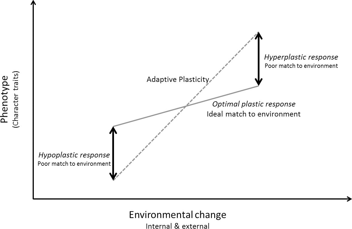
The anatomy of similarly matched individuals may have different sensitivities to adaptation because of dissimilar epistatic and epigenetic profiles and, as environmental change is imposed on the body’s form, the course of mechanobiological homeostasis may result in an optimum, hyperplastic or hypoplastic response; that is to say, whichever tendency individuals possess, it may or may not match the environmental conditions required of it (Fig. 1). Thus, there is natural variation in developmental form and variation in adaptive plasticity through mechanobiological functions. Consequently, it remains unlikely that variation in morphology will accurately or consistently predict any variation in functional traits on its own. It is the physical stressor acting on tissues that evokes a discrete homeostatic response and which, if outside of the parameters of that individual’s capacity for tolerance or adaptation, may induce pathologic change [42]. Significantly, the response may also alter the form-function complex and its (measurable) functional trait. Therefore, it follows that it is the potential stressor force, correlated with the functional trait, that should be prioritized for measurement for diagnosis or treatment planning rather than the morphology of the structure in isolation. The tendency to injury or responses to treatment will also vary despite similar morphology because of unique mechanobiological profiles, although similarities in patterns of injury are likely to emerge [43]. A useful example that serves to illustrate this point is that the normal morphologic deviation of the subtalar joint results in various degrees of deviation of the subtalar joint axis [44]. This offers an opportunity for measurement of a common functional trait (ie, supination resistance of the weightbearing subtalar joint), which is reliably useful in determining the orientation and magnitude of an orthotic reaction force required to offset high (potentially damaging) forces acting on tissues that normally act to resist and control pronation forces [45-47].
Figure 1.
Change in anatomical structure/function complexes (phenotype) may not always match the optimum for a given environment.
It is the combined effects of the developmental alignment of the foot (guided by underlying inherited DNA instructions), influenced by an individual’s capacity for adaptive plasticity and the epigenetic interaction with the environment (through mechanobiological processes) that govern tissue response to the functions required of it. Therefore, it could be contended that an individual with both a medially deviated talus and higher levels of adaptive plasticity would be statistically more likely to demonstrate ever-increasing pronation kinematics over time than an individual with low adaptive plasticity. This would be secondary to mechanobiological processes that continued to deliver a functional trait that would be recognized morphologically as an emerging planovalgus foot. The emerging question for future study is whether this etiology or clinical presentation results from abnormal mechanotransduction [48].
For the older patient, there is a reduction in effective epigenetic processes that is associated with aging and this is considered to be attributable to an accumulation of molecular errors, and has also been linked to osteoporosis and rheumatoid arthritis by means of aberrant gene expression [49]. This serves as a reminder that tissue responses may not always prove beneficial. When the environmental stimulus is novel (eg, altered magnitude, duration, and direction of force) or the evoked response impaired, the outcome may be disruptive and undermine character traits and, by exceeding physiologic thresholds, provoke pathologic change. As to the implications of adaptive plasticity on foot behavior; there are several components requiring consideration regarding the likelihood of foot symptoms emerging for any individual, as follows:
- A common set of anatomical form-function complexes that constitute the foot.
- Normal population and individual genetic variance of form-function complexes.
- Recognizable character performance traits integrated with proximal lower limb function.
- Adaptive plasticity acting through mechanobiological processes that are both epistatic and epigenetic in nature.
- A capacity to retain performance traits without disrupting tissue health.
- Diminishing epigenetic capacity through aging.
Precepts for Foot Typing
It would seem that defining structural and functional foot types to classify the form-function complex is a scientifically worthless taxonomic exercise unless each defined category is explained through its emergence during growth as internal and external forces act on its heterogeneous elements and any impact of differences in function or trait performance can be clearly demonstrated and clarified [50]. Essentially, the predisposition of one defined foot type to develop a specific symptom, when compared to another, remains at the heart of the form-function debate along with the justification of treatment rationales—often orthotic prescription.
Because of the constraining effects of regulated genetic expression in specialized tissues of a broadly common foot structure and the relative consistency in the demands of function, recognizable patterns in form-function complexes are likely to emerge that lend themselves to the taxonomic labeling of reaction norms (ie, foot typing). The fundamental proposition for any model is that genetically driven structure and epigenetic adaptive plasticity work together to produce these form-function complexes of the foot: Resulting shared functional traits emerge, but with differences that might explain predisposition to injury or pathology and be measurable.
Over the past decade, the explanation for the emergence of tissue alteration resulting from physical stress has received much attention from biomedical researchers in an effort to elucidate the biological interactions at different hierarchical levels (ie, at increasing levels of complexity) [51-53]. This knowledge might be used to clarify our understanding of what differences in tissue responses can be associated with a morphologic labeling system and underpin a more scientific basis of foot typing. The effect of aging also serves as a reminder that morphology emerges over time as a response to forces acting on the foot’s component parts. Therefore, legitimate foot typing systems represent only one level (reaction norms) of a hierarchical reductionist explanation for foot problems that must be empirically supported by evidence of tissue responses when delivering common traits. Previous research, notable for its rigorous protocol in differentiating foot postures, has identified a significant difference in muscle activity in people with flat-arched feet and also demonstrated a response to orthotic intervention [54,55]. Further work in this area may also correlate functional traits (eg, supination resistance), note symptoms, and record treatment outcomes, but results would still fail to directly correlate cause and effect because of heterogeneous tissue responses. For foot and ankle biomechanics, capturing supportive evidence for contending paradigms of cause-and-effect relationships is essential to validate treatment pathways. Recent advances in the radiologic imaging of musculoskeletal structures to measure alteration in tissue stiffness seem more likely to offer a robust evaluation of stress-related changes to tendons and fascial tissue [56]. and magnetic resonance elastography for morphologic evaluation of muscle [57], whereas ultrashort echo time sequences represent an applicable magnetic resonance imaging tool that allows quantitative and qualitative assessment of bone and cartilage [58]. Future studies of foot and ankle biomechanics might consider how such radiologic techniques could be used to uncover specific correlations between tissue stress responses and reliably measurable functional traits. In addition, the associated histologic changes driven by cell signaling might help to clarify specific mechanobiological responses [59]. Research directed in this way might represent a valuable contribution to (or remove the need for) a debate that, although durable, seems otherwise irreconcilable. Until such evidence is forthcoming, exemplars of foot typing need to acknowledge that measurable traits and tissue responses must substantiate their contentions. Moreover, all defined foot types represent hypothetical reaction norms, whose predicted form-function traits will overlap with those of other categorized groups, and any extrapolated claims to anticipate tissue responses will be disrupted by variable adaptive plasticity.
Probabilistic Emergence of Foot Pathology
Variability and adaptive plasticity are biological additions to natural selection by which all organisms achieve adaptation to the environment [60]. Clinicians and patients already make judgments and decisions about treatment in the face of uncertainty [61], and the use of such nonstandard terms is a further challenge. However, the inclusion of such concepts is essential to secure an explanatory link for an association between morphology and traits (form-function) and tissue stress reactions (including symptoms). Paradoxically, they also offer the opportunity to develop a more biologically robust approach to foot typing study proposals that themselves may need to be more adaptive in design over longer periods [62].
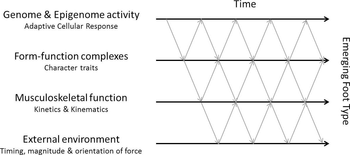
The time lag between morphologic adaptations of a reaction norm driven by adaptive plasticity by means of mechanobiological processes can be expressed in terms of a model that sets a template against which future studies might be considered (Fig. 2). This bidirectional model encapsulates a probabilistic epigenetic element [63]. in that each interaction is not necessarily predictable because of variation in stochastic molecular events, levels of variable plasticity, and changeable functional requirements. What is reflected is the hierarchical nature of the relationship between shared character traits and tissue responses that might form the basis of meaningful claims of functional foot types.
Figure 2.
Epigenetic interaction will alter morphology over time, although age-related decreasing adaptive plasticity should be a factor when deciding interventions.
Conclusions
The reciprocal relationship between musculoskeletal tissues of the foot and the forces resulting from gait are better understood within the framework that includes a mechanobiological explanation of tissue responses. Morphologic (reaction norm) explanations for the causation of lower limb deformity and injury are credible, as studies of shape can characterize developmental and genetic effects and discover their relative importance [64], but this will only be validated through a progressively detailed biological explanation that links tissue responses to any functional trait that a patient exhibits and that makes deformity, dysfunction, and injury more or less likely. Furthermore, differences in the execution of common character traits may be attributable to regulatory genetic factors or variation in adaptive plasticity. This adds to the multifactorial complexity in the modelling of the foot and will require consideration in identifying genetic risk factors that make musculoskeletal injury more likely [65].
Financial Disclosure
None reported
Conflicts of Interest
None reported
References
- BockWJWahlertGV:Adaptation and form-function complex. Evolution330: 269, 1965.
- HillstromHJSongJKraszewskiAP: Foot type biomechanics part 1: structure and function of the asymptomatic foot. Gait Posture37: 445, 2012.
- OwensGK:Role of mechanical strain in regulation of differentiation of vascular smooth muscle cells. Circ Res79: 1054, 1996.
- WilliamsB:Mechanical influences on vascular smooth muscle cell function. J Hypertens16: 1921, 1998.
- RoncesvallesMNWoollacottMHJensenJL:Development of lower extremity kinetics for balance control in infants and young children. J Mot Behav33: 180, 2001.
- WangJHCThampattyBP:An introductory review of cell mechanobiology. Biomech Model Mechanobiol5: 1, 2006.
- EnglerAJSenSSweeneyHL: Matrix elasticity directs stem cell lineage specification. Cell126: 677, 2006.
- LimCTBershadskyASheetzMP:Mechanobiology. J R Soc Interface7(suppl 3): S291, 2010.
- GhibaudoMSaezATrichetL: Traction forces and rigidity sensing regulate cell functions. Soft Matter4: 1836, 2008.
- WenJHVincentLGFuhrmannA: Interplay of matrix stiffness and protein tethering in stem cell differentiation. Nat Mater13: 979, 2014.
- DiMillaPABarbeeKLauffenburgerDA:Mathematical model for the effects of adhesion and mechanics on cell migration speed. Biophys J60: 15, 1991.
- HuttenlocherAGinsbergMHHorwitzAF:Modulation of cell migration by integrin-mediated cytoskeletal linkages and ligand-binding affinity. J Cell Biol134: 1551, 1996.
- WangNTytellJDIngberDE:Mechanotransduction at a distance: mechanically coupling the extracellular matrix with the nucleus. Nat Rev Mol Cell Biol10: 75, 2009.
- Reinhart-KingCADemboMHammerDA:Cell-cell mechanical communication through compliant substrates. Biophys J95: 6044, 2008.
- ZhangQYZhangYYXieJ: Stiff substrates enhance cultured neuronal network activity. Sci Rep4: 6215, 2014.
- VillemureIStokesIAF:Growth plate mechanics and mechanobiology: a survey of present understanding. J Biomech42: 1793, 2009.
- PearsonOMLiebermanDE:The aging of Wolff’s “law”: ontogeny and responses to mechanical loading in cortical bone. Am J Phys Anthropol39(suppl): 63, 2004.
- MorganEFSalisbury PalomaresKTGleasonRE: Correlations between local strains and tissue phenotypes in an experimental model of skeletal healing. J Biomech43: 2418, 2010.
- MuellerMJMalufKS:Tissue adaptation to physical stress: a proposed “physical stress theory” to guide physical therapist practice, education, and research. Phys Ther82: 383, 2002.
- YoungRLBadyaevAV:Evolution of ontogeny: linking epigenetic remodeling and genetic adaptation in skeletal structures. Integr Com Biol47: 234, 2007.
- HellierCAJeffreyN:Morphological plasticity in the juvenile talus. J Foot Ankle Surg12: 139, 2006.
- BuchananCIMarshRL:Effects of long-term exercise on the biomechanical properties of the Achilles tendon of guinea fowl. J Appl Physiol (1985)90: 164, 2001.
- BohmSMersmannFArampatzisA:Human tendon adaptation in response to mechanical loading: a systematic review and meta-analysis of exercise intervention studies on healthy adults. Sports Med Open1: 7, 2015.
- WiesingerHPKostersAMullerE: Effects of increased loading on in vivo tendon properties: a systematic review. Med Sci Sports Exerc47: 1885, 2015.
- ArchambaultJMHartDAHerzogW:Response of rabbit Achilles tendon to chronic repetitive loading. Connect Tissue Res42: 13, 2001.
- RavosaMJKunwarRStockSR: Pushing the limit: masticatory stress and adaptive plasticity in mammalian craniomandibular joints. J Exp Biol210: 628, 2007.
- LynchM:The evolution of genetic networks by non-adaptive processes. Nat Rev Genet8: 803, 207.
- McPoilHGHuntTG:Evaluation and management of foot and ankle disorders: present problems and future directions. J Orthop Sports Phys Ther21: 381, 1995.
- VicenzinoB:Foot orthotics in the treatment of lower limb conditions: a musculoskeletal physiotherapy perspective. Man Ther9: 185, 2004.
- FullerEA:Center of pressure and its theoretical relationship to foot pathology. JAPMA89: 278, 1999.
- WangJLüDMaoD: Mechanomics: an emerging field between biology and biomechanics. Protein Cell5: 518, 2014.
- GroisoJA:Juvenile hallux valgus. A conservative approach to treatment. J Bone Joint Surg Am74: 1367, 1992.
- KilmartinTEBarringtonRLWallaceWA:A controlled prospective trial of a foot orthosis for juvenile hallux valgus. J Bone Joint Surg Br76: 210, 1994.
- QuinnG:Normal genetic variation of the human foot: part 2: population variance, epigenetic mechanisms, and developmental constraint in function. JAPMA102: 149, 2012.
- QuinnG:Normal genetic variation of the human foot: part 1: the paradox of normal anatomical alignment in an evolutionary epigenetic model. JAPMA102: 64, 2012.
- SchmalhausenI:Factors of Evolution: The Theory of Stabilizing Selection, Blakiston, Philadelphia, 1949.
- GauseGF:Problems of evolution. Trans Conn Acad Sci37: 17, 1947.
- ScheinerSMLymanRF:The genetics of phenotypic plasticity. II. Response to selection. J Evol Biol4: 23, 1991.
- HannanMTMenzHBJordanJM: High heritability of hallux valgus and lesser toe deformities in adult men and women. Arthritis Care Res (Hoboken)65: 1515, 2013.
- FragaMFBallestarEPazMF: Epigenetic differences arise during the lifetime of monozygotic twins. Proc Natl Acad Sci U S A102: 10604, 2005.
- WangGYangEBrinkmeyer-LangfordCL: Additive, epistatic and environmental effects through the lens of expression variability QTL in a twin cohort. Genetics196: 413, 2014.
- ChapmanCRTuckettRPSongCW:Pain and stress in a systems perspective: reciprocal neural, endocrine, and immune interactions. J Pain9: 122, 2008.
- RiskowskiJLDufourABHagedornTJ: Associations of foot posture and function to lower extremity pain: results from a population-based foot study. Arthritis Care Res (Hoboken)65: 1804, 2013.
- KirbyKA:Subtalar joint axis location and rotational equilibrium theory of foot function. JAPMA91: 465, 2011.
- KirbyKAGreenDR:“Evaluation and nonoperative management of pes valgus,”inFoot and Ankle Disorders in Children, edited byDeValentine,Sp295, Churchill Livingstone, New York, 1992.
- NoakesHPayneC:The reliability of the manual supination resistance test. JAPMA93: 185, 2003.
- PayneCMunteaunuSMillerK:Position of the subtalar joint axis and resistance of the rearfoot to supination. JAPMA93: 131, 2003.
- IngberDE:Mechanobiology and diseases of mechanotransduction. Ann Med35: 564, 2003.
- HuidobroCFragaMF:A possible role for epigenetics in age-dependent bone diseases. Clin Rev Bone Miner Metab8: 95, 2010.
- NockCAVogtRJBeisnerBE:“Functional Traits”ineLS, John Wiley & Sons, Chichester, United Kingdom, 2016.
- TurnerCHPavalkoFM:Mechanotransduction and functional response of the skeleton to physical stress: the mechanisms and mechanics of bone adaptation. J Orthop Sci3: 346, 1998.
- ThompsonWRRubinCTRubinJ:Mechanical regulation of signalling pathways in bone. Gene503: 179, 2012.
- SunHBSchanielCLeongDJ: Biology and mechano-response of tendon cells: progress overview and perspectives. J Orthop Res33: 785, 2015.
- MurleyGSMenzHBLandorfKB:Foot posture influences the electromyographic activity of selected lower limb muscles during gait. J Foot Ankle Res26: 35, 2009.
- MurleyGSLandorfKBMenzHB:Do foot orthoses change lower limb muscle activity in flat-arched feet towards a pattern observed in normal-arched feet?Clin Biomech25: 728, 2010.
- DrakonakiEEAllenGMWilsonDJ:Ultrasound elastography for musculoskeletal applications. Br J Radiol85: 435, 2012.
- DebernardLRobertLCharleuxF: Characterization of muscle architecture in children and adults using magnetic resonance elastography and ultrasound techniques. J Biomech44: 397, 2011.
- ChangEYDuJChungCB:UTE imaging in the musculoskeletal system. J Magn Reson Imaging41: 870, 2015.
- SpencerGJHitchcockISGeneverPG:Emerging neuroskeletal signalling pathways: a review. FEBS Lett559: 6, 2004.
- NicolausMEdelaarP:Comparing the consequences of natural selection, adaptive phenotypic plasticity, and matching habitat choice for phenotype environment matching, population genetic structure, and reproductive isolation in meta-populations. Ecol Evol8: 3815, 2018.
- CahanAGilonDManorO: Probabilistic reasoning and clinical decision making: do doctors overestimate diagnostic probabilities?Q J Med96: 763, 2003.
- ChowSCChangM:Adaptive design methods in clinical trials: a review. Orphanet J Rare Dis3: 11, 2008.
- GottliebG:Probabilistic epigenesis. Dev Sci10: 1, 2007.
- KlingenbergCP:Evolution and development of shape: integrating quantitative approaches. Nat Rev Genet11: 623, 2010.
- CollinsMRaleighSM:Genetic risk factors for musculoskeletal soft tissue injuries. Med Sport Sci54: 136, 2009.
© 2021 American Podiatric Medical Association