


Abstract
Subungual schwannoma is quite rare and often causes nail deformity and difficulty in wearing shoes. Complete tumor excision is the treatment of choice, and we advocate that restoring the nail appearance should be considered at the same time. We present the case of 43-year-old man with a big toe subungual schwannoma. We designed a zigzag incision method to excise the tumor and also corrected nail-bed deformity. The patient had a smooth recovery, and the nail plate regrew with a good appearance.
Subungual schwannoma is a slowly developing nail tumor. It has grown large with an appearance of a nail protruding mass when found. Complete excision of the tumor is the curative treatment. The transungual approach is the most commonly used method. However, the restoration of nail appearance should also be considered, and good operative exposure is mandatory for complete excision, especially for a large tumor. We report a case of toenail subungual schwannoma that was excised by a zigzag nail-bed flap to correct nail-bed deformity with good prognosis.
Case Report
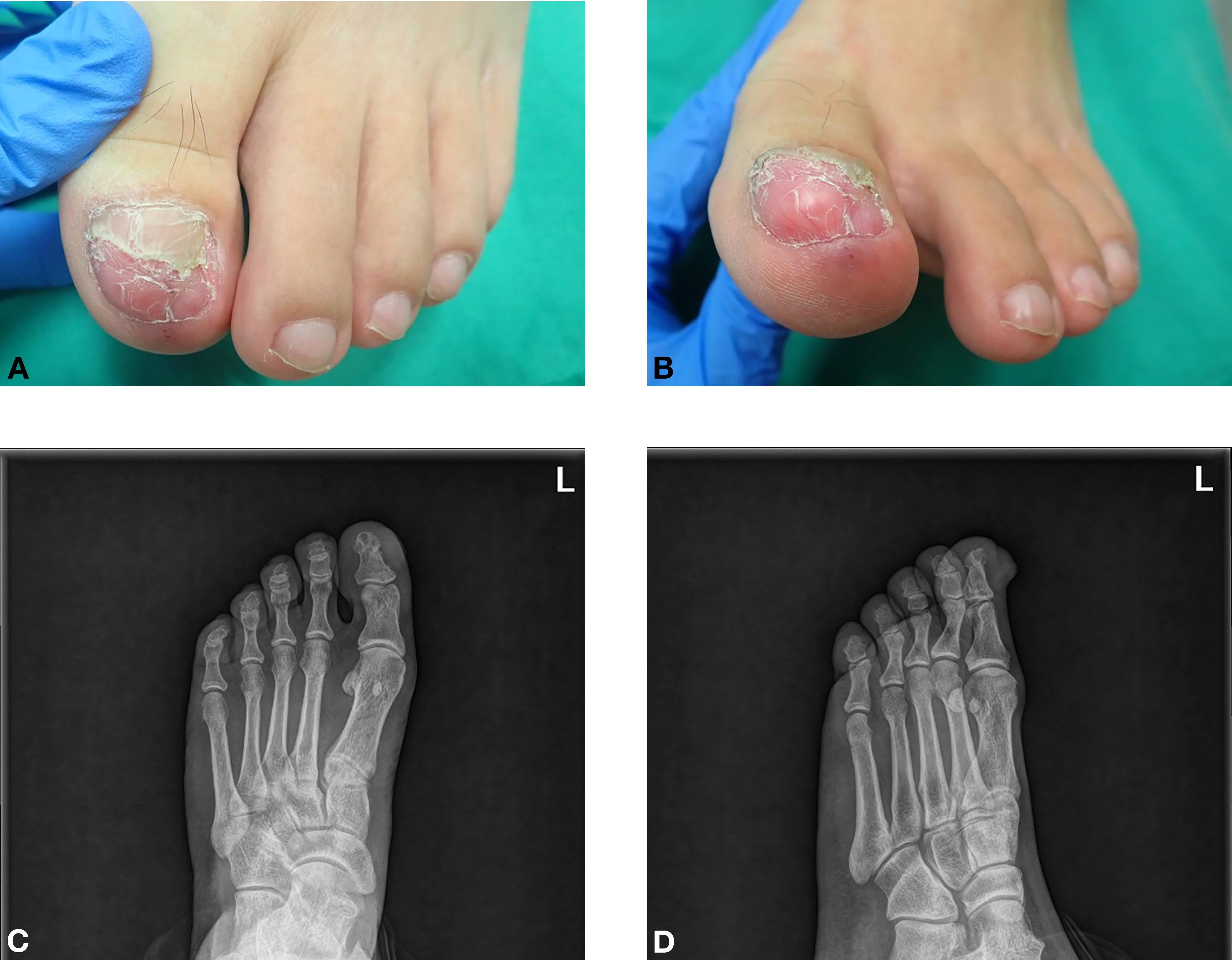
A 43-year-old man presented to the clinic with an indurated mass in the big toe nail of the left foot. The mass had been present for approximately 1.5 years. The mass appeared after the left big toe was crushed by a table leg 2 years prior to the latest presentation. The increase in size resulted in nail deformity and caused discomfort when wearing a shoe. However, the patient denied any pain or tenderness. Physical examination revealed a firm, focal telangiectasia and nonpulsatile mass protruding from the nail bed of the left big toe (Fig. 1 A and B). Plain radiography showed noncalcified soft-tissue thickening without phalangeal bone destruction (Fig. 1 C and D). Therefore, excisional biopsy was advised.
Figure 1.
A and B, A protruding mass causes nail deformity. C and D, Radiographs showing soft-tissue swelling without adjacent phalanx destruction.
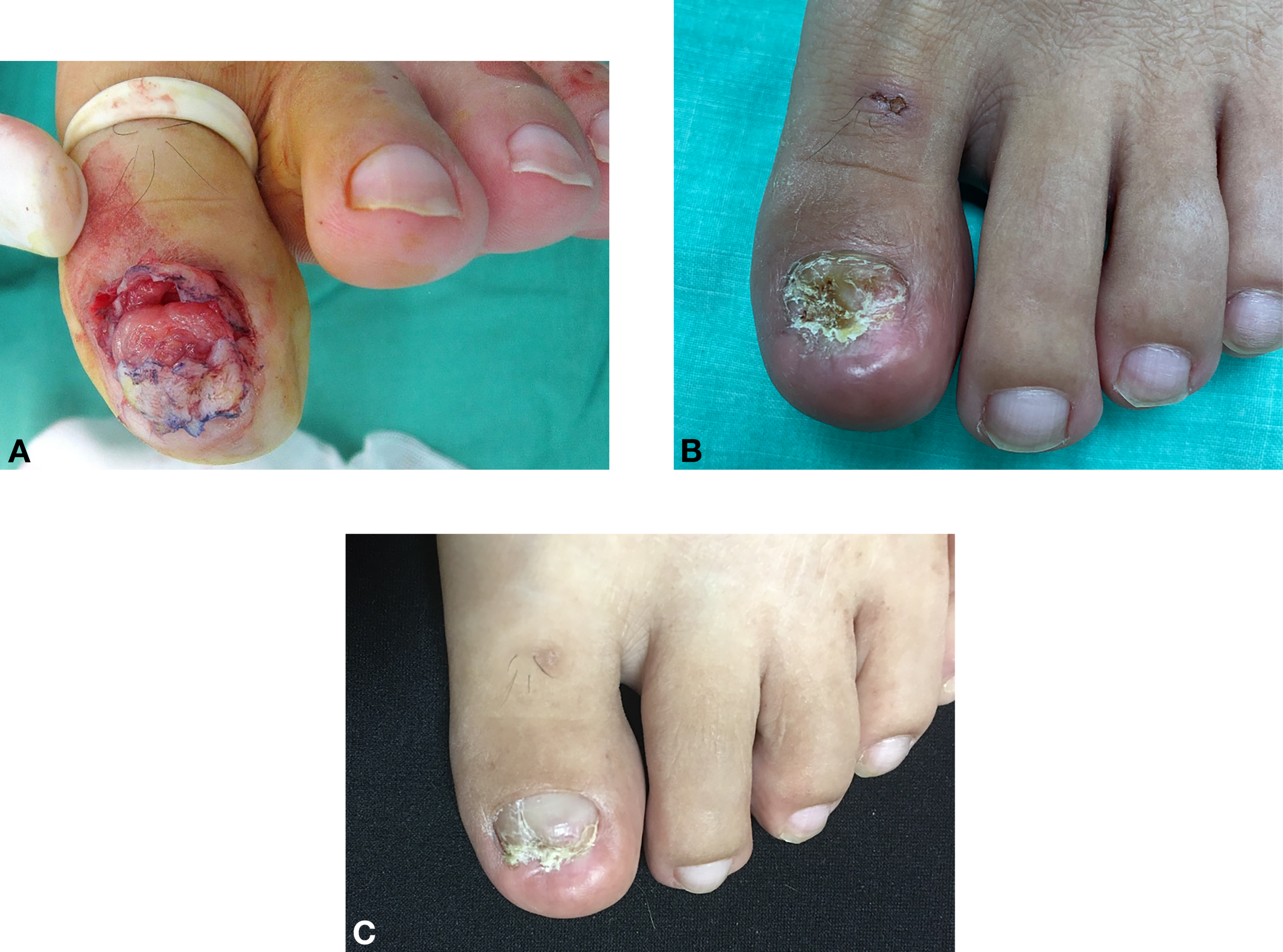
The patient was scheduled for surgery under general anesthesia. A horizontal zigzag incision was designed beneath the hyponychium (Fig. 2). The nail plate was removed, and the nail-bed flap was elevated in a distal-to-proximal direction by meticulously sharp dissection. While dissecting the tumor, the bilateral nail folds were carefully manipulated so as not to detach them from the adjacent skin to preserve the blood supply to the nail bed flap. There was a capsule between the undersurface of the nail bed and tumor (Fig. 3A), and the base of the tumor adhered to the distal phalangeal cortex. The tumor was well encapsulated and excised intact. There was no visible nerve fiber adjacent to the tumor intraoperatively. To lay the preserved nail-bed flap flat as much as possible, we performed deepithelialization on the lower part of the wound edge. The nail-bed flap was simple sutured with 5-0 nylon. A silicone drain was trimmed into a 2 × 2.5-cm sheet as a temporary nail-bed splint, and tie-over fixation for 5 days was prescribed for better fixation of the nail-bed flap. A dressing consisting of a hydrophilic polyurethane matrix was used to cover the nail bed after the tie-over and Penrose sheet were removed on postoperative day 5. The postoperative course was uneventful, and there was no nail-bed ischemia or phalangeal bone exposure. The stitches were removed on postoperative day 14. The patient started to walk with partial weightbearing 2 weeks postoperatively and full weightbearing 1 month postoperatively. The wound healed well and there was no discomfort or pain while wearing leather shoes or exercising (Fig. 3 B and C).
Figure 2.
A schematic diagram of the hyponychium zigzag incision. The gray zone indicates skin overlying the tumor, which was excised together with the tumor. The slant area is the area of deepithelialization.

Figure 3.
A, The tumor and zigzag nail-bed flap. B, Image obtained at follow-up at postoperative month 3. C, By the postoperative month 8 follow-up, the nail had regrown.
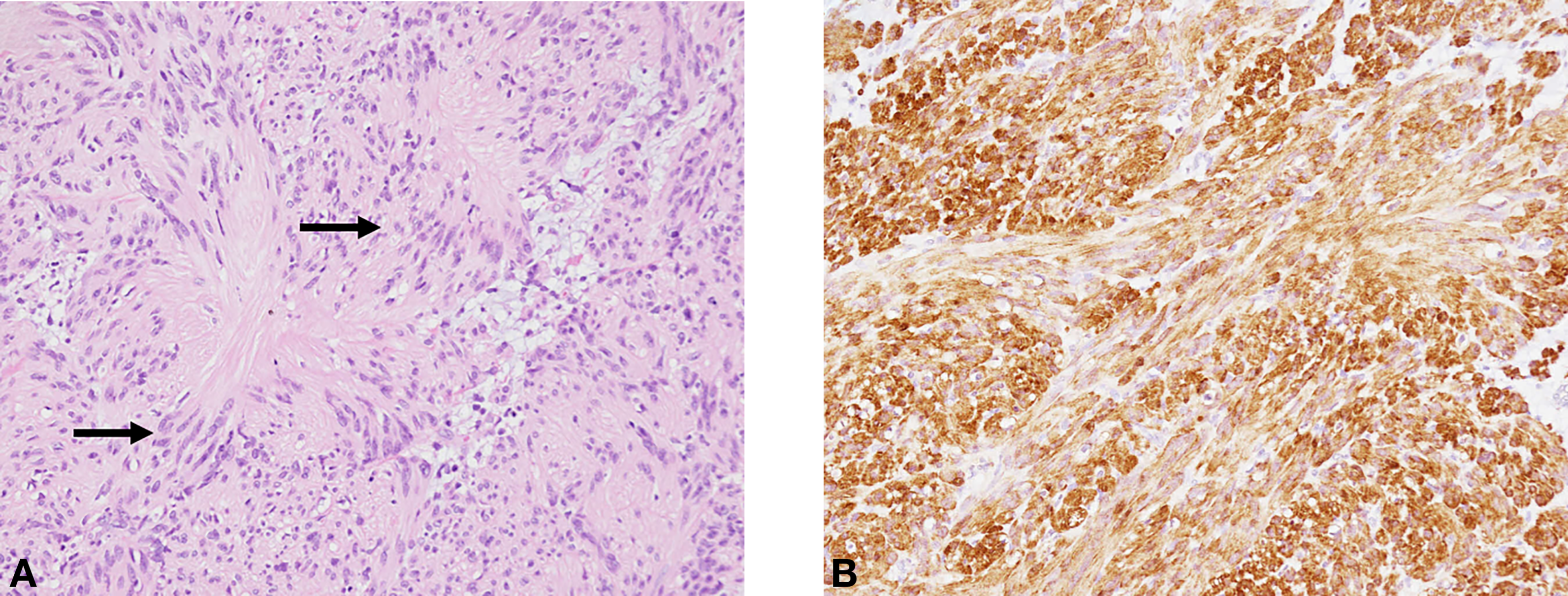
The specimen was measured up to 1.7 cm. The histopathologic examination revealed a typical pattern of schwannoma: a well-defined and biphasic tumor composed of compact areas of spindle cells (Antoni A tissue) alternating with loose foci (Antoni B tissue). The well-differentiated Schwann cells formed parallel rows (Verocay bodies). Cytologically, the tumor cells possess eosinophilic cytoplasm, indistinct cell borders, elongated nuclei, and low mitotic activity. Immunohistochemical staining supports the diagnosis with the evidence of diffused staining for S-100 protein and subcapsular staining for CD34 (Fig. 4).
Figure 4.
Verocay body (arrow) (H&E, ×200). A, Immunohistochemical staining; B, diffuse and strong positive S100 protein.
Discussion
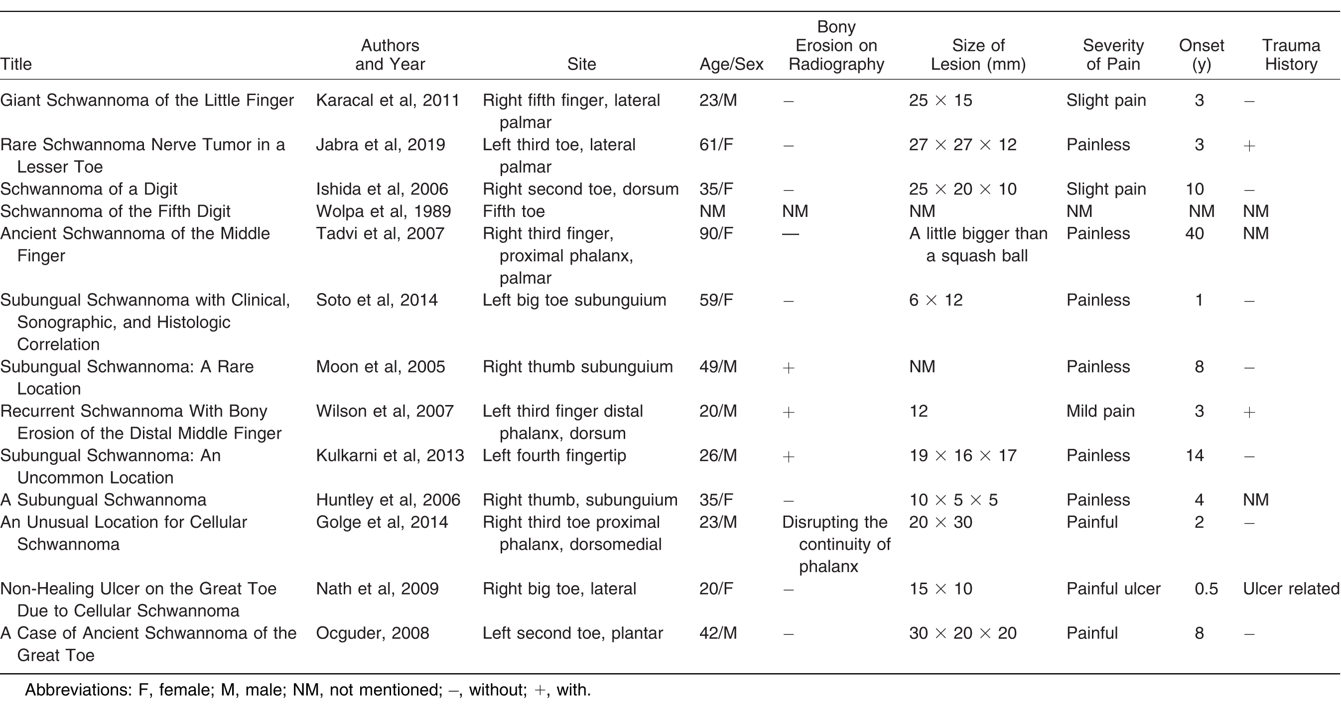
Schwannoma is a benign tumor of the peripheral nerves [1]. Schwannomas generally develop not only in the sensory portion of nerves but also in any peripheral nerves [2]. Schwannoma is often found in the trunk, retroperitoneum, lower extremity, head, and neck. Kransdorf [3] reported that approximately 9% of schwannomas are located in the foot and ankle. Digit involvement is uncommon (Table 1), and schwannomas of the digits are often present on the volar side, because of the larger nerves [4]. Therefore, studies on schwannomas in the finger subunguium are limited [5-7], and those on schwannomas in the toe subunguium are extremely rare [8]. Most schwannomas are idiopathic; however, some studies revealed that trauma or chronic ulcer might be a factor [9-14]. In this case, the big toe was crushed by a table leg and then a subungual mass gradually appeared. Most schwannomas grow in a waxing and waning pattern and mostly are nonpainful tumors [12]. However, digit schwannomas have different characteristics, and patients might feel pain because of inadequate local tissue distensibility and local pressure effects [5]. For this reason, painless lesions have grown larger when found.
Table 1.
Literature Review: Previous Reports of Digit Schwannoma
Subungual schwannoma often causes nail deformity and cosmetic disturbance. In this case, because of the large tumor size, the patient had discomfort in wearing shoes, and their quality of life had declined. Surgical excision was the treatment of choice. The method mostly used is the transungual approach. Because of the concern regarding postoperative nail split deformity, in addition to meticulous repair of the nail bed, Moon et al [5] recommend longitudinal incision in the nail bed and horizontal incision in the nail matrix. Although studies on the effect of the transungual incision on the nail appearance are few, we advocate that the nail bed and the matrix could be preserved intact and protected during surgery because the schwannoma is a well-encapsulated tumor. In this case, the tumor was very large and the nail bed became severely convex. Creating a good operative field is mandatory for excising the tumor completely. Based on our experience with subungual tumors, we found that the hyponychial incision provides an adequate visual field for exploration, especially for large tumors. By elevating the nail bed flap, the subungual tumor could be fully exposed and then completely excised. To maintain the integrity of the nail bed and adequate blood supply, we attempted to not fully detach the bilateral nail folds from the adjacent skin and to not destroy the blood supply from the proximal nail matrix. Although the hyponychial incision wound is larger than the traditional transungual incision and requires more operative time, the former provides space for complete tumor resection.
In this case, the nail bed was expanded and became convex by the large tumor beneath. Therefore, we designed a zigzag incision on the hyponychium [16] and performed deepithelialization on the distal skin wound edge. This method allowed us to lay the nail bed as flat as possible after the tumor was excised. A zigzag incision also creates a multiple–Z-like wound edge, which decreases scar contracture. Moreover, we could adjust the angle and length of the zigzag according to the shape of the tumor, and how much skin we intended to preserve. With regard to reducing the scar contracture and restoring the nail cosmesis, this might increase the operative difficulty but results in better outcomes.
The disadvantage of this study is that more cases and longer follow-up are required to evaluate the benefits of the hyponychial zigzag incision, because subungual tumors are not common and mostly are small. It is difficult to collect an adequate number of cases in a short period. Therefore, we are committed to include more candidates and long-term follow-up data.
To avoid subungual hematoma formation and nail-bed flap floating, the patient's own nail plate could be returned on the nail bed. However, in this case, the deformed and curly nail plate was unsuitable. Instead of the nail plate, we trimmed a Penrose drain into a sheet as a nail-bed protector and temporary spacer. Tie-over fixation was applied on the top of the Penrose sheet to apply a firm fixation. Adequate nail-bed covering could decrease pain and discomfort.17 We used a hydrophilic dressing to cover the nail bed after removal of the tie-over and Penrose sheet. This nonadhesive dressing could keep the nail bed's rough surface moistened and avoid discomfort during daily wound dressing changes.
Conclusions
Subungual schwannoma is extremely rare and will cause nail deformity. Regarding treatment, complete tumor excision is usually adequate. However, restoring nail cosmesis should also be a consideration. A zigzag nail-bed flap with deepithelialization resulted in good prognosis.
Financial Disclosure
None reported.
Conflicts of Interest
None reported.
References
- JacobsRLBarmadaR:Neurilemoma. A review of the literature with six case reports. Arch Surg102: 181, 1971.
- GrahamDILantosPL: Greenfield's Neuropathology, Arnold, London, 2002.
- KransdorfMJ:Benign soft-tissue tumors in a large referral population: distribution of specific diagnoses by age, sex, and location. AJR Am J Roentgenol164: 395, 1995.
- KaracalNSozenEAgdoganO: Giant schwannoma of the little finger. Dermatol Surg37: 1499, 2011.
- MoonSEChoYJKwonOS:Subungual schwannoma: a rare location. Dermatol Surg31: 592, 2005.
- KulkarniJMoholkarAPatilA:Subungual schwannoma: an uncommon location. J Hand Surg Am38: 1258, 2013.
- HuntleyJSDavieRMHooperG:A subungual schwannoma. Plast Reconstr Surg117: 712, 2006.
- SotoRAldunceMJWortsmanX: Subungual schwannoma with clinical, sonographic, and histologic correlation. JAPMA104: 302, 2014.
- HaniaMMannionC:Sporting injuries: an unusual case of a traumatic schwannoma presenting on the upper lip of a professional footballer. A case report. J Med Cases8: 243, 2017.
- KennedyWPBrodyRMLiVolsiVA: Trauma-induced schwannoma of the recurrent laryngeal nerve after thyroidectomy. Laryngoscope126: 1408, 2016.
- WilkinsonJMMcClellandMRBattersbyRD:Spinal cord schwannoma after vertebral trauma: a causal relation?J Neurol Neurosurg Psychiatry59: 358, 1995.
- JabraASGodoyJ:Rare schwannoma nerve tumor in a lesser toe: a case report. JAPMA109: 322, 2019.
- WilsonAGJr,HofmeisterEPThompsonM:Recurrent schwannoma with bony erosion of the distal middle finger. Am J Orthop36: E37, 2007.
- NathAKSanmarkanADD'SouzaM: Non-healing ulcer on the great toe due to cellular schwannoma. Clin Exp Dermatol34: e904, 2009.
- KosakaMAsamuraSWadaY: Pincer nails treated using zigzag nail bed flap method: results of 71 toenails. Dermatol Surg36: 506, 2010.
- HendersonHP:The best dressing for a nail bed is the nail itself. J Hand Surg Br9: 197, 1984.
© 2021 American Podiatric Medical Association