


Abstract
Background: Many people experience gastrocnemius tightness. Few studies demon-strate the relationship between gastrocnemius tightness and forefoot pathology. This study aimed to define the association between intractable plantar keratosis of the second rocker (IPK2) (also known as well-localized IPK or discrete keratosis) and metatarsalgia. Methods: The Silfverskiöld (ST) and lunge (LT) tests, used for measuring ankle dorsiflexion, were applied to diagnose gastrocnemius tightness. An instrument for measuring accurate performance and the force to be applied (1.7–2.0 kg of force to the ankle dorsiflexion) complemented the ST for clinical diagnosis and to obtain repeatedly reliable results (the authors apply force manually, which is difficult to quantify accurately). Results: Of 122 patients studied, 74 were used to devise a prediction model from a logistic regression analysis that determines the probability of presenting gastrocnemius tightness in each test (LT and ST) with the following variables: metatarsalgia, IPK2, and maximum static pressure (baropodometry). The IPK2 plays the principal role in predicting this pathology, with the highest Wald values (6.611 for LT and 5.063 for ST). Metatarsalgia induces a somewhat lower change (66.7% LT and 64.3% ST). The maximum pressure of the forefoot is equally significant (P = .043 LT and P = .025 ST), taking a < .05 as the significance level. Conclusions: The results of this validation report confirm that a model composed of metatarsalgia, IPK2, and maximum pressure in static acts as a predictive method for gastrocnemius tightness.
Gastrocnemius tightness (GT) consists of excessive gastrocnemius muscle shortening, and it is associ-ated with reduced ankle dorsiflexion range or even equine foot. It is a relatively common issue with a 20% prevalence [1]. Commonly present in spastic and neurologic patients, congenital equine foot can be defined as plantarflexion deformity with limited ankle dorsiflexion of 908. However, some evidence gives support to the existence of a functional equine foot in nonspastic patients with limited ankle dorsiflexion [2,3].
Some studies relate equine ankle with foot and lower-limb pathologies [2]. Normal gait generally includes from 108 dorsiflexion to 208 plantarflex-ion [2,3]. In this sense, DiGiovanni et al. [2] described two types of equines depending on the muscular tightening: gastrocnemius equine, defined by dorsi-flexion of 58 or less with extended knee, and gastrocnemius-soleus equine, defined by dorsiflex-ion of 108 or less with extended knee, just when the heel leaves the floor (heel-off). Gastrocnemius tightness causes tensile forces on the calcaneus tendon and leads to reactive forces in the metatarsal heads, leading to heel flattening [4] and resulting in metatarsal pain [2,5,6].
The Silfverskiöld (ST) [7] and lunge (LT) [8,9] tests are two validated scientific tests. The LT is frequently used to assess dorsiflexion range of movement at the ankle joint, and the ST is used to assess whether the isolated contracture of the soleus muscle can reduce ankle dorsiflexion with either extended or flexed knee [10].
Little is known about the relations between GT and forefoot alterations. DiGiovanni et al. [2] applied a force of 10N (1.7–2.0 kg) with an electrogoniometer. Others, such as Barouk and Maceira [11,12], applied a slight force using the hand under the head of the second metatarsal, but they did not measure the force, so this value is only indicative.
Therefore, this study aims to quantify the tightness of the gastrocnemius muscle and to establish its connection with metatarsalgia and intractable plantar keratosis of the second rocker (IPK2) as a feasible and reproducible tool to identify the presence of GT in the initial phases of forefoot alterations. The IPK is a focused, painful lesion that commonly takes the form of a discrete, focused callus on the plantar aspect of the forefoot (under a metatarsal head) in response to increased shearing forces applied to the skin [13].
Materials and Methods
This is a level 2 investigational diagnostic study adopting a case-control design in which patient groups are separated by the presence or absence of GT. The study protocol was reviewed and approved by the ethics committee of Parc Taulı’ Sabadell Hospital, Barcelona, Spain, at the July 26, 2016, session; the study was performed in accordance with the principles of the Declaration of Helsinki, and all of the participants signed an informed consent document.
We analyzed 122 patients (age range, 24–65 years), 37 men and 85 women with a mean weight of 67.03 kg (range, 49–92 kg). All of the patients were recruited from Parc Taulı’ Sabadell Hospital. They were informed, in writing, of the research protocol, and they signed an informed consent document. They had to be older than 18 years and healthy.
Patients were excluded if they were younger than 18 years; had undergone surgery of a lower limb; or had a neurologic disease, diabetes mellitus, subtalar joint axis medialized or lateralized [14,15,16], hallux limitus/rigidus, dorsal pain in the Chopart and Lisfranc joints (rheumatoid disease, impingement), or tibialis posterior muscle dysfunction because these pathologic disorders limit the windlass mechanism and may distort the results.
The patients were given a full physical examina-tion in which the hyperkeratosis pattern and the presence or not of metatarsalgia and IPK2 were observed. Ankle dorsiflexion with the knee flexed and extended was evaluated for the diagnosis of GT using the ST and the LT.
The centers of pressure and hyperpressures (maximum hyperpressure forefoot/hindfoot in static posture and medium hyperpressure in dynamic posture) were studied when the patients were both stationary and in movement by means of comput-erized digital baropodometry. A digital pressure plate system was used (Podoprint platform; Medi-capteurs Co, Balma, France). In static baropodo-metric footprint studies the patient stands on the platform in a relaxed position with his or her own Fick angle. In dynamic baropodometric studies the patient starts the first step with the left foot and we record the next left foot support. We repeat the protocol with the right foot.
Instruments
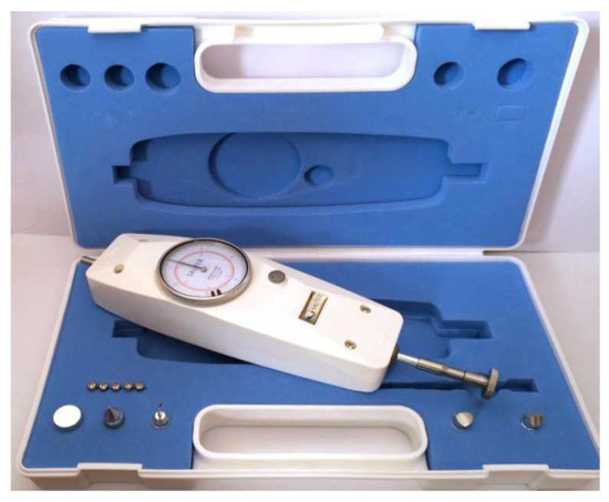
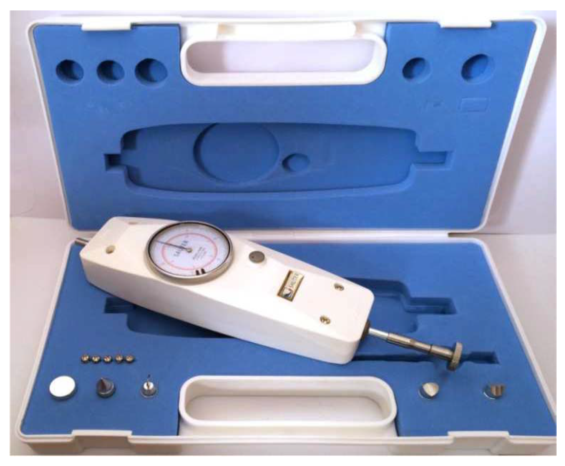
A dynamometer (Sauter FA 30; Sauter AG, Basel, Switzerland) (capacity, 30N; d = 0.0200N) (Figure 1) was used to verify the ST by applying exactly 10N (1.7–2.0 kg) of force to the ankle dorsiflexion as proposed by different authors [2,11], and a clinometer app, iPhone 5s TiltMeter, was used to measure the dorsiflexion angle [17].
Figure 1.
Dynamometer used to verify the Silfver-skiöld test.
Protocol
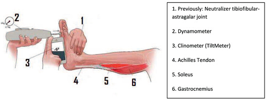
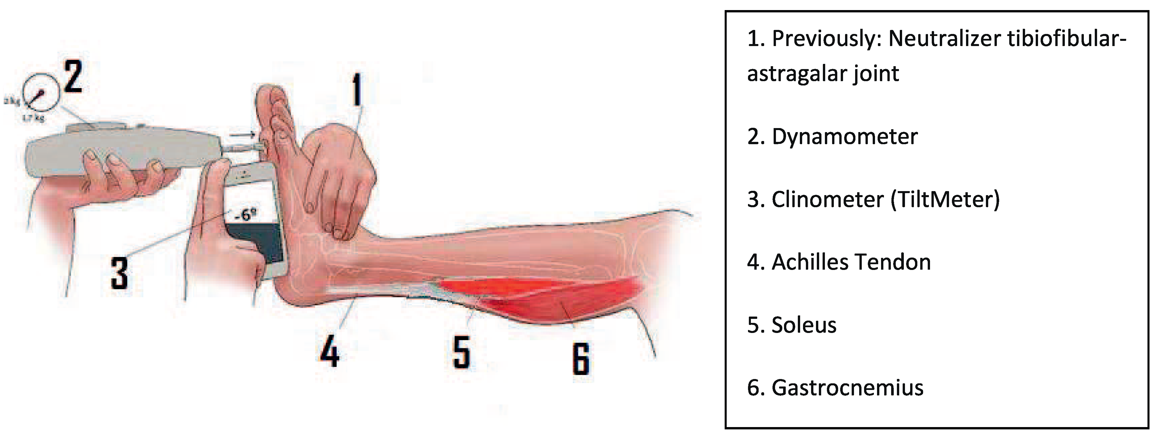
The ST [7] was performed with the patient in the supine position, flexing and extending the knee. Although patients had a neutral subtalar joint axis, in patients with slight pronation the ankle was neutralized by slightly inverting the foot, especially by blocking the tibiofibular-astragalar joint, thereby correcting the pronation, which would give a false result (Figure 2).
Figure 2.
Silfverskiöld test showing the dynamometer and clinometer procedure. 1 indicates (previously) neutralizer tibiofibular-astragalar joint; 2, dynamometer; 3, clinometer; 4, Achilles tendon; 5, soleus; 6, gastrocnemius.
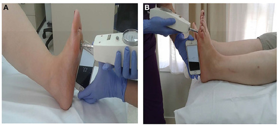
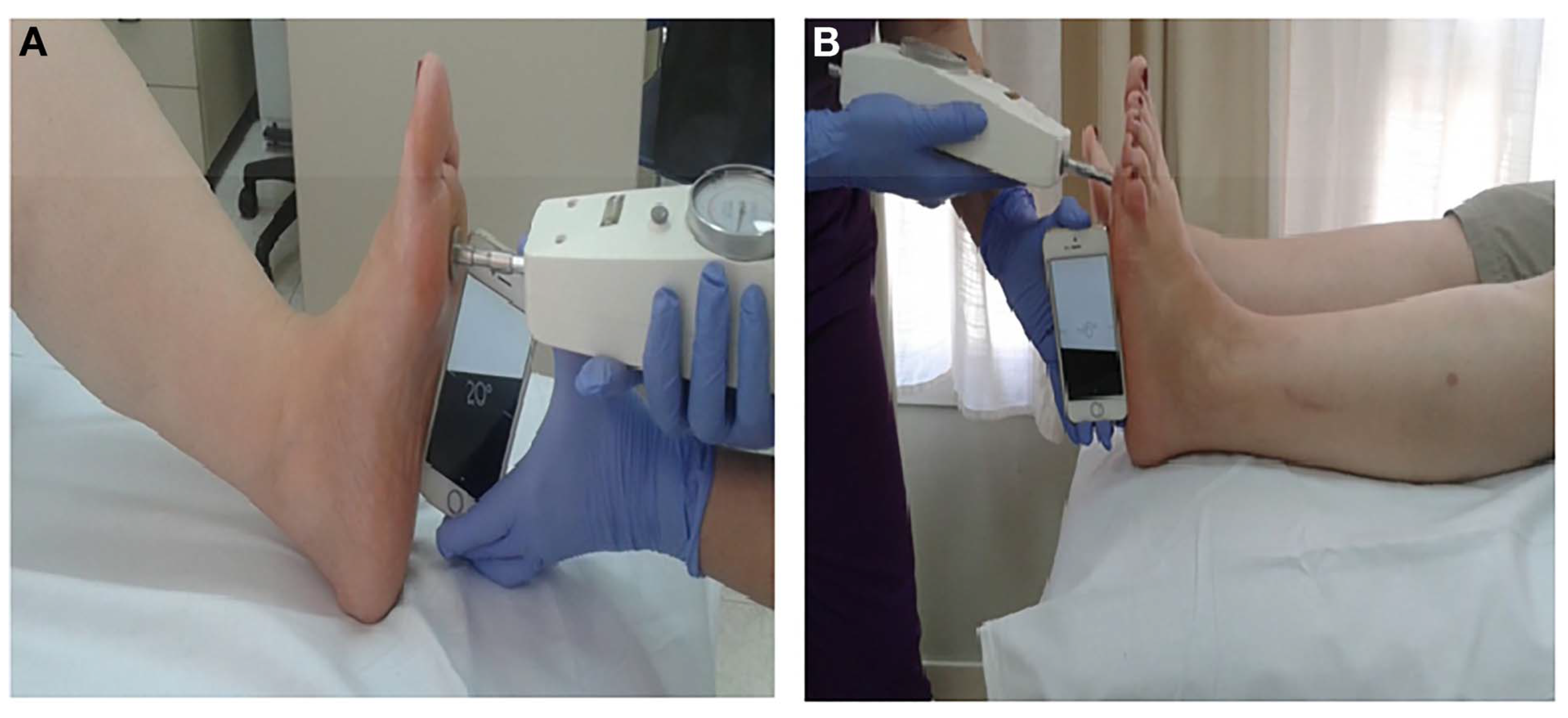
We applied an exact force of 10N (1.7–2.0 kg) to evaluate dorsiflexion (Figure 3). For this we used a dynamometer, positioning the pressure plate on the head of the second metatarsal. Thus, we obtained a reliable ankle dorsiflexion result because applying different forces would change the diagnosis. To obtain the degrees of dorsiflexion, we placed the TiltMeter app [18] on the exterior edge of the foot on the axis of the fifth metatarsal, which can easily be located.
Figure 3.
Ankle dorsiflexion angle measurements during the Silfverskiöld test with flexed knee (A) and extended knee (B).
Patients who presented negative dorsiflexion of the ankle or equal to zero when extended were deemed to present GT. This dorsiflexion normalized when flexed, with a minimum of a 158 difference.
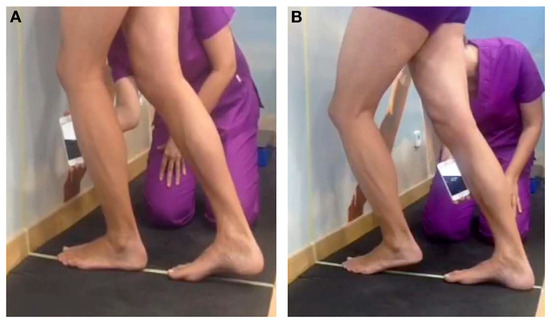
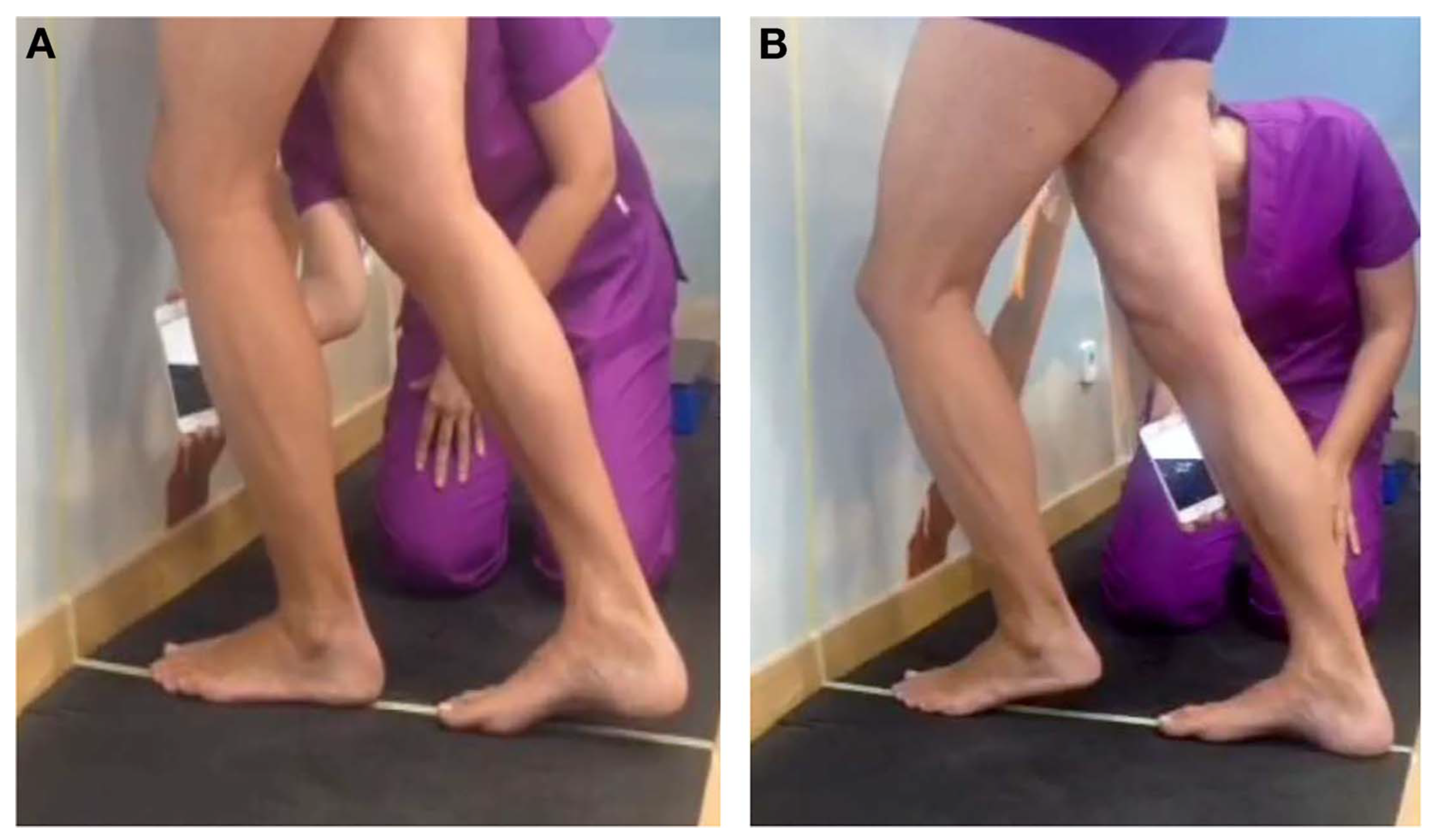
The LT [9] was performed with the patient standing with the knee flexed and extended (Figure 4). The patient stood with the leg that was to be evaluated in a forward position. The second toe and the center of the heel of the limb to be evaluated were positioned on a line drawn on the floor that was also perpendicular to the wall (drawing these lines on the floor and wall helped to neutralize the tibiofibular-astragalar joint, thereby avoiding prona-tion, which would give a false result).
Figure 4.
Clinometer used during the lunge test with flexed knee (A) and extended knee (B).
In the LT, a TiltMeter clinometer [17] was positioned under the tuberosity of the tibia to measure dorsiflexion. The patient was asked to put the flexed/extended knee toward the wall in the direction of the perpendicular line until feeling maximum tightness at the back of the leg. Just before lifting the calcaneus off the floor, the angle between the anterior surface of the tibia and the vertical surface was measured. Patients who pre-sented an angle lower than 358 were considered to have reduced range of motion.
Statistical Analysis
A descriptive analysis was made of all of the factors that could influence the presence of GT (gastrocne-mius recession). A bivariate analysis was performed by contrasting hypotheses that would determine the association of the factors with the pathology for both tests (LT and ST), thereby obtaining the odds ratio and 95% confidence interval. Contrasting was performed by parametric tests (t test) and nonpara-metric tests (Pearson v2 test and Mann-Whitney U test) according to the scale of the variable, and it is fit to a normal distribution. An independence analysis of the factors was performed by factorial techniques of principal components and Pearson correlation so as not to impinge on the basic assumptions that rule logistic regression (LR). The binary LR of the multivariate model was applied, including the independent factors that were statis tically significant. The LR model was built for each test with 122 patients and was subsequently validated with a prospective sample of 74 patients. Receiver operating characteristic curves and statis-tical goodness of fit of the LR function were obtained for each model with the evaluation data of the pathology by using the LT and the ST. A 5% (a < .05) significance level, an 80% (β = 0.20) power, and bilateral approximation were used in all of the analyses.
Results
The prevalence of GT was 39.3% as measured by the LT and 34.3% by the ST. Despite the fact that 70% of the patients were women, no significant differences could be attributed to sex (P = .839 LT; P > .99 ST) and GT presence (Table 1). Forefoot pain was significant (P < .001). The odds ratio presented differences depending on the test used: 22.67 versus 12.60 by the LT and the ST, respectively. An IPK2 promotes the presence (66.7%) of GT. In contrast, GT was absent in 93.2% of participants without hyperkeratosis.
Table 1.
Analysis of Association (Inference) of the Variables with the Presence of Gastrocnemius Tightness According to the Lunge and Silfverskiöld Tests.
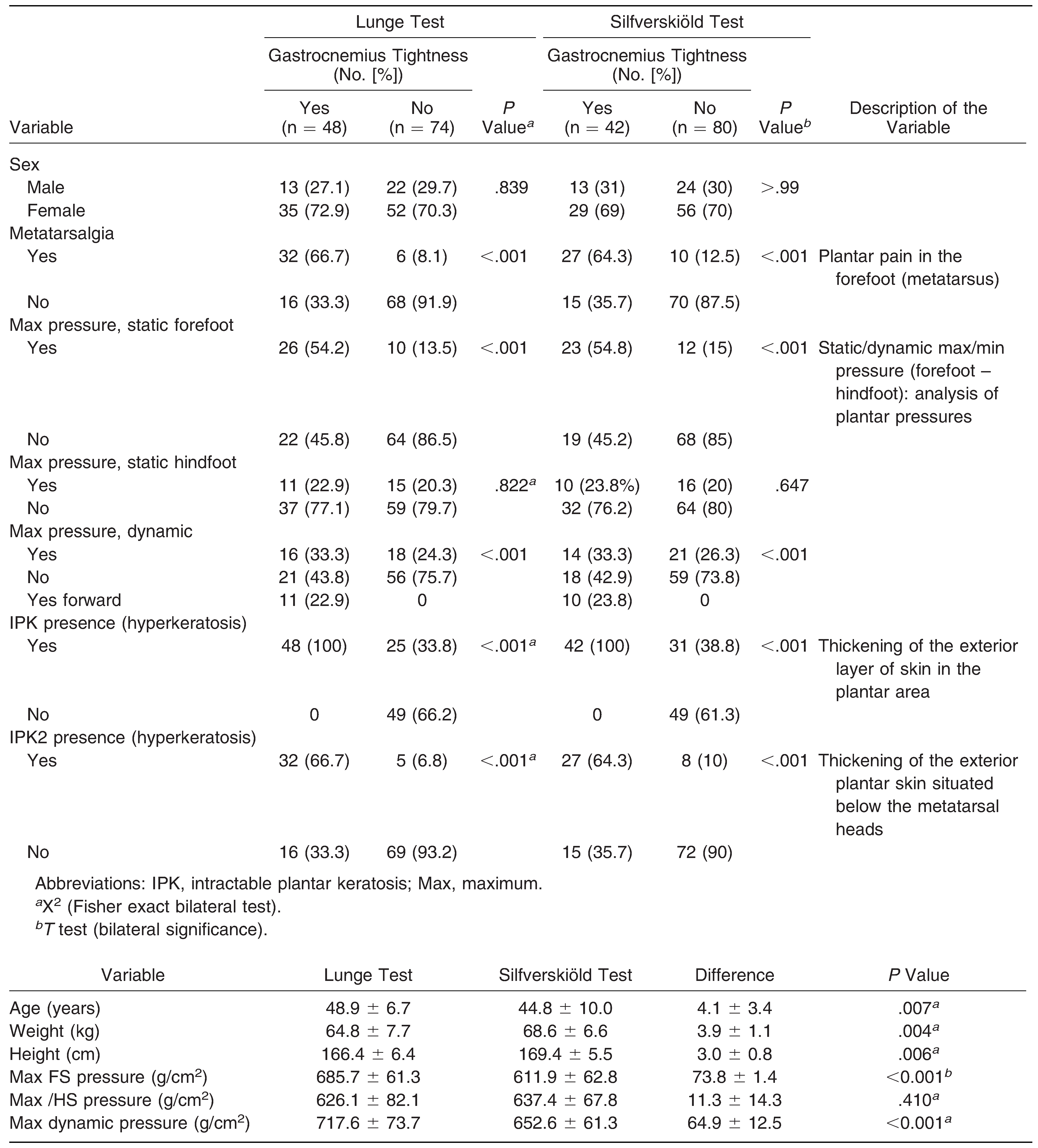
Static and dynamic forefoot pressures were both significant (P = .006 and P < .001, respectively). Greater mean 6 SD pressure values were found in participants with versus without GT (LT: 685.7 6 61.3 g/cm2 versus 611.9 6 62.8 g/cm2; ST: 683.5 6 61.6 g/cm2 versus 613.4 6 62.6 g/cm2).
The multivariate model of LR was used to identify the biomechanical parameters involved in the development of GT. The variables with statistical significance in the bivariate analysis were included in the regression model (Table 2). Forefoot pain, IPK2, and maximal static forefoot pressure were finally selected. Hyperkeratosis was the most relevant variable predicting GT, independently of the test, showing Wald values of 6.611 (LT) and 5.063 (ST). In summary, the probability of present-ing GT in patients with hyperkeratosis is 4.46- to 6.1-fold higher than in patients without this alteration. In addition, the probability of presenting GT in patients with forefoot pain is 4.46- to 5.0-fold higher than in control subjects.
Table 2.
Coefficients, Statistical Parameters, and Classification Matrix of the Logistic Regression for the Lunge Test and Silfverskiöld Test Models.
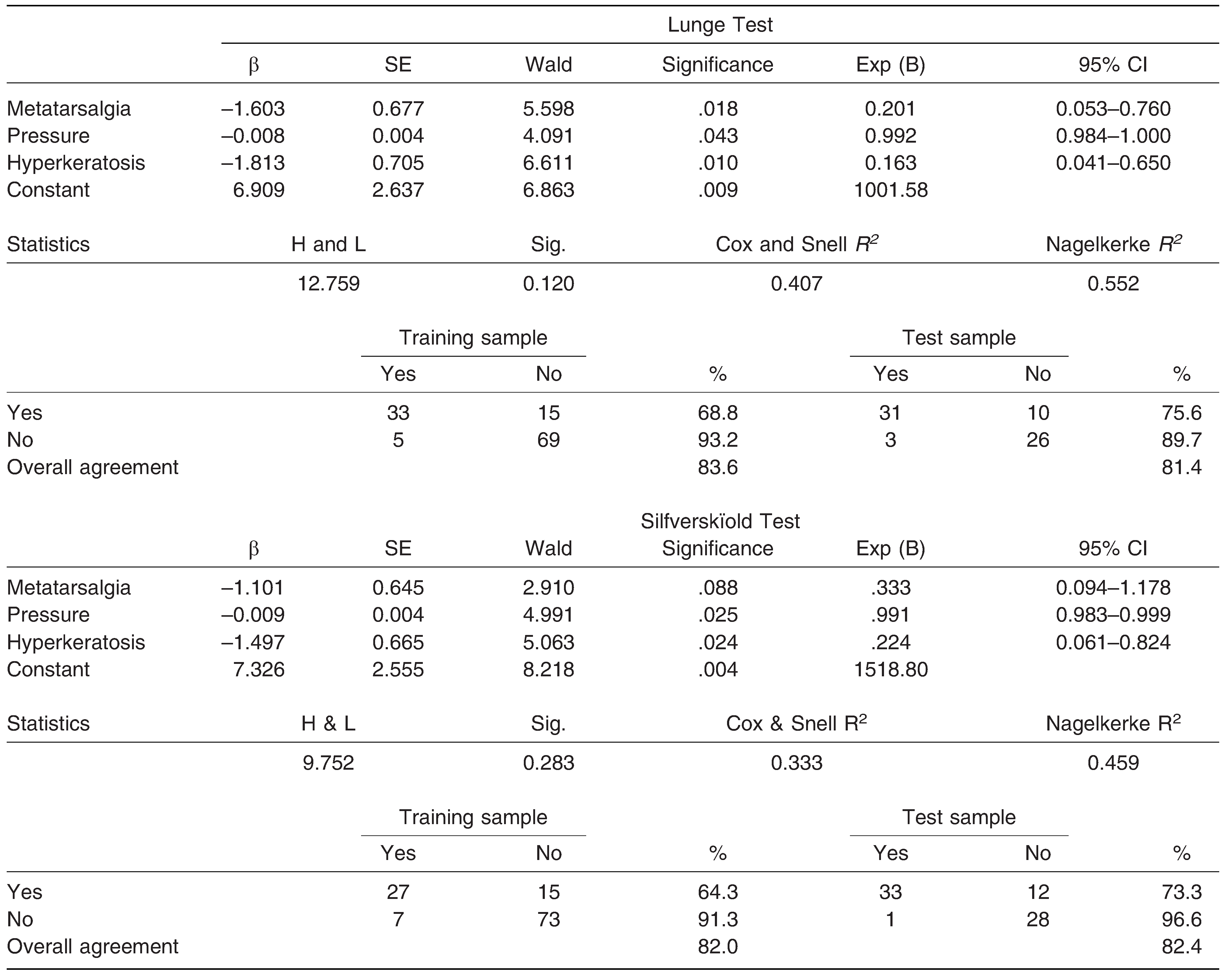
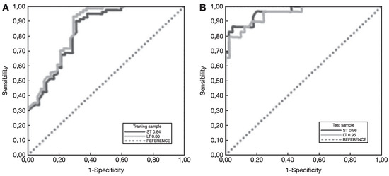
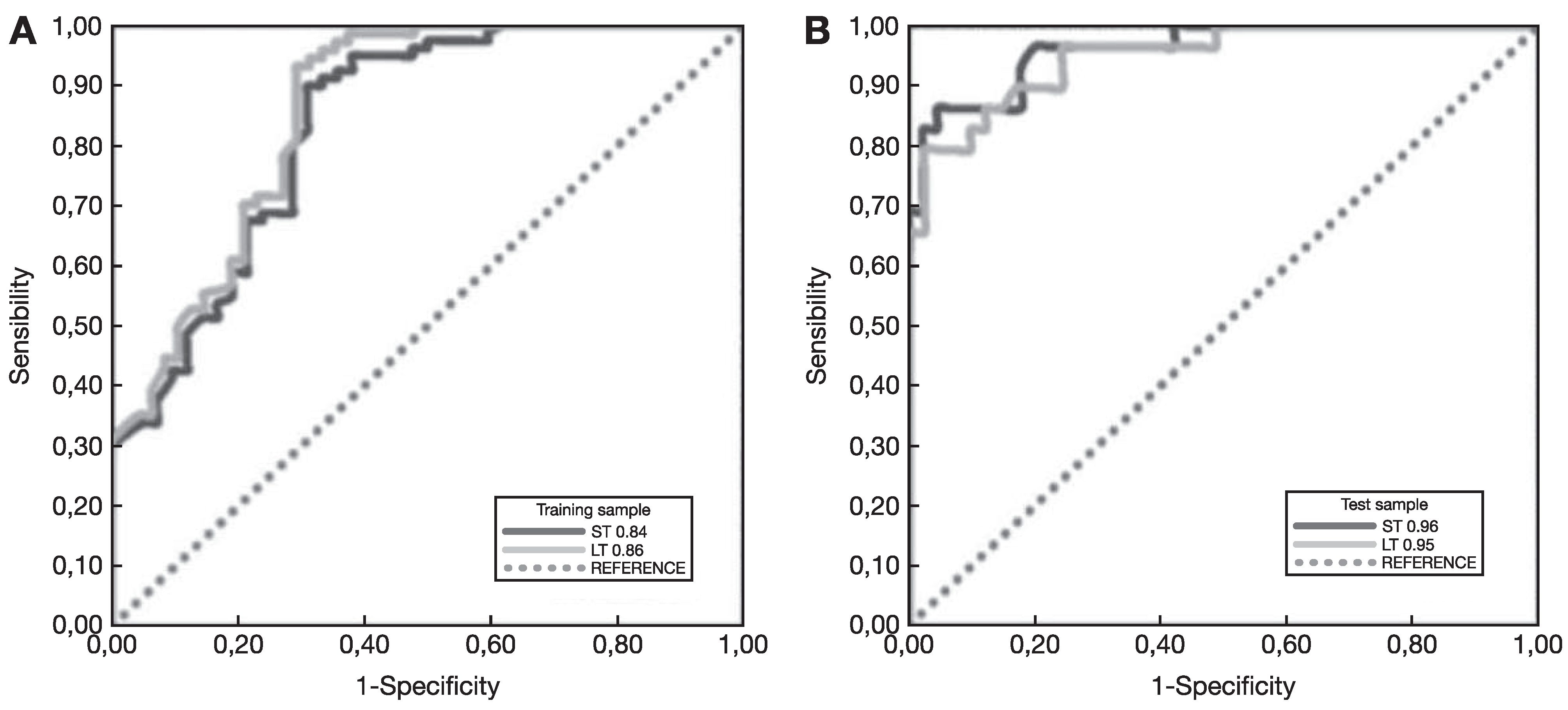
Validation of the LR was performed to assess the quality of both tests. The accuracy statistics indicate overall agreement of 83.6% on the LT and 82.0% on the ST and, as shown in Table 2 and Figure 5, through the construction and analysis of receiver operating characteristic curves (the method used for measuring the predictive functioning mod-el [18,19]), the sensitivity and specificity statistics indicate good capacity of both predictive models (LT and ST) for GT diagnosis.
Figure 5.
Receiver Operating Characteristic Curves (ROC) and Areas under the curve (AUC) of the Silfverskiöld test/ Lunge test ratio (ST/LT) from: (a) the training sample: (ST/LT = 0.84/0.86) the values denote an excellent discrimination the presence or absence of gastrocnemius tightness. (b) the test sample: (ST/LT = 0.96/0.95) the values denote an outstanding discrimination the presence or absence of gastrocnemius tightness.
Discussion
It is known that the percentage of individuals with GT is high, and it significantly higher in individuals with ankle and foot impairment. Gastrocnemius tightness is a relatively common issue with a 20% prevalence [1], although the present data indicate a prevalence of 34%.
As a previous consideration, we developed a reliable method to apply a force of 10N (1.7–2 kg) by using a dynamometer and the Tiltmeter app [17], avoiding intraoperator variability. This new proce-dure (force and angle) could be a reliable solution to the indicated limitations previously reported by several authors [2,3,4,11]. Typically the ST [11] is the most used test for GT diagnosis, but the LT can also be used in light of the differences found between tests (62%).
Regarding the associations between forefoot alterations and GT, DiGiovanni et al. [2] observed that 65% of the cases were associated with metatarsal-gia. Fitting with this, we found that 66.7% and 64.3% of the cases were associated with metatarsalgia as measured by the LT and the ST, respectively. This research establishes the direct relationship between GT and metatarsalgia, being threefold greater than in patients without this alteration. This report confirms a direct connection between GT and the presence of metatarsalgia. Likewise, the probability of presenting GT in patients with IPK2 is 4.5-fold greater than the probability of not manifesting it. This study confirms that there is a direct connection between GT and the presence of IPK2.
Conclusions
The test results confirm that the model presented acts as a prediction method of GT; the model comprises the following variables: metatarsalgia, IPK2, and maximum pressure when static. There-fore, the possibility of determining the probability of presenting GT is confirmed by the prediction model. In short, the results obtained in the test report indicate that the model is reliable, robust, and suitable for predicting the problem of GT. Never-theless, the present situation shows only an obvious ineffectiveness in clinical diagnosis, which is hindered by the fact that priority and focus is given to more serious equinus conditions (the case of patients with cerebral palsy) without first address-ing the more common issue (muscular tightness of a nonneurologic cause), ie, the most moderate and minor degrees.
Acknowledgments
Parc Taulı’ Sabadell Hospital, Barcelona, Spain, for participating in the study.
Financial Disclosure
None reported.
Conflicts of Interest
None reported.
References
- BAROUK, LS; BAROUK, P. Gastrocnemios cortos. Rev Pie Tobillo 2012, 26, 7. [Google Scholar] [CrossRef] [Scilit]
- DIGIOVANNI, CW; KUO, R; TEJWANI, N. ET AL: Isolated gastrocnemius tightness. J Bone Joint Surg Am 2002, 84, 962. [Google Scholar] [CrossRef] [Scilit]
- MEYER, DC; WERNER, CM; WYSS, T; ET, AL. A mechanical equinometer to measure the range of motion of the ankle joint: interobserver and intraobserver reliability. Foot Ankle Int 2006, 27, 202. [Google Scholar] [CrossRef] [Scilit] [PubMed]
- CHARLES, J; SCUTTER, SD; BUCKLEY, J. Static ankle joint equinus: toward a standard definition and diagnosis. JAPMA 2010, 100, 195. [Google Scholar] [CrossRef] [Scilit] [PubMed]
- DOWNEY, MS; BANKS, AS. Gastrocnemius recession in the treatment of nonspastic ankle equinus: a retrospective study. JAPMA 1989, 79, 159. [Google Scholar] [CrossRef] [Scilit] [PubMed]
- HILL, RS. Ankle equinus: prevalence and linkage to common foot pathology. JAPMA 1995, 85, 295. [Google Scholar] [CrossRef] [Scilit] [PubMed]
- SINGH D: NILS SILFVERSKIÖLD (1888-1957) and gastrocnemius contracture. Foot Ankle Int 2011, 32, 764.
- HALL, EA; DOCHERTY, CL. Validity of clinical outcome measures to evaluate ankle range of motion during the weight-bearing lunge test. J Sci Med Sport 2017, 20, 618. [Google Scholar] [CrossRef] [Scilit] [PubMed]
- BENNELL, KL; TALBOT, RC; WAJSWELNER, H. ET AL: Intra-rater and inter-rater reliability of a weight-bearing lunge measure of ankle dorsiflexion. Aust J Physiother 1998, 44, 175. [Google Scholar] [CrossRef] [Scilit] [PubMed]
- DONLEY, BG; PINNEY, SJ. HOLMES J: Gastrocnemius reces-sion. Tech Foot Ankle Surg 2003, 2, 35. [Google Scholar] [CrossRef] [Scilit]
- BAROUK, P. BAROUK LS: Clinical diagnosis of gastrocnemi-us tightness. Foot Ankle Clin 2014, 19, 659. [Google Scholar] [CrossRef] [Scilit] [PubMed]
- MONTEAGUDO, M; MACEIRA, E; GARCIA-VIRTO, V. ET AL: Chronic plantar fasciitis: plantar fasciotomy versus gastrocne-mius recession. Int Orthop 2013, 37, 1845. [Google Scholar] [CrossRef] [Scilit] [PubMed]
- Garcia Carmona, FJ; Pascual Huerta, J. HERNANDEZ TOLEDO J: Plantar epidermoid cyst as a possible cause of intractable plantar keratosis lesions. JAPMA 2009, 99, 148. [Google Scholar] [CrossRef] [Scilit] [PubMed]
- KIRBY KA: Rotational equilibrium across the subtalar joint axis. JAPMA 1989, 79, 1.
- KIRBY KA: Methods for determination of positional variations in the subtalar joint axis. JAPMA 1987, 77, 228. [CrossRef] [Scilit] [PubMed]
- KIRBY KA: Subtalar joint axis location and rotational equilibrium theory of foot function. JAPMA 2001, 91, 465. [CrossRef] [Scilit] [PubMed]
- WILLIAMS, CM; CASERTA, AJ; HAINES, TP. The TiltMeter app is a novel and accurate measurement tool for the weight bearing lunge test. J Sci Med Sport 2013, 16, 392. [Google Scholar] [CrossRef] [Scilit] [PubMed]
- Van Den Eeckhaut, M; Vanwalleghem, T; Poesen, J. : Prediction of landslide susceptibility using rare events logistic regression: a case-study in the Flemish Ard-ennes (Belgium). Geomorphology 2006, 76, 392. [Google Scholar] [CrossRef] [Scilit]
- YESILNACAR E, TOPAL T: Landslide susceptibility mapping: a comparison of logistic regression and neutral net-works methods in a medium scale study, Henkek region (Turkey). Eng Geol 2006, 79, 392.
This is an open-access article distributed under the terms of the Creative Commons Attribution Non-Commercial No Derivatives (CC BY-NC-ND) 4.0 License.