


Abstract
Medial forefoot pain, or midarch pain, is usually attributed to plantar fasciitis. The authors present their findings of a previously unreported nerve entrapment of the medial proper plantar digital nerve (MPPDN). Ten fresh-frozen cadaveric specimens were analyzed for anatomical variance in the nerve distribution of the MPPDN. In addition, clinical results from a retrospective review of nine patients who underwent surgical nerve decompression of the MPPDN are presented. Significant anatomical variance was found for the MPPDN in the cadaveric dissection of 10 fresh-frozen specimens. Nine patients with a clinical diagnosis of entrapment of the MPPDN all obtained excellent pain relief with surgical external neurolysis. Only one complication occurred: a hypertrophic scar formation that was successfully treated with intralesional steroid injections. The authors believe that this MPPDN entrapment is often overlooked or misdiagnosed as plantar fasciitis. Surgical peripheral nerve decompression of this nerve can provide positive outcomes for patients suffering from midarch foot pain caused by this pain generator.
A patient complaint of “pain in the midarch region of the foot” can be difficult to both understand and evaluate. Most patients voicing this complaint, almost by default, are thought to have plantar fasciitis [1,2,3], despite the fact that many of these patients do not have poststatic dyskinesia and most have their pain worsen with the day's activities. Usually, normal radiographic imaging studies of the foot and ankle are present and the plantar fascia ultrasound evaluation is normal. By history, there is no complaint of heel pain or of toe numbness, although there may be some paresthesias to the medial hallux pulp. Patients may have a negative Tinel sign over the tibial nerve in the tarsal tunnel and not have pain at the medial calcaneal tubercle. These patients uniformly have pain with pressure over the medial arch, just below the navicular and medial cuneiform bones. Therefore, these patients do not have tarsal tunnel syndrome [4]. These patients have not had surgery near their distal great toe, such as sesamoid excision or osteotomy, so a neuroma of the distal proper digital nerve to the hallux, as described by Joplin, does not exist [5].
This article reports a previously undescribed, clinical entrapment neuropathy in the midarch of the foot found as the medial plantar nerve exits the medial plantar tunnel and gives off two branches: one to the skin of the medial arch, which does not have a unique name, and one to the medial side of the hallux, variously named the proper digital nerve of the hallux or the medial digital nerve to the hallux [6,7]. It is our belief that physical activity with sufficient medial loading of the arch, or sudden stretch/traction, results in swelling of the abductor hallucis muscle within the medial compartment of the foot, which may lead to compression of these nerve branches to create the symptoms referred to as the midarch.
Diagnosis
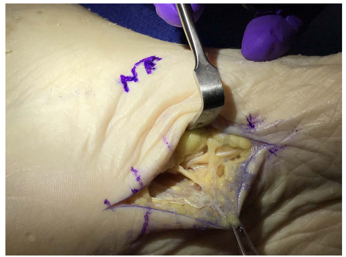
Often, when there is pain in the medial arch area, the immediate default diagnosis of plantar fasciitis is given. A more comprehensive workup is needed to ensure an accurate diagnosis, therefore predicting a successful outcome for the patient. After sonographic evaluation of the plantar fascia, that fasciitis diagnosis can be definitively excluded with a normal image. Thorough lower-extremity peripheral nerve examination will evaluate the known sites of entrapment, including the tarsal tunnel and forefoot intermetatarsal spaces. In our experience, a provocation sign along the course of the medial proper plantar digital nerve (MPPDN) is pathognomonic for entrapment of this nerve at this location (Fig. 1). Most patients with this nerve entrapment cannot tolerate a rigid or semirigid orthotic device, which might be prescribed and can exacerbate the condition. Peripheral nerve blocks with lidocaine are diagnostic if the local pain is eliminated or greatly reduced after infiltration. If there is suspicion of a coexistent medial plantar nerve entrapment at the level of the tarsal tunnel, the diagnostic block must be performed distally at the maximal site of provocation along the MPPDN because proximal blockade at the tarsal tunnel would potentially eliminate pain and/or sensation distally. If there is still a provocation sign, and one present as well at the tarsal tunnel, a proximal block needs to be implemented. This, in most cases, would indicate a diagnosis of medial plantar nerve entrapment at the level of the tarsal tunnel and entrapment of the MPPDN, a double-crush phenomenon. This might require neurolysis of both sites of entrapment.
Figure 1.
In this photograph, the medial proper plantar digital nerve is clearly seen to be substantially narrower under the scissors than 4 mm proximally, where some remaining bands of fibrous tissue remain.
Methods
The authors investigated 10 fresh-frozen cadaveric specimens to determine the anatomical variability and course of this nerve in the medial compartment of the foot. Evidence from every specimen showed that this nerve is approximately 1 mm in diameter and that there are numerous fibrous septal bands consistently present in the medial compartment that could lead to local compression or entrapment of the MPPDN proximal to the level that has been described as Joplin's neuroma [2]. After institutional review board approval from Kennesaw State University, Kennesaw, Georgia, for a retrospective review, the results and findings from nine patients are included in this report.
Surgical Technique
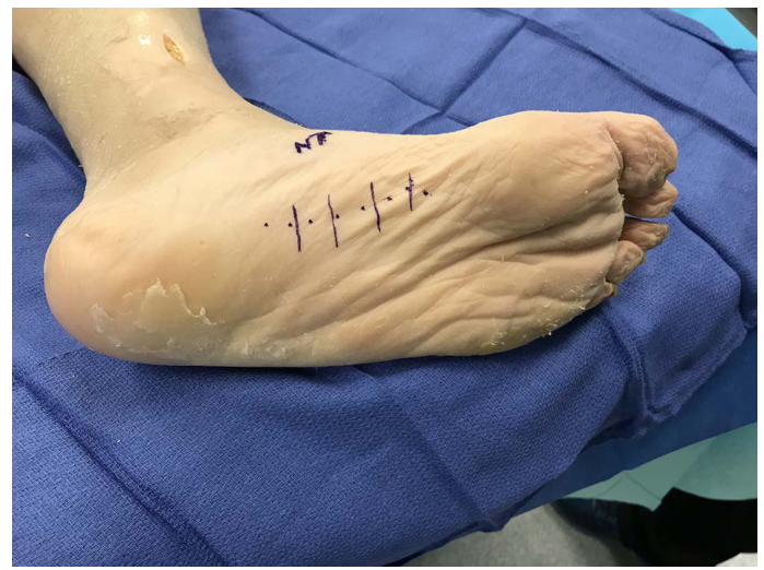
As can be seen in Figure 2, the incision is placed in the medial arch located in correlation with the most significant area of provocation elicited along the course of the nerve. Initially, the incision is topographically mapped with the least amount of invasiveness possible but often will need to be extended based on the level and extent of entrapment along the course of the MPPDN. The procedure is performed under 3× to 4× power loupe magnification. Blunt dissection is carried down to the fascial plane of the medial compartment overlying the abductor hallucis muscle. A fasciotomy is then made the length of the incision and the muscle belly is bluntly reflected inferiorly, without damage to the muscle itself, until the nerve is identified. Invariably, there is visible entrapment of the nerve, close to the level of the marked site of maximum provocation. Neurolysis is performed proximally and distally until there is no evidence of entrapment. Because this nerve can be relatively superficial, we occasionally implement the placement of an amniotic tissue adhesion barrier over the nerve and deep to the dermis. No deep sutures are used and the skin is closed with 5-0 nylon, simple interrupted sutures. Care should be taken in meticulous wound closure so that scarring can be kept to a minimum. Patients are allowed full weightbearing the same day of surgery but are cautioned to avoid unnecessary standing or walking to minimize local edema, adhesions, and pain.
Figure 2.
In this cadaveric specimen, a usual incision approach is mapped. Note that this is medial to the main substance of the plantar fascia and more proximal than the site of Joplin's neuroma.
Results
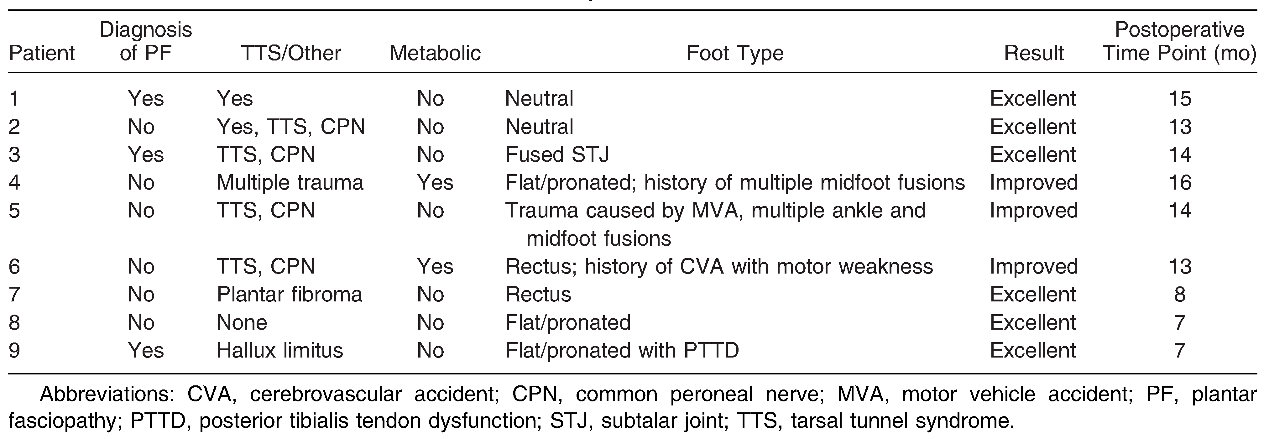
Anatomical variability was seen in the cadaveric dissection of 10 fresh-frozen, randomly selected specimens. In all 10 specimens, the nerve was found and seen originating as a branch of the medial plantar nerve, continuing distally to the medial plantar aspect of the first metatarsal phalangeal joint, where the traditional Joplin's neuroma is located. In four of the 10 specimens, the diameter of the nerve was greater than 1.5 mm at the midarch/navicular level, and in all of these specimens there were multiple sites of entrapment noted from fascial connective tissue bands along the course of the nerve just deep to the fascia of the abductor hallucis muscle. These nerves were anatomically located within the medial compartment and exited distally to the subcutaneous plane, 2 to 3 cm proximal to the first metatarsophalangeal joint. In the remaining six specimens, the nerve was also identified with the same origination off the medial plantar nerve. None of the nerves at the midarch/navicular level measured more than 1.5 mm, but nerves were also identified within the medial compartment, exiting the medial compartment more proximally. A retrospective review was completed for nine patients who underwent surgery for peripheral nerve decompression of the MPPDN. As can be seen in Table 1, all nine patients who had the procedure had either an improved or excellent outcome, based on their own reporting. Because this was not a prospectively designed study, a preoperative visual analog scale score or other metrics were not gathered for this cohort. All patients who reported that they did not have any pain after complete postoperative healing based on this self-report were categorized as having an “excellent” outcome, and those categorized as improved were not completely pain free but individually reported that they were significantly improved. The longest follow-up has been 1.5 years, and the shortest 6 months, with a mean of approximately 1 year. The only complication was hypertrophic scar formation in one patient and was effectively managed with postoperative infiltration with a steroid/local anesthetic combination.
Table 1.
Medial Division of Medial Plantar Nerve Decompression
Discussion
In patients who present with midarch pain, entrapment of the MPPDN should be considered in the differential diagnosis, especially if there is sonography that shows normal plantar fascia. This nerve entrapment has not been well described or documented previously in the literature and is probably more common than we believe.
Accurate diagnosis is predicated on a good history of present illness and complete lower-extremity physical examination. In addition, sonography and diagnostic nerve blocks can strongly support the diagnosis. Tarsal tunnel compression of the medial plantar nerve must be evaluated, as this is commonly coexistent with this nerve entrapment, being found in four of our nine patients. The use of neurosensory testing can solidify the diagnosis. As with all lower-extremity peripheral nerve entrapments, this entrapment is more common in patients with metabolic disease, such as diabetes, where nerve diameter is enlarged. This nerve entrapment is distinctly different than what has been described as Joplin's neuroma, because the nerve is focally entrapped more proximally in the arch, and sometimes at several foci (Figure 3 and Figure 4). It is our experience that this is a common pain generator that is either overlooked or misdiagnosed as plantar fasciitis. With an understanding of this nerve entrapment, and how to effectively diagnose it, more patients will have improved outcomes. If a patient presents with a sudden onset of medial arch pain after exertion, a medial foot compartment syndrome should be considered in the differential diagnosis. Two such cases have been described [8,9], but this too may be more common than reported. A similar compartment syndrome has been reported for an intrinsic muscle in the hand [10]. In the posterior compartment of the leg, compression of the tibial nerve at the Soleal sling gives symptoms similar to those of compartment syndrome [10,11]. It is likely that our surgical approach to decompression of the MPPDM, which requires release of the fascia over the abductor muscle compartment, is also releasing pressure on the nerve and may be, in fact, addressing the same mechanism causing an exertional type of compartment syndrome. Indeed, it may be that the cases of compartment syndrome were in fact cases of compression of the MPPDM, not unlike other commonly known sites of peripheral nerve entrapment in the lower extremity such as the common and superficial fibular nerves, as the same operative approach is used. The authors believe that this pathology is more prevalent than thought at this time, and further studies need to be conducted to correlate the relationships to the metabolic status of the patient, and whether there is coexistent proximal nerve entrapment such as tarsal tunnel syndrome.
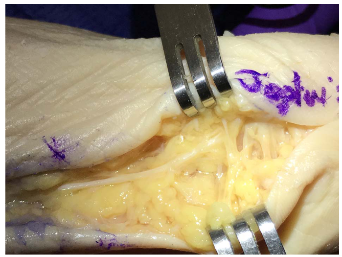
Figure 3.
Here the dissection illustrates the different anatomical locations of Joplin's neuroma and the medial proper plantar digital nerve entrapment.
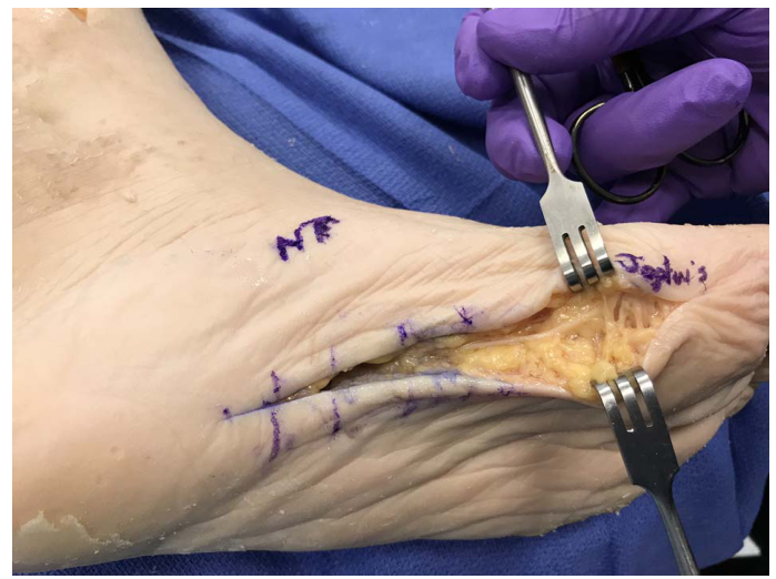
Figure 4.
This cadaveric dissection demonstrates the typical entrapment site of Joplin's neuroma, and this illustrates why this nerve can become entrapped after bunion surgery with normal postoperative edema.
Financial Disclosure
None reported.
Conflict of Interest
None reported.
References
- Barrett, SJ; O'Malley, R. Plantar fasciitis and other causes of heel pain. Am Fam Physician 1999, 59, 2200. [Google Scholar]
- Barrett, SL. Practical Pain Management for the Lower Extremity Surgeon, 1st Ed 2015.
- Park, SY; Bang, HS; Park, DJ. Potential for foot dysfunction and plantar fasciitis according to the shape of the foot arch in young adults. J Exerc Rehabil 2018, 14, 497. [Google Scholar]
- Mackinnon, SE; Dellon, AL. Surgery of the Peripheral Nerve 1988.
- Melendez, MM; Patel, A; Dellon, AL. The diagnosis and treatment of Joplin's neuroma. J Foot Ankle Surg 2016, 55, 320. [Google Scholar]
- Ferner, H; Monsen,, H. Eduard Pernkopf's Atlas of Topographical and Applied Human Anatomy, Vol, 2 1964.
- Rochen, JW; Yokochi, C; Lütjen-Drescoll, E. A Photographic Study of the Human Body 2006.
- Blacklidge, DK; Kurek, JB; Soto, AD; et al. Acute exertional compartment syndrome of the medial foot. J Foot Ankle Surg 1996, 35, 19. [Google Scholar]
- Izadi, FE; Richie, DH. Jr: Exertional compartment syndrome of the medial foot compartment—diagnosis and treatment: a case report. JAPMA 2014, 104, 417. [Google Scholar]
- Dellon, AL; Fine, IT. A noninvasive technique for diagnosis of chronic compartment syndrome in the first dorsal interosseous muscle. J Hand Surg Am 1990, 15, 1008. [Google Scholar]
- Williams, EH; Williams, CG; Rosson, GD; et al. Anatomic site for proximal tibial nerve compression: a cadaver study. Ann Plast Surg 2009, 62, 322. [Google Scholar]
© 2021 American Podiatric Medical Association