


Abstract
Tarsal tunnel syndrome (TTS), resulting from compression of the posterior tibial nerve (PTN) within the tarsal tunnel, is a relatively uncommon entrapment neuropathy. Many cases of tarsal tunnel syndrome are idiopathic; however, some causes, including space-occupying lesions, may lead to occurrence of TTS symptoms. Schwannoma, the most common tumor of the sheath of peripheral nerves, is among these space-occupying lesions, and may cause TTS when it arises within the tarsal tunnel, and it may mimic TTS even when it is located outside the tarsal tunnel and cause a significant delay in diagnosis. The possibility of an occult space-occupying lesion compressing the PTN should be kept in mind in the differential diagnosis of TTS, and imaging studies that are usually not used in entrapment neuropathies may be of importance in such patients. This case report presents a 65-year-old woman with TTS symptoms and neurophysiologic findings secondary to an occult schwannoma of the PTN proximal to the tarsal tunnel. Avoidance of delay in diagnosis in secondary cases is emphasized.
Tarsal tunnel syndrome (TTS) results from the compression of the posterior tibial nerve (PTN) and/or its branches within the tarsal tunnel on the medial side of the ankle [1]. It is a relatively uncommon entrapment neuropathy, although the published data contain no precise estimate of its prevalence. Pain, altered sensation, paresthesia affecting the plantar aspect of the foot, and motor weakness of the intrinsic foot muscles are the chief complaints. Tarsal tunnel syndrome is commonly underdiagnosed, frequently because of false-negative results from neurophysiologic tests [2,3]. Although many cases of TTS are idiopathic, some causes such as trauma, foot deformities, and space-occupying lesions are well-known causes that may lead to TTS [2]. Space-occupying lesions such as ganglia, lipomas, accessory muscles, tenosynovitis of the flexors tendons, and varicose veins are among the commonly cited causes [2].
The possibility of an occult space-occupying lesion compressing the PTN should be kept in mind in the differential diagnosis of TTS, and imaging studies that are usually not used in TTS may be of importance in such patients. Further imaging is of importance. An experienced ultrasonographer/radiologist can identify the presence of a mass in the tarsal tunnel. If a mass is identified, magnetic resonance imaging (MRI) is advised preoperatively to locate the tumor (within or around the nerve) and differentiate a schwannoma or other soft-tissue tumor [4]. In TTS with a space-occupying lesion, MRI is useful for localizing lesions within the tarsal tunnel and for determining the lesion extent and relationship to the PTN and its branches [1].
Schwannomas of the posterior tibial nerve and/or its branches also have been reported to cause TTS [5,6,7,8,9]. Schwannomas are the most common tumors of the sheath of peripheral nerves [5,10,11]. They are benign, slow-growing, encapsulated, and noninvasive tumors that may cause nerve compression symptoms, resulting in pain and discomfort. A neurologic deficit may occur when the tumor arises in a confined space [5]. Schwannomas usually occur in the head and neck, commonly involving the brachial plexus and spinal nerves [5,10].
Treatment of schwannoma is excision of the tumor without damaging the nerve bundles. Schwannomas are eccentrically located and do not involve the main nerve; the lesion spreads the nerve fibers apart without anatomical interruption. Because of these peculiarities, excision of the tumor does not necessitate partial or complete removal of nerves, resulting in nerve deficit. Adequate exposure of normal nerve proximal and distal to the tumor is essential. A longitudinal plane is developed between the nerve bundles, and the encapsulated tumor is removed. The outcome of schwannoma excision is very favorable, and nerve damage or neurologic deficits are uncommon.
This case report presents a 65-year-old woman with TTS secondary to an occult schwannoma of the posterior tibial nerve with neurophysiologic, MRI, and operative findings. Avoidance of delay in diagnosis in secondary cases is emphasized.
Case Report
A 65-year-old woman presented with pain and paresthesia affecting the medial aspect of the right ankle and the plantar aspect of the foot, exacerbated by walking, of 2 months' duration with a sudden onset. She had no history of trauma and was otherwise healthy. On physical examination, the medial side of the right ankle was very tender on palpation, a sensory deficit on the plantar aspect of the foot was present, and the Tinel sign over and proximal to the tarsal tunnel was significantly positive. There was slight weakness of the intrinsic foot muscles elicited by the reduction of toe flexor and abduction strength when compared to the contralateral foot. She did not have heel valgus or hypermobility of the ankle, and the lumbar examination findings were normal. The patient reported a score of 80 on the visual analog scale (ranging from 0–100, with 0 being pain free and 100 being horrible pain). Anteroposterior and lateral radiographs of the ankle were normal, and the patient had a clinical diagnosis of TTS.
Neurophysiologic tests were performed that included nerve conduction studies and needle electromyography of both feet. An amplitude of sensory nerve action potential of 3 mV (normal, 5–15 mV), a sensory nerve conduction velocity of 33 m/sec (normal, 40 m/sec), a distal motor latency of 7 ms (normal, <6.2 ms) of the medial plantar nerve to the abductor hallucis muscle and 7.6 ms (normal, <7 ms) of the lateral plantar nerve to the abductor digiti quinti muscle, and an amplitude of motor nerve action potential of 3.7 mV (normal, 5–15 mV) were found with nerve conduction studies. Electromyography showed fibrillation activity, decreased motor unit recruitment, and positive sharp waves, and abnormalities in the configuration of the motor unit action potential in the abductor hallucis and abductor digiti quinti muscles. The posterior tibial muscle above the tarsal tunnel demonstrated normal findings. The contralateral lower extremity was neurophysiologically intact and the neurophysiologic test findings confirmed the clinical diagnosis of TTS.
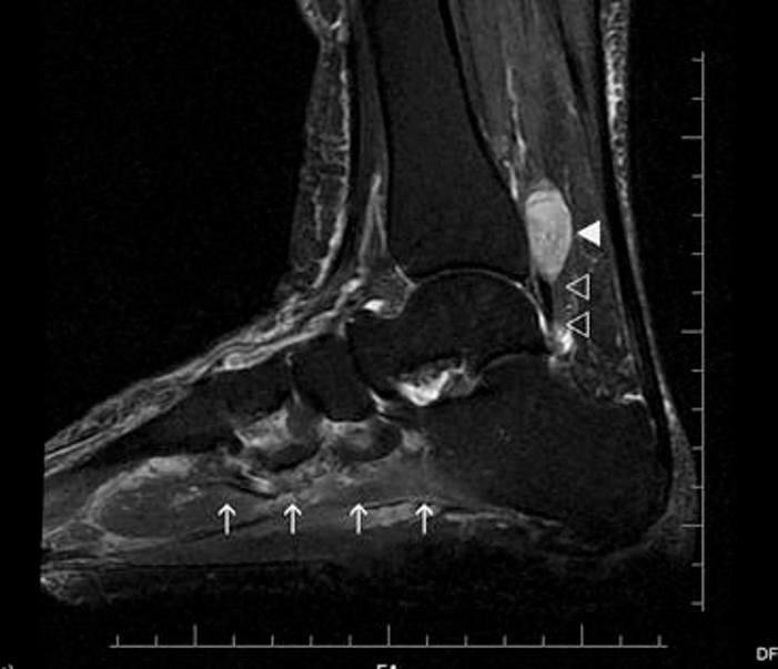
Given the rapid progression, severity of symptoms, and unilateral occurrence, MRI of the right ankle was performed to reveal a morphologic etiology, and showed a 23 × 11 × 16-mm, fusiform-shaped, space-occupying lesion with well-defined margins between the flexor hallucis longus and the posterior tibial tendons, originating from the posterior tibial nerve just proximal to the tarsal tunnel (Fig. 1). Increased signal intensity of the posterior tibial nerve implying intraneural edema and increased signal intensity of the adductor hallucis and quadratus plantae muscles, both of which are innervated by the lateral plantar nerve (a main branch of the posterior tibial nerve), implying denervation, were also present (Fig. 2). The lesion showed isointense signal intensity on T1-weighted and hyperintense signal intensity on T2-weighted images and homogenous contrast fixation after gadolonium injection. A radiologic diagnosis of schwannoma of the posterior tibial nerve causing TTS symptoms was reached.
Figure 1.
(A) T1-weighted, sagittal magnetic resonance imaging (MRI) scan and (B) T2-weighted, fat-saturated coronal MRI scan of the left ankle showing a 23 × 11 × 16-mm, fusiform-shaped, space-occupying lesion with well-defined margins between the flexor hallucis longus and the posterior tibial tendons, originating from the posterior tibial nerve just proximal to the tarsal tunnel (arrows).
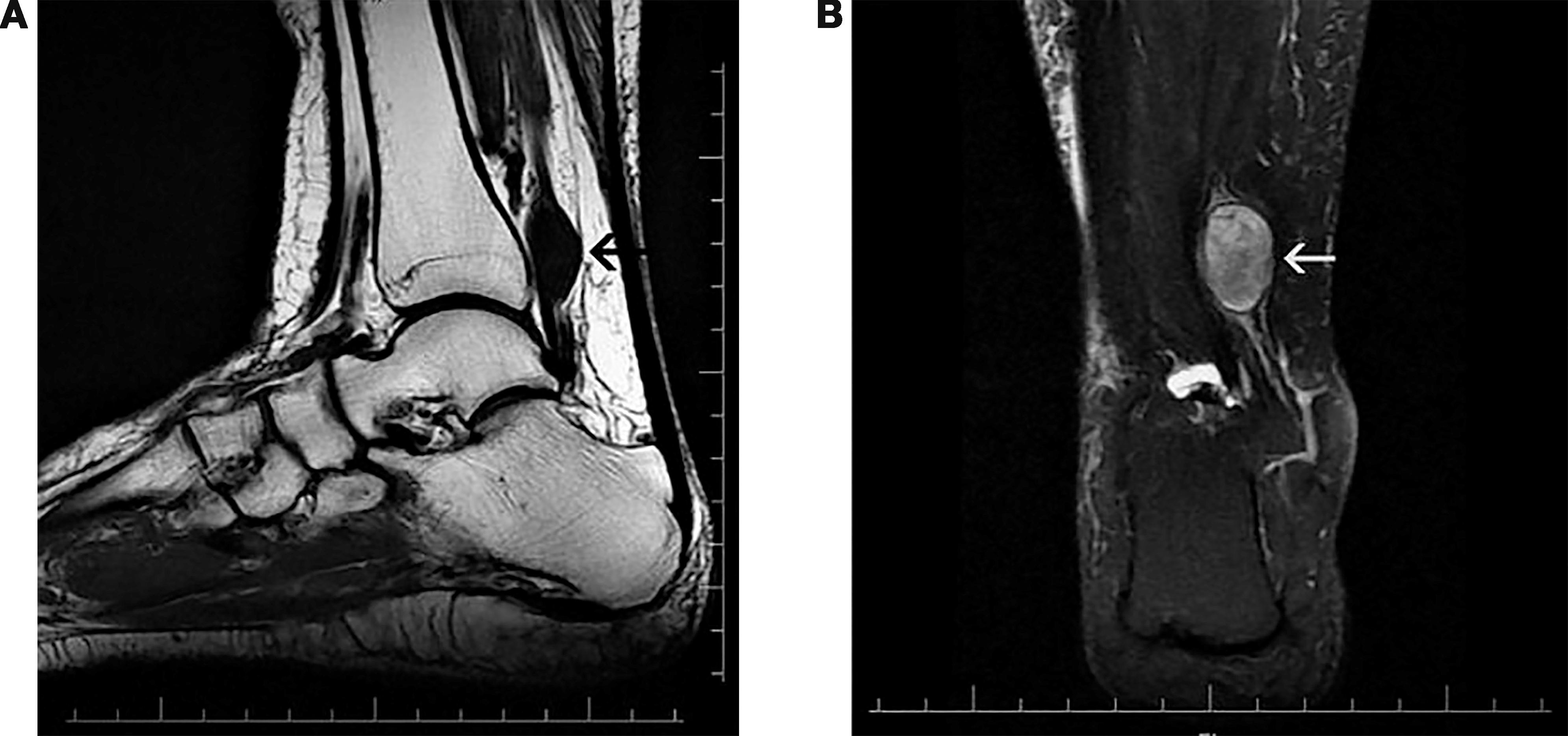
Figure 2.
T2-weighted, fat-saturated sagittal magnetic resonance image scan showing a 23 × 11 × 16-mm, fusiform-shaped, space-occupying lesion (filled arrowhead), increased signal intensity of the posterior tibial nerve implying intraneural edema (open arrowheads), and increased signal intensity of the adductor hallucis and quadratus plantae muscles, implying denervation (arrows).
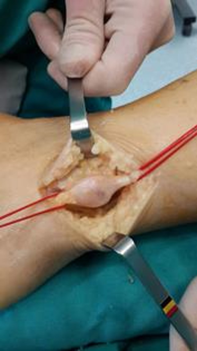
The patient was operated on under general anesthesia with pneumatic tourniquet hemostasis. The mass within the posterior tibial nerve was easily identified, and adequate exposure of normal nerve proximal and distal to the tumor was obtained (Fig. 3). A longitudinal plane was developed between the nerve bundles, and the encapsulated tumor was removed. Tarsal tunnel release was not performed.
Figure 3.
Intraoperative photograph showing the tumor within the posterior tibial nerve and normal nerve proximal and distal to the tumor.
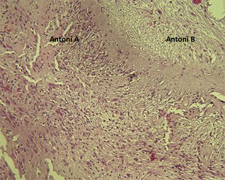
Histologic examination revealed hypercellular Antoni A tissue consisting of spindle cells with marked palisading and myxomatous, degenerative, and hypocellular Antoni B tissue with thick-walled blood vessels consistent with a schwannoma (Fig. 4). Diffuse positive staining for S-100 protein by immunohistochemical analysis confirmed the diagnosis of schwannoma.
Figure 4.
Histomicrograph of schwannoma (H&E, ×40). Histologic examination revealed hypercellular Antoni A tissue consisting of spindle cells with marked nuclear palisading and myxomatous, degenerative, and hypocellular Antoni B tissue with thick-walled blood vessels consistent with a schwannoma.
The patient reported immediate relief of all symptoms, the neuropathic pain was abolished, and at 4 weeks postoperatively she was asymptomatic and fully returned to her normal activities. At the latest follow-up at 2 years, physical examination showed normal findings, and the patient reported a score of 0 points on the visual analog scale. The outcome was assessed according to subjective patient satisfaction criteria for postoperative grading of tarsal tunnel decompression as excellent, which is considered to indicate complete symptom resolution, no functional limitations, and no analgesic medication requirements [12].
Discussion
This case report presents a 65-year-old woman with TTS secondary to an occult schwannoma of the posterior tibial nerve with neurophysiologic, MRI, and operative findings. At first, it was evaluated as an idiopathic TTS by the surgeons; however, positive Tinel sign proximally and over the tarsal tunnel caused a suspicion of a secondary nature of the disorder.
Although schwannomas are the most common tumors of the sheath of peripheral nerves, only 2% to 3% are localized in the foot and ankle [10]. A problem of delay up to 30 years in diagnosis of a schwannoma of the posterior tibial nerve has been reported in several studies [5,9,13,14,15]. The symptoms were often attributed to entrapment neuropathy or to lumbosacral radiculopathy, and rarely to chronic regional pain syndrome, psychogenic pain, or diabetic peripheral neuropathy. Clinical diagnosis of schwannoma in an extremity is usually simple; a painful, tender mass with a positive Tinel sign is pathognomonic. However, neoplasms of peripheral nerves can be obscured, especially during the early phase [14,15]. A deep-seated lump may not be palpable, and neuropathic pain and paresthesia affecting the plantar aspect of the foot and motor weakness of the intrinsic foot muscles may mislead the clinician to suspect radiculopathy or entrapment neuropathy [5,9,15]. Delay of diagnosis, especially in foot and ankle schwannomas, is quite common [5,13] and surgeons should consider schwannoma as a cause of TTS despite the rarity of the disease.
Although malignant transformation of benign schwannoma is rare, it is important to emphasize that malignant peripheral nerve sheath tumors, also known as malignant schwannomas, do exist and account for approximately 5% to 10% of all malignant soft-tissue tumors [11]. Less than 1% of schwannomas become malignant, and only 2% to 3% are localized in the foot and ankle [10]. There is a limited number of case reports related to malignant schwannomas of the PTN [16,17].
Although many cases of TTS are idiopathic, a secondary nature as in our patient might be suspected in patients with an atypical history, severe unilateral symptoms, and severe unilateral neurophysiologic impairment, especially in nondiabetic patients. Neurophysiologic tests cannot differentiate between idiopathic and secondary disease. However, imaging provides direct visualization of the compressed posterior tibial nerve and the tarsal tunnel content, depicting structural abnormalities.
On suspicion of a secondary nature of TTS, the reported patient was evaluated in this regard and MRI revealed an intraneural lesion compressing the PTN and causing intraneural edema and denervation of the abductor hallucis muscle. Imaging of the lesion in the current patient led to prompt surgical intervention, skipping a period of conservative treatment, which would have been the initial choice of treatment in idiopathic TTS.
In contrast, histologic examination of the tumor did not reveal “ancient changes.” Ancient schwannoma is a subtype characterized by distinctive degenerative tumor features such as cystic degeneration, fibrosis, stromal edema, xanthomatous change, and perivascular hyalinization, which have been attributed to the duration of growth (ie, aging of the tumor) [5]. We believe that this feature points out the short duration of occurrence of the lesion. Revealing denervation of the abductor hallucis muscle also was alarming for the progressive feature of the entrapment process. Imaging of the intraneural lesion also led to removal of the lesion only, avoiding a formal tarsal tunnel release.
Conclusions
Tarsal tunnel syndrome is a rare compression neuropathy, in general, with an idiopathic cause but a positive Tinel test over the PTN, and suspicion of a space-occupying lesion around the nerve may lead one to recognize a schwannoma, which is the most common tumor of the sheath of peripheral nerves, with a rare localization rate in the foot and ankle. After diagnosing the tumor mass by means of further radiologic investigation such as MRI, a delay in both diagnosis and treatment can be avoided by performing an excisional biopsy as the definite treatment of choice in such a case mimicking TTS.
Financial Disclosure: None reported.
Conflict of Interest: None reported.
References
- BoyaHOzcanOOztekinHH:Tarsal tunnel syndrome associated with a neurilemoma in posterior tibial nerve: a case report. Foot (Edinb)18: 174, 2008.
- AhmadMTsangKMackenneyPJ: Tarsal tunnel syndrome: a literature review. Foot Ankle Surg18: 149, 2012.
- DellonAL.The four medial ankle tunnels: a critical review of perceptions of tarsal tunnel syndrome and neuropathy. Neurosurg Clin North Am19: 629, 2008.
- TladiMJSaragasNPFerraoPN: Schwannoma and neurofibroma of the posterior tibial nerve presenting as tarsal tunnel syndrome: review of the literature with two case reports. Foot (Edinb)32: 22, 2017.
- NawabiDHSinisiM.Schwannoma of the posterior tibial nerve: the problem of delay in diagnosis. J Bone Joint Surg Br89: 814, 2007.
- HallahanKVinokurJDemskiS: Tarsal tunnel syndrome secondary to schwannoma of the posterior tibial nerve. J Foot Ankle Surg53: 79, 2014.
- KwokKBLuiTHLoWN.Neurilemmoma of the first branch of the lateral plantar nerve causing tarsal tunnel syndrome. Foot Ankle Spec2: 287, 2009.
- MaruiTYamamotoTAkisueT: Neurilemmoma in the foot as a cause of heel pain: a report of two cases. Foot Ankle Int25: 107, 2004.
- MilnesHLPavierJC:Schwannoma of the tibial nerve sheath as a cause of tarsal tunnel syndrome—a case study. Foot (Edinb)22: 243, 2012.
- JuddTJonesTThornberryL:Schwannoma of the posterior tibial nerve: case study. JAPMA104: 539, 2014.
- AlbertPPatelJBadawyK: Peripheral nerve schwannoma: a review of varying clinical presentations and imaging findings. J Foot Ankle Surg56: 632, 2017.
- PfeifferWHCracchioloAIII.Clinical results after tarsal tunnel decompression. J Bone Joint Surg Am. 76: 1222, 1994.
- SmithWAmisJ:Neurilemmona of the tibial nerve: a case report. J Bone Joint Surg Am74: 443, 1992.
- GhalyRF:A posterior tibial nerve neurilemmoma unrecognized for 10 years: case report. Neurosurgery48: 668, 2001.
- KellnerCPSussmanEBar-DavidT: Schwannomas of the foot and ankle: a technical report. J Foot Ankle Surg53: 505, 2014.
- ZivotMLPitzerSPantig-FelixL: Malignant schwannoma of the medial plantar branch of the posterior tibial nerve [abstract]. J Foot Surg29: 130, 1990.
- ViolaESolàMStroppaS: Malignant schwannoma in the posterior tibial nerve. Foot16: 216, 2016.
© 2021 American Podiatric Medical Association