


Abstract
Background:Different closed kinematic tasks may present different magnitudes of knee abduction, foot pronation, and foot plantar pressure and area. Although there are plenty of studies comparing knee abduction between different tasks, the literature lacks information regarding differences in foot pronation and foot plantar pressure and area. We compared foot angular displacement in the frontal plane and foot plantar pressure and area among five closed kinematic tasks. Methods: Forefoot and rearfoot angular displacement and foot plantar pressure and area were collected in 30 participants while they performed the following tasks: stair descent, single-leg step down, single-leg squat, single-leg landing, and drop vertical jump. Repeated-measures analyses of variance were used to investigate differences between tasks with α = 0.05. Results: Single-leg squat and stair descent had increased foot total plantar area compared with single-leg landing (P = .005 versus .027; effect size [ES] = 0.66), drop vertical jump (P = .001 versus P = .001; ES = 0.38), and single-leg step down (P = .01 versus P = .007; ES = 0.43). Single-leg landing and single-leg step down had greater foot total plantar area compared with drop vertical jump (P = .026 versus P = .014; ES = 0.54). There were differences also in rearfoot and midfoot plantar area and pressure and forefoot plantar pressure. Conclusions: Differences in foot-striking pattern, magnitude of ground reaction force, and task speed might explain these findings. Clinicians should consider these findings to improve decisions about tasks used during rehabilitation of patients with foot conditions.
Increased foot pronation has been related to the development of lower-limb injuries such as patellofemoral pain syndrome [1,2] and knee anterior cruciate ligament tear. [3] This relationship might be explained by the coupling between increased foot pronation and knee abduction during closed kinematic tasks, [4,5] which might strain soft tissues that limit knee abduction (eg, medial patellofemoral and anterior cruciate ligaments) and consequently contribute to the occurrence of knee injuries. [6,7] Although increased foot pronation and knee abduction might occur during any closed kinematic task, it is possible that different tasks have different magnitudes of foot pronation and knee abduction. For example, Herrington [8] demonstrated that individuals have increased knee abduction during single-leg landing compared with single-leg squat. This finding might be explained by the faster movements and increased ground reaction force related to the former. Although there are plenty of studies comparing knee abduction magnitude between different tasks, [9-11] there is a lack of literature regarding possible differences in the magnitude of foot pronation. [12]
Different closed kinematic tasks might also have different foot plantar pressure and plantar area patterns. For example, Rathleff et al [13] showed that individuals with patellofemoral pain syndrome have increased forefoot medial plantar pressure and area during drop vertical jump compared with single-leg squat. These findings suggest that drop vertical jump is more stressful than single-leg squat for this population because foot medially oriented plantar pressure is associated with increased knee abduction moment, [14] a known risk factor for patellofemoral pain syndrome. [15] The pattern of foot plantar pressure and area might also help explain the relationship between sports practice and the occurrence of foot injuries. For example, previous studies have demonstrated that long-distance running increases forefoot plantar pressure, [16,17] which might help explain the occurrence of metatarsalgia and metatarsal stress fractures in long-distance runners.
Better understanding of foot kinematics and foot plantar pressure and area during different tasks might also improve movement evaluation and treatment in clinical settings. In a patient with heel pain, for example, tasks that have increased rearfoot plantar pressure should be avoided during acute-phase treatment. [18] In addition, it is possible that tasks that require faster movements, [19,20] higher ground reaction forces, [12] and unilateral limb support [21] have increased foot plantar pressure and foot pronation. Therefore, performing these tasks might strain soft tissues that absorb increased foot plantar pressure and limit foot pronation and consequently contribute to the development of foot injuries. In this context, this study compared forefoot and rearfoot frontal plane angles and foot plantar pressure and area at the moment of occurrence of peak knee abduction among the following tasks: stair descent, single-leg step down, single-leg squat, single-leg landing, and drop vertical jump.
Methods
Participants
Thirty participants (15 females and 15 males) with a mean ± SD age of 19 ± 5 years, height of 163 ± 49 cm, mass of 61 ± 19 kg, and body mass index (BMI) of 22.8 ± 4.7 participated in this study. The inclusion criteria were being physically active (ie, ≥60 min of physical activity per week), no cardiovascular or neuromuscular disorders, and no history of surgeries or injuries to the lower limbs during the past 6 months. The exclusion criterion was the report of pain or discomfort during data collection. The volunteers were screened and recruited for convenience at the campuses of the Medical School of Ribeirão Preto and the School of Physical Education and Sports of Ribeirão Preto, University of São Paulo (São Paulo, Brazil). Participants signed a consent form approved by the university's ethical research committee.
Procedures
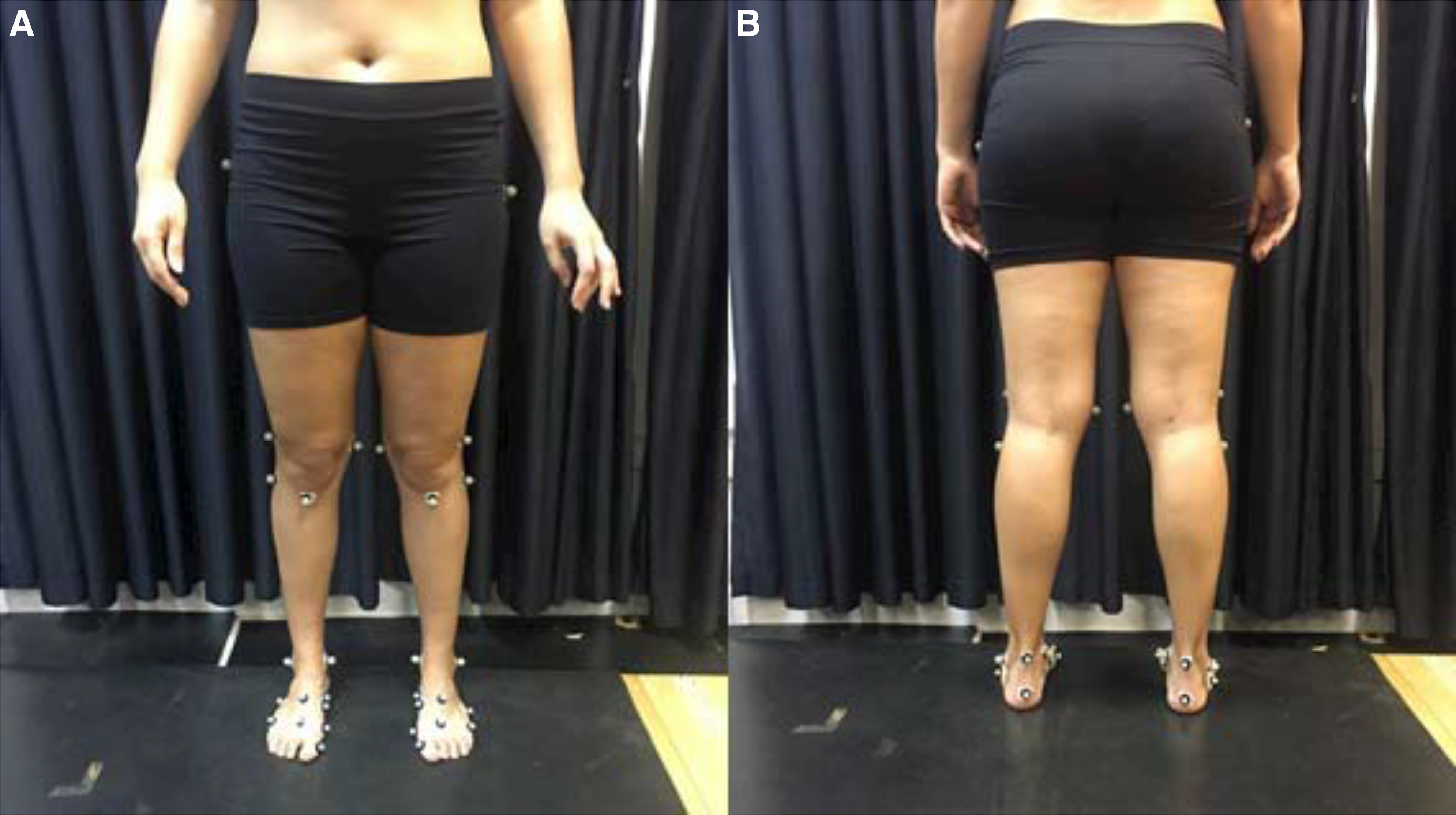
Kinematic data during the different tasks were recorded at 250 Hz using an eight-camera motion capture system (model MX-T-40S; Vicon Motion Systems Inc, Centennial, Colorado) synchronized with two force plates, one Bertec (Bertec Corp, Columbus, Ohio) and one AccuGait (AMTI, Watertown, Massachusetts). Anatomical and tracking markers were used to determine the coordinates of the thigh, shank, [22] and feet [23] with the participant in the orthostatic position (static trial) (Fig. 1).
Figure 1.
Marker placement, anterior (A) and posterior (B) views.
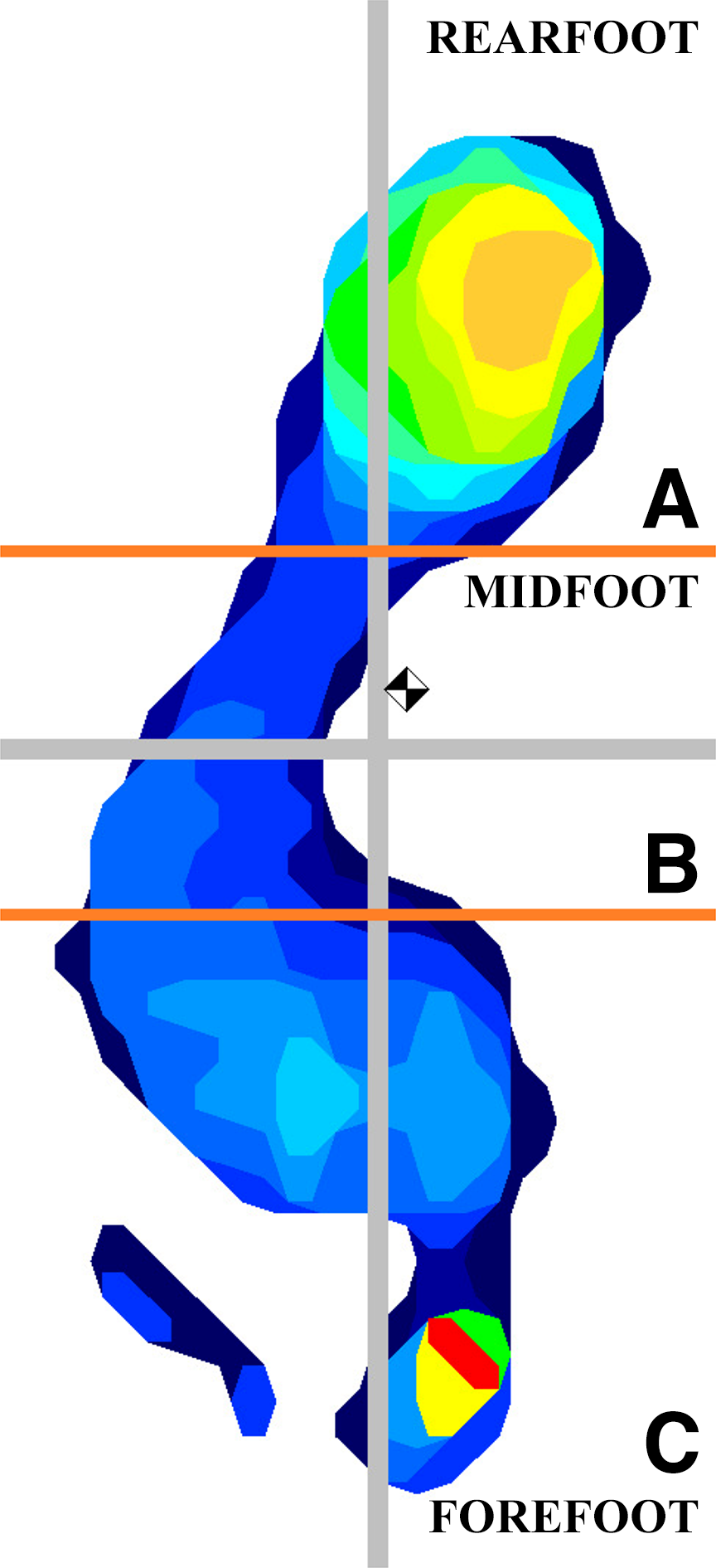
Foot plantar pressure and area data were collected using a 100-Hz baropodometer (MatScan system; Tekscan Inc, South Boston, Massachusetts; Fig. 2). Data from the baropodometer and the force platform were synchronized; this procedure guaranteed synchronization of baropodometric and kinematic data because the latter were collected simultaneously with the force platform data.
Figure 2.
Plantar area and pressure of the rearfoot (A), midfoot (B), and forefoot (C).
Participants performed approximately four repetitions of each task before data collection for familiarization. The order of data collection was as follows: stair descent, single-leg step down, single-leg squat, single-leg landing, and drop vertical jump. The participants performed three trials of the different tasks. The dominant lower limb was analyzed during the first valid trial of each task. The participants rested for 30 sec between each trial to avoid muscle fatigue during data collection.
During the stair descent task, participants were instructed to walk at a self-selected pace in a step over step fashion starting with the nondominant limb. The stairs had three steps, each 20 cm high, and the force plate (AMTI) was positioned on the step closer to the ground (Fig. 3A). [24,25] The baropodometer was firmly laid on the force plate.
Figure 3.
The five tasks in sequence: stair descent (A), single-leg step down (B), single-leg squat (C), single-leg landing (D), and drop vertical jump (E).
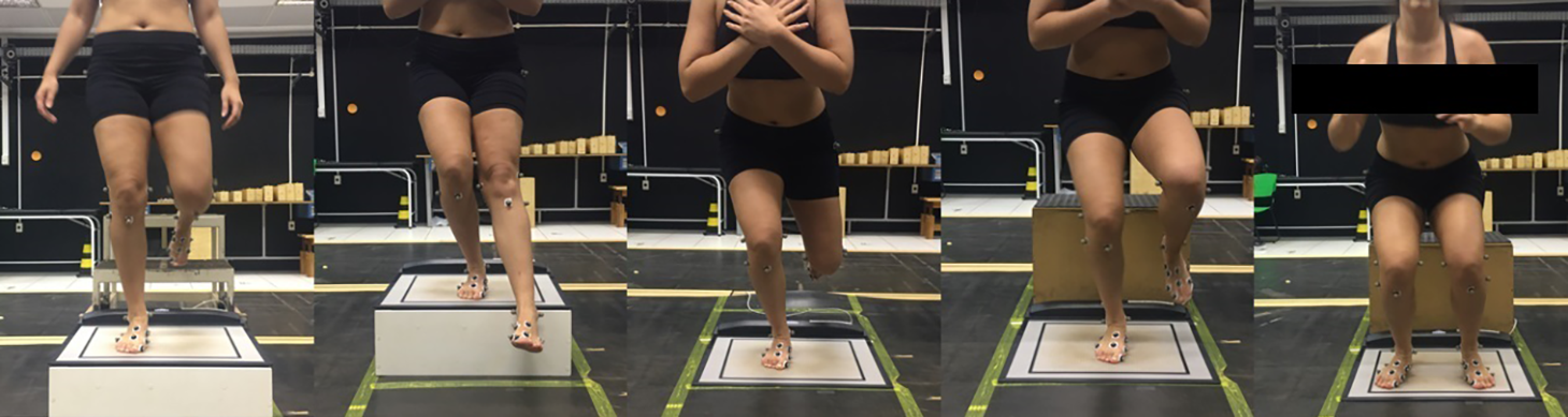
During the single-leg step down task, participants stood with arms crossed on top of a 20-cm-high box. They were instructed to “stand on your dominant leg and slowly lower the nondominant foot to lightly touch the floor with your heel and return to standing on both feet” (Fig. 3B). [9] The baropodometer was firmly laid on the box.
Participants performed the single-leg squat with the dominant lower limb at a rate of one squat per 4 sec: 2 sec for the eccentric phase and 2 sec for the concentric phase. The arms were crossed on the chest (Fig. 3C). [26]
Participants were instructed to perform the single-leg landing task from a 40-cm-high block. The force plate (Bertec) was on the ground, with the baropodometer firmly laid on it. The goal was to drop from the block and land with the dominant lower limb. Participants were asked to keep the arms crossed throughout the trials (Fig. 3D). [27]
During the drop vertical jump, participants were instructed to drop off of a 40-cm-high box with both feet, followed by a maximum vertical jump, moving their arms above the head as if they were trying to touch an object with both upper limbs (Fig. 3E). [28] The force plate (Bertec) was on the ground, with the baropodometer firmly laid on it. A trial was not valid if the participants jumped off of the box instead of just dropping or if they clearly fell or lost balance. [29]
Data Reduction
Data were processed using MATLAB software (The MathWorks Inc, Natick, Massachusetts). Raw kinematic data were filtered using a low-pass fifth-order Butterworth filter with a cutoff frequency set at 8 Hz. The following joint kinematics were calculated during the different tasks: 1) forefoot inversion-eversion, represented by the motion of the forefoot relative to the midfoot (X-axis) [23]; 2) rearfoot inversion-eversion, represented by the motion of the rearfoot relative to the shank (X-axis) [23]; and 3) knee flexion-extension and adduction-abduction, represented by the motion of the shank relative to the thigh. Kinematic data were computed in the joint coordinate system. [30]
For the foot plantar pressure and area data extraction, the foot was divided into three segments: forefoot, midfoot, and rearfoot. [31] In addition, the total plantar pressure area was used for data analysis, and foot plantar pressure and area are reported in kilopascal and square centimeters, respectively.
For the stair descent and drop vertical jump tasks, kinematic and baropodometric data were computed between first contact of the foot on the force plate until toe-off using the vertical component of the ground reaction force. For the single-leg step down and single-leg squat tasks, kinematic and baropodometric data were computed from the beginning of knee flexion until maximum knee flexion angle. For the single-leg landing task, kinematic and baropodometric data were computed from the first contact of the foot on the force plate until the maximum knee flexion angle. After this, the moment of occurrence of the peak value of the knee abduction angle, or knee valgus, during each task was determined. Then, the forefoot and rearfoot angle values in the frontal plane of motion and the forefoot, midfoot, and rearfoot plantar pressure and plantar area values at the moment of occurrence of the knee valgus peak were extracted for the statistical comparisons among the five tasks.
Statistical Analysis
The joint angle values and the plantar pressure and area values extracted for analysis were tested for normal distribution and homogeneity of variance using the Shapiro-Wilk and Mauchly tests, respectively, and then were compared among the five tasks using one-way repeated-measures analysis of variance (ANOVA), with significance set at α = 0.05. Greenhouse-Geisser and lower-bound corrections were used for the variables that violated the assumption of sphericity. [32] For variables with significant differences in ANOVA, preplanned contrasts were performed between the joint angles, plantar pressures, and plantar areas of the five different tasks, with α set at 0.05. The analyses were performed using SPSS software version 20 (SPSS Inc, Chicago, Illinois). The effect sizes (ESs) (ie, r values) of the contrasts with statistically significant differences were calculated as follows [33]: 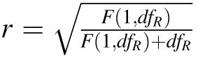 . [34]
Results
The mean (95% confidence interval) joint angle values and foot plantar pressure and area values during the five different tasks and the results of the repeated-measures ANOVAs are presented in Table 1. There were no differences in forefoot and rearfoot angles. There were differences between tasks for the total plantar area (Table 1). For the preplanned contrasts, single-leg squat and stair descent had increased total plantar area compared with single-leg landing (P = .005 and P = .027; mean ES = 0.47), drop vertical jump (P = .001 and P = .001; mean ES = 0.75), and single-leg step down (P = .01 and P = .007; mean ES = 0.47). In addition, the single-leg landing and single-leg step down tasks had greater total plantar area than the drop vertical jump task (P = .026 and P = .014; mean ES = 0.42).
Table 1.
Summary of Dependent Variables During the Five Different Tasks with the Results of the Repeated-Measures Analyses of Variance.
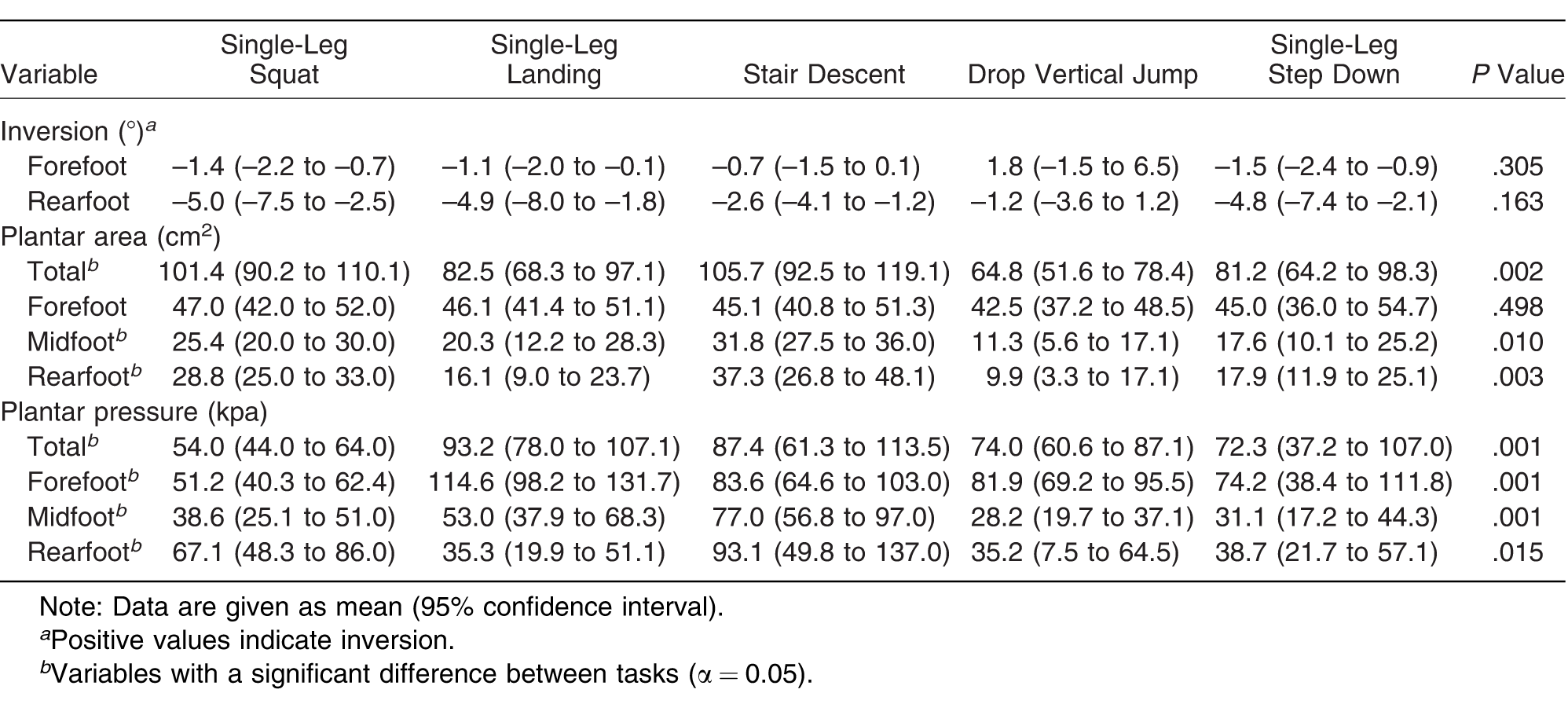
There were no differences among tasks for forefoot plantar area. There were differences among tasks for the midfoot and rearfoot plantar area (Table 1). Single-leg squat and stair descent had increased midfoot area compared with single-leg landing (P = .037 and P = 0.003; mean ES = 0.46), drop vertical jump (P = .001 and P = .001; mean ES = 0.70), and single-leg step down (P = .03 and P = .001; mean ES = 0.51). In addition, drop vertical jump had a smaller midfoot plantar area compared with the other tasks (P < .05; mean ES = 0.54). Finally, single-leg squat and stair descent had increased rearfoot area compared with single-leg landing (P = .019 and P = .007; mean ES = 0.86) and drop vertical jump (P = .001 and P = .011; mean ES = 0.89).
There were differences in foot total plantar pressure among tasks (Table 1). Single-leg landing and stair descent had increased total plantar pressure compared with the other tasks (P < .05; mean ES = 0.35 and mean ES = 0.28). In addition, the single-leg squat had reduced total plantar pressure compared with single-leg landing (P = .001; ES = 0.82), stair descent (P = .001; mean ES = 0.59), and drop vertical jump (P = .001; mean ES = 0.64).
There were differences in the forefoot, midfoot, and rearfoot plantar pressures among tasks (Table 1). Single-leg landing had increased forefoot plantar pressure compared with the other tasks (P < .001; mean ES = 0.96), and single-leg squat had reduced forefoot plantar pressure compared with the other tasks (P < .021; mean ES = 0.94). In addition, stair descent had increased midfoot plantar pressure compared with the other tasks (P < .05; mean ES = 0.59), and drop vertical jump had greater midfoot plantar pressure compared with single-leg landing (P = .006; ES = 0.49) and single-leg squat (P = .028; ES = 0.40). Finally, drop vertical jump had smaller rearfoot plantar pressure compared with stair descent (P = .014; ES = 0.70).
Discussion
The participants demonstrated no differences in forefoot and rearfoot inversion at the moment of occurrence of peak knee abduction during stair descent, single-leg step down, single-leg squat, single-leg landing, and drop vertical jump. However, participants had increased total plantar area during single-leg squat and stair descent compared with the other tasks. In addition, the participants had increased total plantar pressure during single-leg landing and stair descent compared with the other tasks. These results might be explained by differences in foot-striking pattern, magnitude of ground reaction force, and task speed.
The increased total and midfoot plantar area during stair descent might be related to the fact that this task is performed with forefoot striking. [35] Forefoot striking initially loads the arch in a three-point bending: 1) upward ground reaction force at the metatarsal heads, 2) downward body force through the talus, and 3) upward force in the triceps surae tendon. [36] Therefore, the plantar fascia is stretched and the foot medial arch is plantarflexed, which might help explain the increased rearfoot, midfoot, and total plantar area values during stair descent. Although drop vertical jump and single-leg landing are also forefoot-striking activities, they require faster movements than stair descent, therefore not allowing the plantar fascia to fully stretch by the time of knee abduction peak.
Participants had greater total plantar area during single-leg landing and single-leg step down compared with drop vertical jump. In addition, participants had reduced midfoot plantar area during drop vertical jump. These findings confirm the hypothesis that unipedal closed kinematic tasks, such as single-leg landing and single-leg step down, have increased total plantar area independently of increased forefoot and rearfoot eversion, probably by means of increased midfoot plantarflexion and consequently increased midfoot plantar area, as demonstrated in this study.
Similar to stair descent, single-leg squat had increased rearfoot, midfoot, and total plantar area. This is a unipedal and slow execution task. Therefore, it increases ground reaction force at the foot, but its slow rhythm of execution allows the plantar fascia and midfoot ligaments to stretch. Increased total plantar area suggests that the participants had a more balanced pressure distribution among the forefoot, midfoot, and rearfoot during single-leg squat and stair descent. This might be beneficial for people with heel pain or metatarsalgia. [16,37]
On the other hand, increased plantar area of the midfoot has been related to lower-limb injuries, such as patellofemoral pain syndrome. [13] Previous studies have suggested that a medially oriented foot pattern increases the lateral forces acting on the patella. [38,39] Therefore, the present study indicates that for the activities of descent of step and unilateral squatting there is a need for greater support and greater area of the foot, which must be taken into account during the rehabilitation process. Although the objectives of the present study are not objective, changes in pressure and contact areas of the foot during these tasks must also observe changes in the proximal chain for greater clinical relevance. Participants increased total plantar pressure in the forefoot during unilateral landing, increased total and median plantar pressure during ladder descent, and reduced forefoot plantar pressure during unilateral squatting.
These findings might be related to the increased ground reaction forces associated with single-leg landing and stair descent. [35,40] Although participants had increased total plantar area during stair descent, this was not sufficient to reduce plantar pressure, especially at the midfoot. Increased midfoot plantar pressure may increase the amount of loading transmitted through the plantar fascia, and consequently contribute to the development of plantar fasciitis, [41,42] which is still speculative at this point. Finally, participants had reduced total plantar pressure during single-leg squat compared with the other tasks, except for single-leg step down. Therefore, the increased total plantar area demonstrated during single-leg squat had the positive effect of reducing total plantar pressure, especially at the forefoot.
The fact that this study found differences between tasks in foot plantar area and pressure and did not find differences in forefoot and rearfoot eversion shows that baropodometric data should not be used as a surrogate of foot kinematic data, at least for the five tasks evaluated in this study. Note that this study investigated only forefoot and rearfoot frontal plane motion. It is possible that foot segment motion in the sagittal and transverse planes have the closest relationship with plantar pressure and area data. For example, Giacomozzi et al [43] demonstrated that during gait, peak pressure at the different foot regions is not correlated with rearfoot or forefoot frontal plane motion. However, they found high correlations between forefoot and rearfoot sagittal and transverse plane motion and peak plantar pressure data, such as a negative correlation between forefoot range of motion in the sagittal plane and forefoot peak pressure. Future studies should investigate these relationships for the five tasks included in the present study.
The order of data collection between the different tasks was not randomized, and this is a limitation of the study. It is possible that some learning effect and/or muscle fatigue occurred during data collection, compromising or improving performance during the last tasks (single-leg landing and drop vertical jump). Nonetheless, randomization was not possible for practical reasons. In addition, the dependent variables were extracted at the moment of occurrence of knee abduction peak, which might have hidden some differences in forefoot and rearfoot frontal plane motion and plantar pressure and area between tasks. For example, it is possible that forefoot and rearfoot segments respond to the different demands of the different tasks at sooner or later moments than the knee. [27] However, we chose the knee abduction peak as a reference because it is the most clinically and scientifically investigated motion pattern during the tasks evaluated in this study. [9,44]
Conclusions
There were no differences in forefoot and rearfoot eversion angles at the moment of occurrence of knee abduction peak among stair descent, single-leg step down, single-leg landing, single-leg squat, and drop vertical jump. On the other hand, the participants had increased total and midfoot plantar area during single-leg squat and stair descent, increased total plantar pressure during single-leg landing and stair descent, increased forefoot plantar pressure during single-leg landing, and increased midfoot plantar pressure during stair descent. These findings might be related to differences in foot-striking pattern, magnitude of ground reaction force, and execution speed among the five tasks. Finally, these results can help in the selection of tasks that are more appropriate for use at certain times of rehabilitation and for patients with different conditions of contact and coupling of the foot with the ground during the rehabilitation of several lower-limb dysfunctions in different types of tasks performed.
Financial Disclosure
None reported.
Conflicts of Interest
None reported.
References
- BartonCJLevingerPMenzHB: Kinematic gait characteristics associated with patellofemoral pain syndrome: a systematic review. Gait Posture30: 405, 2009.
- BolingMCPaduaDAMarshallSW: A prospective investigation of biomechanical risk factors for patellofemoral pain syndrome: the Joint Undertaking to Monitor and Prevent ACL Injury (JUMP-ACL) cohort. Am J Sports Med37: 2108, 2009.
- AllenMKGlasoeWM:Metrecom measurement of navicular drop in subjects with anterior cruciate ligament injury. J Athl Train35: 403, 2000.
- GrossMT:Lower quarter screening for skeletal malalignment: suggestions for orthotics and shoewear. J Orthop Sports Phys Ther21: 389, 1995.
- PowerVCliffordAM:The effects of rearfoot position on lower limb kinematics during bilateral squatting in asymptomatic individuals with a pronated foot type. J Hum Kinet31: 5, 2012.
- FarrokhiSKeyakJHPowersCM:Individuals with patellofemoral pain exhibit greater patellofemoral joint stress: a finite element analysis study. Osteoarthritis Cartilage19: 287, 2011.
- LiGDeFrateLEZayontzS: The effect of tibiofemoral joint kinematics on patellofemoral contact pressures under simulated muscle loads. J Orthop Res22: 801, 2004.
- HerringtonL:Knee valgus angle during single leg squat and landing in patellofemoral pain patients and controls. Knee21: 514, 2014.
- EarlJEMonteiroSKSnyderKR:Differences in lower extremity kinematics between a bilateral drop vertical jump and a single leg step down. J Orthop Sports Phys Ther37: 245, 2007.
- KristianslundEKrosshaugT:Comparison of drop jumps and sport-specific sidestep cutting: implications for anterior cruciate ligament injury risk screening. Am J Sports Med41: 684, 2013.
- TaylorJBFordKRNguyenAD: Biomechanical comparison of single- and double-leg jump landings in the sagittal and frontal plane. Orthop J Sports Med4: 2325967116655158, 2016.
- RathMEStearneDJWalkerCR: Effect of foot type on knee valgus, ground reaction force, and hip muscle activation in female soccer players. J Sports Med Phys Fitness56: 546, 2016.
- RathleffMSRichterCBrushøjC: Increased medial foot loading during drop jump in subjects with patellofemoral pain. Knee Surg Sports Traumatol Arthrosc22: 2301, 2014.
- FerrignoCWimmerMATrombleyRM: A reduction in the knee adduction moment with medial thrust gait is associated with a medial shift in center of plantar pressure. Med Eng Phys38: 615, 2016.
- MyerGDFordKRDi StasiSL: High knee abduction moments are common risk factors for patellofemoral pain (PFP) and anterior cruciate ligament (ACL) injury in girls: is PFP itself a predictor for subsequent ACL injury?Br J Sports Med49: 118, 2015.
- NagelAFernholzFKibeleC: Long distance running increases plantar pressures beneath the metatarsal heads: a barefoot walking investigation of 200 marathon runners. Gait Posture27: 152, 2008.
- WillemsTMDe RidderRRoosenP:The effect of a long-distance run on plantar pressure distribution during running. Gait Posture35: 405, 2012.
- PhillipsAMcClintonS:Gait deviations associated with plantar heel pain: a systematic review. Clin Biomech (Bristol, Avon)42: 55, 2017.
- HuangHKeijsersNHoremansH: Anterior cruciate ligament rupture is associated with abnormal and asymmetrical lower limb loading during walking. J Sci Med Sport20: 432, 2017.
- MokhtarzadehHYeowCHHong GohJC: Contributions of the soleus and gastrocnemius muscles to the anterior cruciate ligament loading during single-leg landing. J Biomech46: 1913, 2013.
- AraiTMiakiH:Influence of static alignment of the knee, range of tibial rotation and tibial plateau geometry on the dynamic alignment of “knee-in” and tibial rotation during single limb drop landing. Clin Biomech28: 642, 2013.
- CappozzoADella CroceULeardiniA: Human movement analysis using stereophotogrammetry: part 1. Theoretical background. Gait Posture21: 186, 2005.
- PortinaroNLeardiniAPanouA: Modifying the Rizzoli foot model to improve the diagnosis of pes-planus: application to kinematics of feet in teenagers. J Foot Ankle Res7: 57, 2014.
- BrechterJHPowersCM:Patellofemoral joint stress during stair ascent and descent in persons with and without patellofemoral pain. Gait Posture16: 115, 2002.
- CowanSMBennellKLCrossleyKM: Physical therapy alters recruitment of the vasti in patellofemoral pain syndrome. Med Sci Sports Exerc34: 1879, 2002.
- CrossleyKMZhangWJSchacheAG: Performance on the single-leg squat task indicates hip abductor muscle function. Am J Sports Med39: 866, 2011.
- LeeSPPowersCM:Fatigue of the hip abductors results in increased medial–lateral center of pressure excursion and altered peroneus longus activation during a unipedal landing task. Clin Biomech28: 524, 2013.
- FordKMyerGTomsH: Gender differences in the kinematics of unanticipated cutting in young athletes. Med Sci Sport Exerc37: 124, 2005.
- StensrudSMyklebustGKristianslundE: Correlation between two-dimensional video analysis and subjective assessment in evaluating knee control among elite female team handball players. Br J Sports Med45: 589, 2011.
- SuntayWJGroodESHefzyMS: Error analysis of a system for measuring three-dimensional joint motion. J Biomech Eng105: 127, 1983.
- RodgersM:Dynamic biomechanics of the normal foot and ankle during walking and running. Phys Ther68: 1822, 1988.
- HinkleDJursSWiesmaW:Applied Statistics for the Behavioral Sciences,2nd Ed,Houghton Mifflin, Boston, 1988.
- MorrisSBDeShonRP:Combining effect size estimates in meta-analysis with repeated measures and independent-groups designs. Psychol Methods1: 105, 2002.
- FieldA:Discovering Statistics Using SPSS, 2nd Ed, Sage, London, 2006.
- LeeJHongYGShinCS:Effects of mid-foot contact area ratio on lower body kinetics/kinematics in sagittal plane during stair descent in women. Gait Posture48: 89, 2016.
- PerlDPDaoudAILiebermanDE:Effects of footwear and strike type on running economy. Med Sci Sports Exerc447: 35, 2012.
- SilvaRSMacielCDSerrãoFV:The effects of forefoot varus on hip and knee kinematics during single-leg squat. Man Ther20: 79, 2015.
- AlibertiSCostaMSPassaroAC: Influence of patellofemoral pain syndrome on plantar pressure in the foot rollover process during gait. Clinics66: 367, 2011.
- AlibertiSCostaMSPassaroAC: Medial contact and smaller plantar loads characterize individuals with patellofemoral pain syndrome during stair descent. Phys Ther Sport11: 30, 2010.
- KawakamiYOgasawaraIYonetaniY: Difference in dynamic body balance between forward and lateral single-leg hop landing. Kurume Med J63: 2, 2016.
- MS,Chan ParkLeeSKimS: The effects of the application of low-dye taping on paretic side plantar pressure among patients with plantar fasciitis. J Phys Ther Sci27: 3555, 2015.
- KimTHKohEKJungDY:The effect of arch support taping on plantar pressure and navicular drop height in subjects with excessive pronated foot during 6 weeks. J Korean Soc Phys Med6: 489, 2011.
- GiacomozziCLeardiniACaravaggiP:Correlates between kinematics and baropodometric measurements for an integrated in-vivo assessment of the segmental foot function in gait. J Biomech47: 2654, 2014.
- DonohueMREllisSMHeinbaughEM: Differences and correlations in knee and hip mechanics during single-leg landing, single-leg squat, double-leg landing, and double-leg squat tasks. Res Sports Med23: 394, 2015.
© 2020 American Podiatric Medical Association