


Abstract
Malignant transformation of wounds is a rare complication that if missed can lead to loss of life or limb. This case report presents a rare invasive variant of squamous cell carcinoma presenting in the setting of a chronic wound complicated by osteomyelitis. This aggressive form of squamous cell carcinoma has a high growth rate and a high propensity for metastasis and recurrence. Early intervention greatly decreases the risk of metastasis and recurrence. We present the systematic evaluation and surgical management of an aggressive primary tumor occurring in the forefoot.
Squamous cell carcinoma (SCC) of the skin is the second most common form of skin cancer in the United States, and recently the incidence has been rising.[1] Squamous cell carcinoma commonly develops in people with fair skin in areas that are subject to sun exposure.[2-6] Histopathologic examination of SCC is characterized by keratinocyte dysplasia throughout the epidermis extending into the dermis. Excessive mitoses are seen, and the keratinocytes are pleomorphic with hyperchromatic nuclei.[7]
In a small percentage of patients, SCC can develop as an invasive variant with a higher propensity to metastasize. The development of this aggressive form of SCC is often unrelated to sun exposure.[8] Tumor size greater than 2 cm, depth of invasion greater than 6 mm, poor degree of differentiation, perineural invasion, tumor occurring in the site of a previous burn or chronic wound, and stage of disease are prognostic factors for recurrence, death, and metastasis in these patients.[9-12] Invasive cutaneous SCC is a rare soft-tissue neoplasm that can be difficult to manage due to its aggressive growth and propensity for metastasizing. Reports of metastasis of these SCC variants can be up to 42%.[8,12] The risk of recurrence is 3.8 times higher in patients with tumors larger than 2 cm or invading into deep dermal structures. Full-body work-up is warranted owing to the increased risk of metastasis with the invasive SCC variant. Aggressive surgical resection of locally or regionally advanced SCC may lower the risk of recurrence.[13]
Edwards et al[14] found that tumors occurring in sites of chronic or previous wounds have a 58% chance of recurrence and lead to 5-year mortality of 48%. Mullen et al[13] demonstrated that 12% of the invasive SCC variant can develop at a site of previous skin injury or wound, termed a scar carcinoma. This study also found that primary tumors larger than 2 cm in diameter and/or invading deep structures led to a recurrence rate of 84%, 45% greater than tumors lacking these characteristics.[13] However, this same study found that aggressive surgical resection, including amputation, reduced the recurrence rate to 29%.
Management of low-risk SCC includes surgical excision, cryotherapy, electrodessication and curettage, photodynamic therapy, radiotherapy, and topical 5-fluorouracil for low-risk lesions.[15-17] However, early and aggressive management of high-risk lesions is necessary due to their high chance of recurrence and metastasis.
Malignancies of the foot and ankle are rare, and late diagnosis of these lesions increases the propensity for metastasis. Early biopsy and aggressive management, including surgical resection, improves outcomes by preventing metastases and recurrence. This case highlights the need for foot and ankle surgeons to have a high suspicion for malignancy.
Case Report
This case study describes a 65-year-old woman with a chronic, nonhealing wound of the plantar forefoot despite previous third metatarsal head resection. The patient presented to the University of Cincinnati outpatient clinic (Cincinnati, Ohio) in October 2016. Third metatarsal head resection was performed at an outside facility due to osteomyelitis of this location before 2013.
Her medical history was significant for hyperlipidemia, hypertension, type 2 diabetes mellitus with a hemoglobin A1C level of 6.0%, and depression. She was taking atorvastatin for her hyperlipidemia, lisinopril for her hypertension, metformin for her diabetes mellitus, and sertraline for her depression. She had no known allergies to medication. Her family history was unremarkable, and she had no history of malignancy. Her surgical history was noncontributory aside from previous third metatarsal head resection for osteomyelitis, as mentioned previously herein. She was a former smoker and denied any drug use.
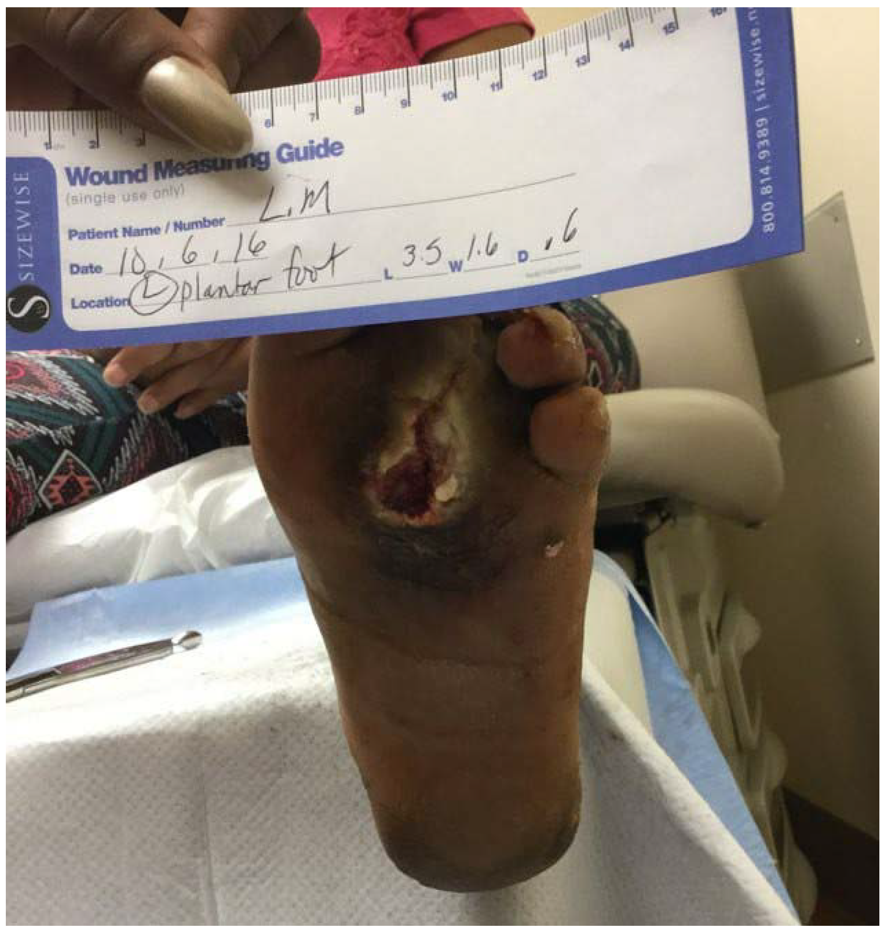
Her physical examination findings were unremarkable aside from a central wound near previous surgery for removal of the third metatarsal head. At this time the wound measured 3.5 × 1.6 × 0.6 cm and did not probe to bone or display acute signs of infection (Fig. 1).
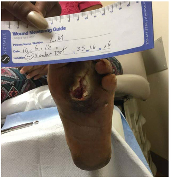
Figure 1.
Initial presentation of the chronic wound to the plantar left third metatarsal head.
Radiographic examination revealed previous third metatarsal head resection without evidence of soft-tissue emphysema, soft-tissue or osseous neoplasm, fracture, or dislocation (Fig. 2). A magnetic resonance image (MRI) was obtained due to the chronicity and irregularity of the wound and concern for residual osteomyelitis or neoplasm of the third metatarsal stump and wound. The MRI report at this time was significant for abscess near the third proximal phalanx, findings concerning for osteomyelitis of the third metatarsal stump, and dorsal foot cellulitis (Fig. 3). The patient was taken to the operating room for third-digit amputation and more proximal resection of the third metatarsal. Pathology evaluation from this procedure reported invasive SCC. The distance of the carcinoma from the unaffected margin was within 1 mm, indicating that the carcinoma clearance margin was not adequate.
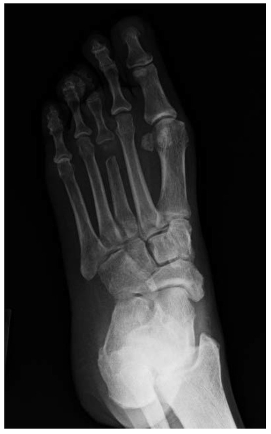
Figure 2.
Medial oblique radiograph of the left foot demonstrating no soft-tissue emphysema, neoplasm, or fracture.
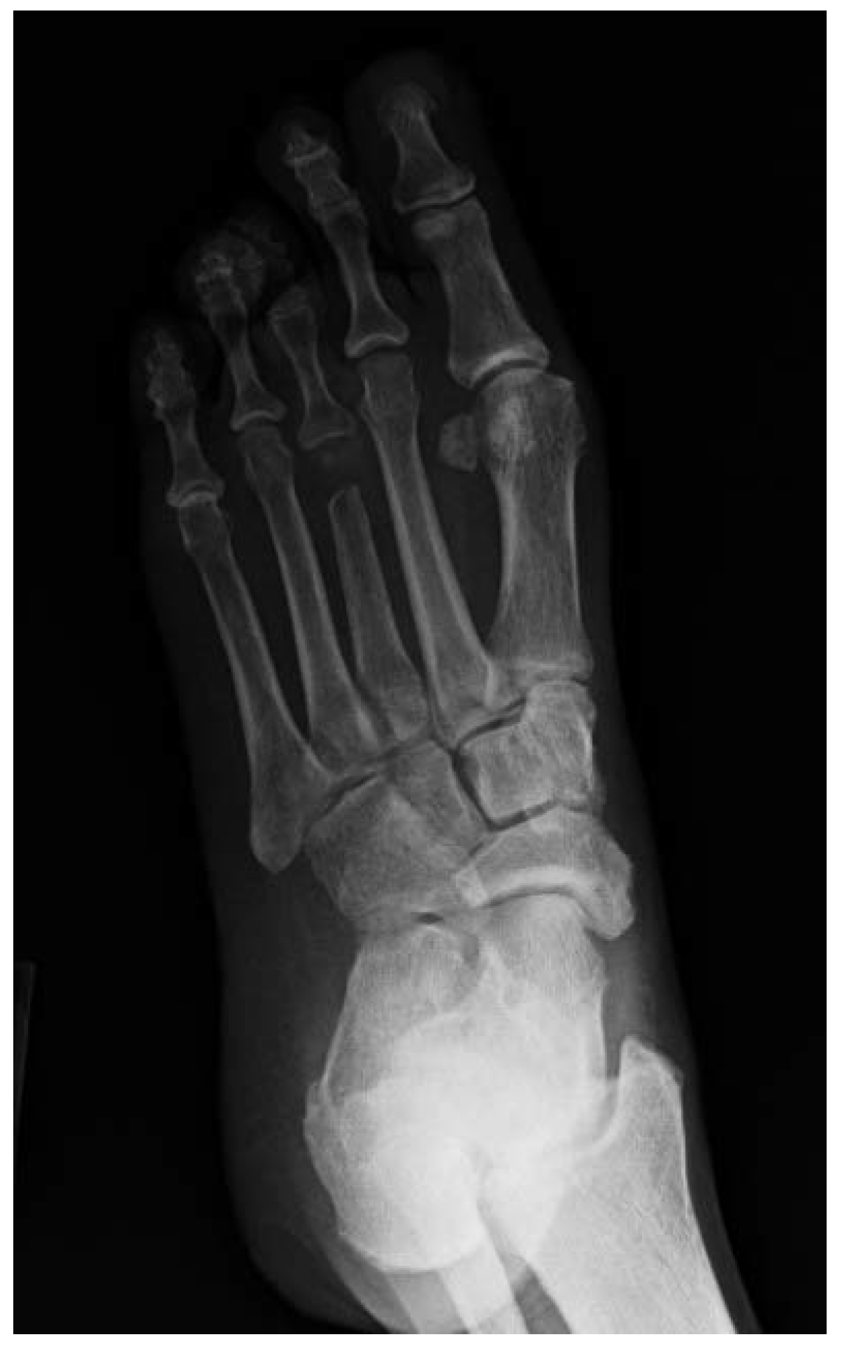
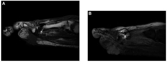
Figure 3.
Representative sagittal T1-weighted (A) and T2-weighted (B) magnetic resonance imaging slices demonstrating the irregular appearance of the distal third metatarsal stump with surrounding cellulitis.
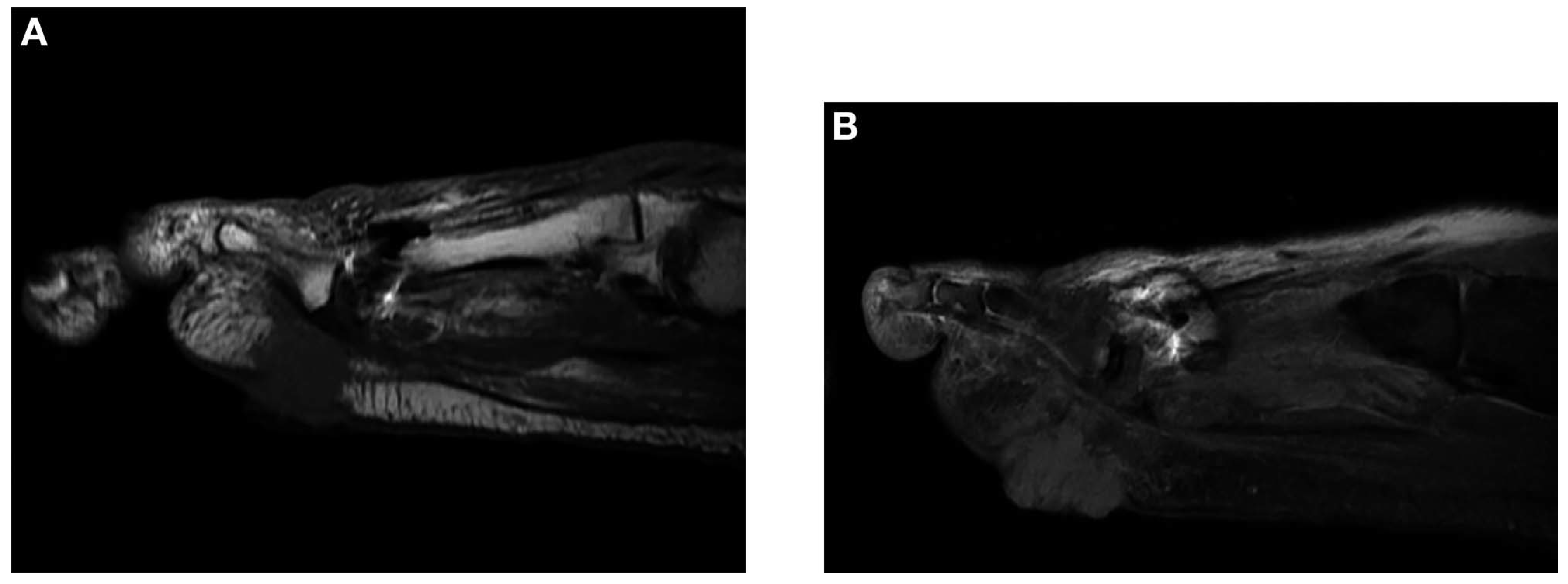
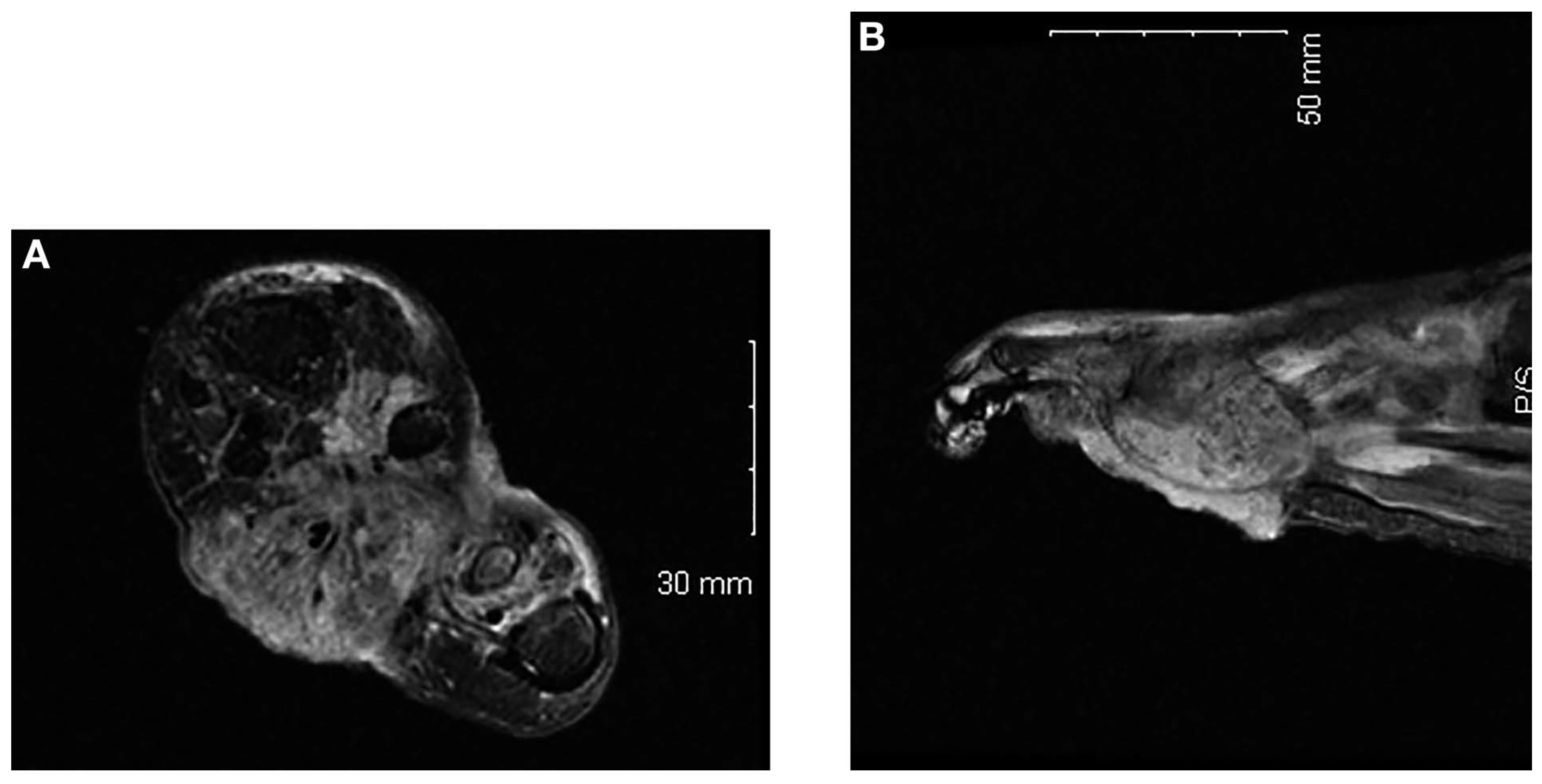
The patient was intermittently lost to follow-up and presented to the clinic 3 months later with worsening soft-tissue ulceration and an enlarging soft-tissue mass (Fig. 4). Repeated MRI demonstrated a soft-tissue mass measuring 5 × 3.4 × 3.5 cm infiltrating digits 1 through 4 and involving the flexor muscles, tendons, interossei muscles, pathologic fracture of the fourth proximal phalanx, and subcutaneous region extending to the skin (Fig. 5). The hematology and oncology service, along with the surgical oncology service, were consulted and recommended wide excision of the carcinoma.
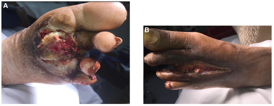
Figure 4.
Clinical plantar (A) and dorsal (B) appearance of an enlarging soft-tissue mass after third-digit amputation.
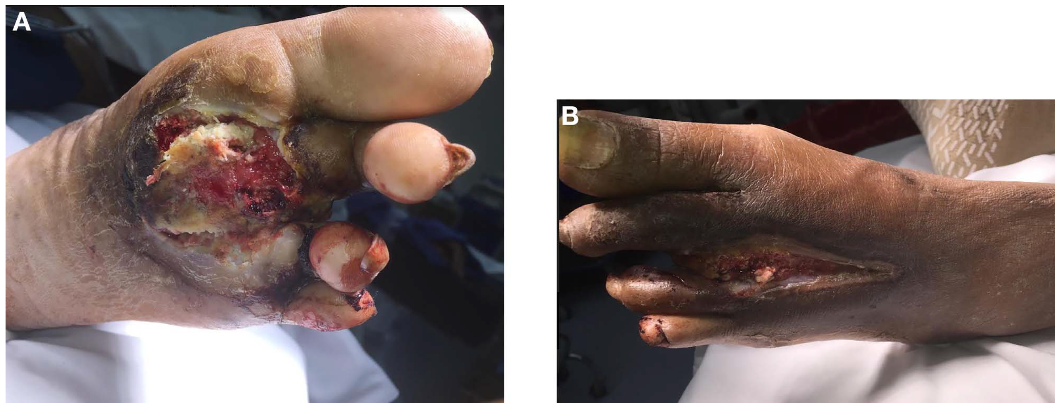
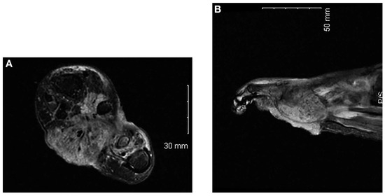
Figure 5.
Axial (A) and sagittal (B) T2-weighted magnetic resonance images showing extensive deep invasion of squamous cell carcinoma.
Owing to the invasive nature of SCC, a full-body work-up for metastasis was completed in coordination with the surgical oncology service and the hematology and oncology service, including positron emission tomography–computed tomography, which demonstrated increased uptake consistent with tumor in the left foot, mildly increased uptake in the left inguinal lymph nodes consistent with infection, and a lung nodule without increased uptake (Fig. 6). Biopsy of a lung nodule was subsequently performed and was negative for malignancy.

Figure 6.
Positron emission tomography–computed tomography showing increased uptake in the left forefoot.

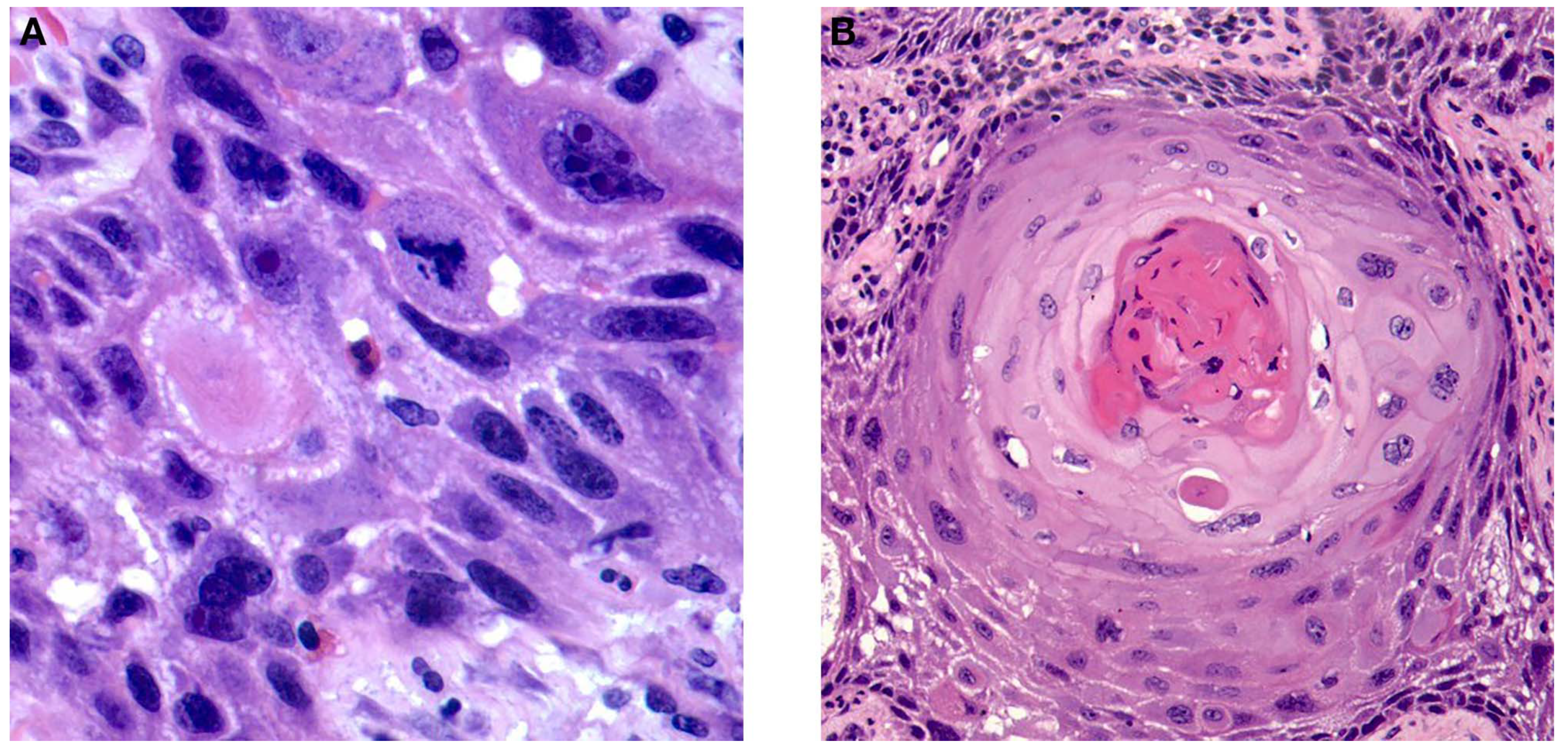
The patient was scheduled for surgical excision of SCC under general anesthesia with wide resection. A transverse incision was made proximal to the area of the tumor located by MRI. Dissection was continued deep until bone was encountered, and the first, second, fourth, and fifth metatarsals were transversely osteotomized, leaving approximately 1 cm of bone. The third metatarsal was completely excised at the tarsometatarsal joint. Intraoperative frozen section examination of the clearance margin 1 cm proximal to the soft-tissue mass was negative for carcinoma or another pathologic tissue. Surgical pathologic examination of the resected mass revealed a 6.0 × 4.5 × 3.8-cm mass of invasive, well-differentiated SCC with all margins free of tumor by more than 5 mm. Histopathologic examination revealed atypical cells, prominent nucleoli, and nuclear atypia consistent with SCC (Fig. 7).
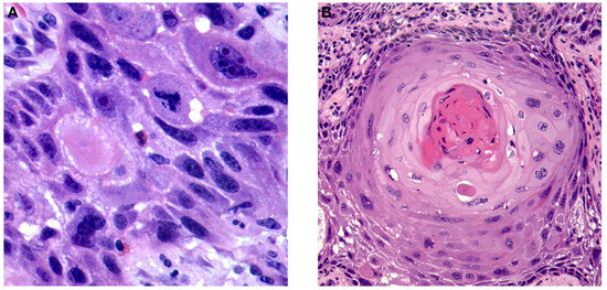
Figure 7.
Representative slides of well-differentiated squamous cell carcinoma demonstrating nuclear atypia and prominence. Hematoxylin-eosin stain: ×60 (A) and ×20 (B).
A follow-up visit 1 month postoperatively revealed no local recurrence of pathologic tissue. Repeated chest radiography and chest computed tomography showed no worsening of the lung nodule. More than 1 year postoperatively, the patient had no local recurrence (Fig. 8). She follows up with the hematology and oncology service along with the pulmonology service for monitoring of the lung nodule, which is stable.
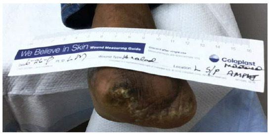
Figure 8.
Clinical picture of the healed surgical site without local recurrence 1 year postoperatively.
Discussion
Squamous cell carcinoma of the lower extremity has been described in the past by different authors; however, the typical presentation is either SCC in situ (Bowen disease) or slow malignant transformation of chronic wounds (Marjolin ulcer).[18,19] The present case differs from these previous reports due to the rapid growth and presentation of this malignancy as an aggressive, invasive variant in a postsurgical wound. This case report demonstrates the need for foot and ankle surgeons to have a high suspicion of malignancy in irregular or chronic wounds. Delay in diagnosis of aggressive, rapidly growing tumors greatly increases the chance for metastasis and puts the limb at risk, whereas prompt diagnosis may lead to limb salvage and reduce morbidity and mortality. In addition, the present case reports the systematic work-up and multidisciplinary approach to aggressive tumors occurring in the lower extremity and the need for consultation with the oncology specialty services that commonly manage malignancy. Close follow-up of patients with invasive SCC is necessary to address recurrence, new lesions, and metastases.
Financial Disclosure
None reported.
Conflicts of Interest
None reported.
References
- KariaPSHanJSchmultsCD:Cutaneous squamous cell carcinoma: estimated incidence of disease, nodal metastasis, and deaths from disease in the United States, 2012. J Am Acad Dermatol68: 957, 2013.
- BrantschKDMeisnerCSchönfischB: Analysis of risk factors determining prognosis of cutaneous squamous-cell carcinoma: a prospective study. Lancet Oncol9: 713, 2008.
- SamarasingheVMadanVLearJT:Management of high-risk squamous cell carcinoma of the skin. Expert Rev Anticancer Ther11: 763, 2011.
- FarasatSYuSSNeelVA: A new American Joint Committee on Cancer staging system for cutaneous squamous cell carcinoma: creation and rationale for inclusion of tumor (T) characteristics. J Am Acad Dermatol64: 1051, 2011.
- RoweDECarrollRJDayCLJr: Prognostic factors for local recurrence, metastasis, and survival rates in squamous cell carcinoma of the skin, ear, and lip: implications for treatment modality selection. J Am Acad Dermatol26: 976, 1992.
- BroughamNDDennettERCameronR: The incidence of metastasis from cutaneous squamous cell carcinoma and the impact of its risk factors. J Surg Oncol106: 811, 2012.
- YanofskyVRMercerSEPhelpsRG:Histopathological variants of cutaneous squamous cell carcinoma: a review. J Skin Cancer2011: 210813, 2011.
- CherpelisBSMarcusenCLangPG:Prognostic factors for metastasis in squamous cell carcinoma of the skin. Dermatol Surg28: 268, 2002.
- AmesFCHickeyRC:Squamous cell carcinoma of the skin of the extremities. Int Adv Surg Oncol3: 179, 1980.
- JohnsonTMRoweDENelsonBR: Squamous cell carcinoma of the skin (excluding lip and oral mucosa). J Am Acad Dermatol26: 467, 1992.
- BarksdaleSKO'ConnorNBarnhillR:Prognostic factors for cutaneous squamous cell and basal cell carcinoma: determinants of risk of recurrence, metastasis, and development of subsequent skin cancers. Surg Oncol Clin N Am6: 625, 1997.
- NorthJHJr,SpellmanJEDriscollD: Advanced cutaneous squamous cell carcinoma of the trunk and extremity: analysis of prognostic factors. J Surg Oncol64: 212, 1997.
- MullenJTFengLXingY: Invasive squamous cell carcinoma of the skin: defining a high-risk group. Ann Surg Oncol13: 902, 2006.
- EdwardsMJHirschRMBroadwaterJR: Squamous cell carcinoma arising in previously burned or irradiated skin. Arch Surg124: 115, 1989.
- LansburyLBath-HextallFPerkinsW: Interventions for non-metastatic squamous cell carcinoma of the skin: systematic review and pooled analysis of observational studies. BMJ347: f6153, 2013.
- Bath-HextallFJMatinRNWilkinsonD: Interventions for cutaneous Bowen's disease. Cochrane Database Syst Rev2013: CD007281, 2013.
- LansburyLLeonardi-BeeJPerkinsW: Interventions for non-metastatic squamous cell carcinoma of the skin. Cochrane Database Syst Rev4: CD007869, 2010.
- HolgadoRDWardSCSuryaprasadSG:Squamous cell carcinoma of the hallux. JAPMA90: 309, 2000.
- PavlovicSSurowiecDBranieckiM:Marjolin's ulcer of the hallux. JAPMA103: 250, 2013.
© 2020 American Podiatric Medical Association