


Abstract
Background: Pregnancy is a period when a woman's body undergoes changes. The purpose of this study was to analyze the mechanisms of gait adaptation in overweight pregnant women regarding spatiotemporal gait parameters, ground reaction forces, and plantar pressure distribution. Methods: The tests were performed in 29 normal-weight pregnant women and 26 pregnant women who were overweight before pregnancy. The measurements included spatiotemporal gait parameters, in-shoe plantar pressure distribution, and ground reaction forces during gestation. Results: The results indicate that both normal-weight and overweight pregnant women make use of the same spatiotemporal gait parameters to increase body stability and safety of movement during pregnancy. The double-step duration in the third trimester of pregnancy was higher in normal-weight and overweight pregnant women compared with in the first trimester (P < .05). A significant change in pressure amplitude was found under all anatomical parts of the foot in the third trimester (P < .05). The results also suggest a higher increase in the maximum amplitude of force in overweight pregnant women in the third trimester compared with the normal-weight group. Conclusions: This study suggests that both normal-weight and overweight pregnant women use different mechanisms of gait adaptation during pregnancy. In practice, understanding the biomechanical changes in women's gait can protect the musculoskeletal system during gestation.
The most substantial change during pregnancy is weight gain, where the anterior mass on the trunk is increased by an average of 0.29 kg per week in the lower trunk segment. [1] In normal pregnancy, the body mass increases and a significant change in body mass distribution is observed during the second and the third trimesters of pregnancy. [1-3] The increasing body mass in pregnant women can change the position of the center of gravity and have an effect on unstable postural balance, incorrect positioning of the feet, and the biomechanics of gait. [4-11]
In normal-weight pregnant women, some researchers have analyzed spatial and temporal parameters such as stride length, step length, stride width, and single and double support time during pregnancy. They found a significant decrease in the step length [5,12-14] and the length of the gait cycle. [5,13,14] A significant increase, on the other hand, was found for double support time. [4,7,13] Other studies have reported a significant reduction in single support time [4,7] and a significant increase in step width, by approximately 10%. [14-16] Tridimensional adaptation of kinematic parameters in pregnant women was also analyzed in several studies. [5,7,17] Branco et al [18] analyzed the adaptations of kinetic parameters between pregnant women and a control group. In the joint moments they found a decrease in hip flexors, knee extensors, ankle dorsiflexors, and ankle evertors' moment and an increase in the participation of hip external rotators during the loading response phase in pregnant women. Few studies have evaluated the kinetic parameters of gait during pregnancy. [16,19] Lymbery and Gilleard [16] found no significant differences in ground reaction forces (GRFs) in women in the last trimester of pregnancy compared with the postpartum period. In the sagittal plane, a significant decrease was found in the knee extensor moment and under the plantar flexor moment. Some studies have also demonstrated changes in plantar overload on the feet during pregnancy. [20-22] Nyska et al [21] found that in the static position, pregnant women displayed significantly lower maximal forefoot pressures and higher rearfoot pressures. Goldberg et al [23] also found that peak pressures increase in the rearfoot and decrease in the forefoot. However, Karadag-Saygi et al [22] observed increased peak pressures in the forefoot.
An increasingly more pronounced problem in pregnant women is obesity. With more than 40% of women gaining weight in excess of Institute of Medicine recommendations, [24] pregnancy is now emerging as an important risk factor for excessive weight gain [17,25,26] and an important target for obesity prevention studies. [27] Throughout pregnancy, women with a body mass index (BMI; calculated as weight in kilograms divided by height in meters squared) lower than 19.8 (underweight) should increase their weight by 12.5 to 18 kg; women with a BMI of 19.8 to 25.9 (normal weight) by 11.5 to 16 kg; women with a BMI of 26.0 to 28.9 (overweight) by 7 to 11.5 kg; and women with a BMI equal to or greater than 29 (obese) by at least 6.8 kg. [24]
Obesity has a negative effect on the musculoskeletal system, leading to knee and ankle overexertion, balance difficulties, and functional disability. Pregnant women experience various complications, including gestational diabetes, hypertensive disorders, or Cesarean deliveries. [17,28-30] To our knowledge, no studies have been performed to investigate gait parameters during gestation and adaptation of the musculoskeletal system due to the fact that physiologic changes in overweight pregnant women are not fully recognized. The purpose of this study was to analyze changes in spatiotemporal gait parameters, GRFs, and plantar pressure distribution in overweight pregnant women. We hypothesized that for every increase in mass, significant changes in gait parameters occur. Understanding how women adapt to the increased anterior mass during pregnancy can help us understand the modifications that may improve their stability.
Methods
Participants
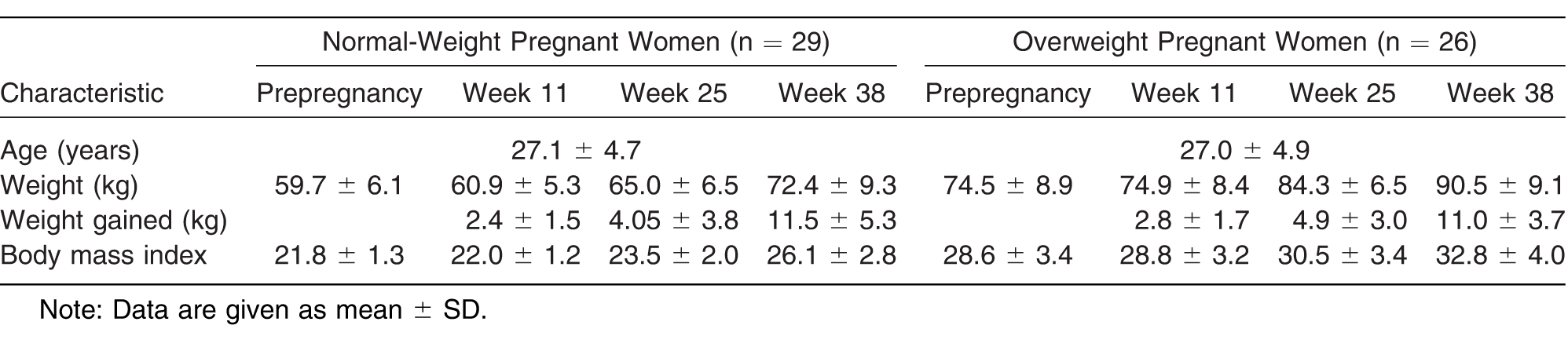
The tests were performed in 29 normal-weight pregnant women and 26 pregnant women who were overweight before pregnancy. Both normal-weight and overweight pregnant women were randomly selected from a population of 152 women. The inclusion criteria were specified as follows: women must be aged 18 to 40 years, of normal body weight or overweight based on prepregnancy BMI, and own sports shoes to be worn during data collection. The exclusion criteria were high-risk pregnancy, complications of pregnancy, and disorders that may affect the woman's gait and plantar pressure distribution, such as those connected with musculoskeletal abnormalities, diabetes, pelvic girdle pain, neuromuscular trauma or disease, or cardiovascular problems. The local ethics committee (Institute of Mother and Child, Warsaw, Poland) approved the study. All of the women received full information about the study before granting their signed consent. Weight was recorded on an electronic scale, and height was measured with a stadiometer. Normal weight was defined as a BMI of 18.5 to 24.99. Similarly, overweight was defined as a BMI of 25 to 29.9 based on pregestational weight (Table 1).
Table 1.
Characteristics of the 55 Study Participants
Measurement of Spatiotemporal Gait Parameters and Plantar Pressure Distribution
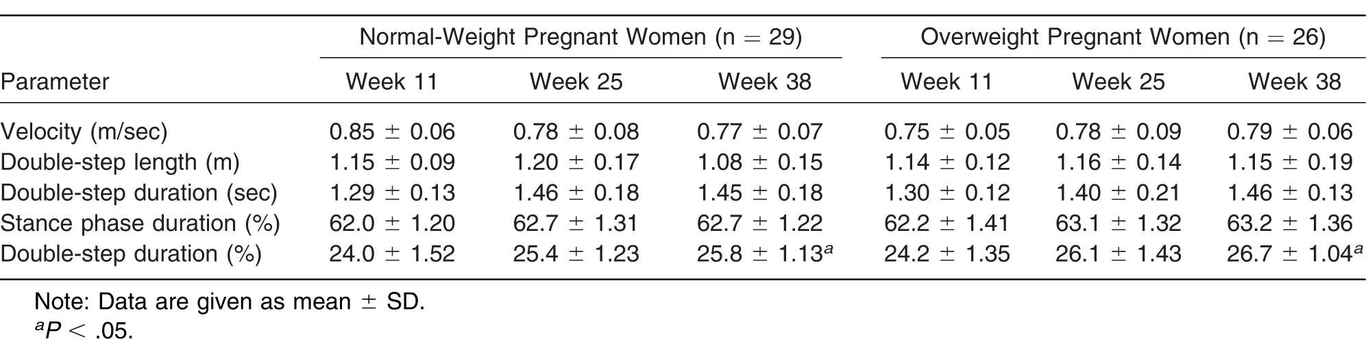
To measure plantar pressure distribution, pregnant women were instructed to walk a distance of approximately 10 m at their habitual speed inside of a consulting room. Plantar pressure distribution and spatiotemporal gait parameters during walking were measured with a pedobarograph (T&T Medilogic Medizintechnik GmbH, Munich, Germany) by means of shoe insoles with capacitive sensors (maximum of 240 SSR sensors per insole), with the participants wearing their own shoes. Gait data were sampled at a rate of 60 Hz. Each measurement was repeated three times per foot, and the women had to take a 3-minute rest between each trial. The mean values of parameters such as gait velocity (m/sec), double-step length (m), double-step duration (sec), stance phase duration (%), and double-step duration (%) were calculated. The gait data are summarized in Table 2.
Table 2.
Spatiotemporal Gait Parameters in 55 Pregnant Women
To quantify plantar pressure distribution, the maximum magnitude of plantar pressure under five anatomical masks was measured. These masks represent the anatomical plantar regions: toes, metatarsal heads, lateral arch, medial arch, and heel. [31] The maximum pressure under each anatomical region was measured per each individual step.
Measurement of Vertical GRF
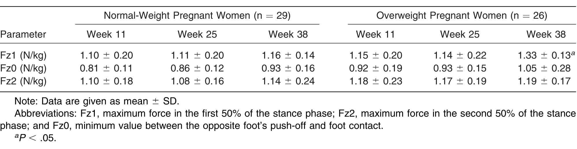
Ground reaction force was measured at the same time as plantar pressure distribution and spatiotemporal gait parameters using two force platforms (model 9286AA-A; Kistler Instrumente AG, Winterthur, Switzerland). Force data were sampled at a rate of 1,000 Hz. To assess GRF, the participants were asked to walk a 10-m path barefoot at their habitual speed. The measurement was repeated to obtain three valid walking trials with a 3-minute rest between each of them. The three recorded trials were averaged for the purpose of data analysis. The vertical force was characterized by a double-bump pattern. The first peak is related to body weight loading, and the second one is the result of push-off. Vertical GRF (Fz) was quantified by Fz1 (maximum force in the first 50% of the stance phase), Fz2 (maximum force in the second 50% of the stance phase), and Fz0 (minimum value between the opposite foot's push-off and foot contact). The measured forces were normalized to the participant's body mass at the time of data collection.
Statistical Analysis
Descriptive statistics were used for sample characterization. Data distribution was assessed using mean ± SD for quantitative variables. The presumptions of data normality were verified using the Shapiro-Wilk test. Repeated-measures analysis of variance was applied. Post hoc tests were used to compare groups if the within-subject (analysis of variance) test showed significant results. The statistical tests were performed with Statistica software, Version 13.1 (StatSoft, Krakow, Poland). The significance level was set at P < .05.
Results
Participant Demographic Characteristics
The research group consisted of 29 normal-weight pregnant women (mean ± SD age, 27.1 ± 4.7 years) and 26 overweight pregnant women (mean ± SD age, 27.0 ± 4.9 years). The participants' demographic characteristics were measured before pregnancy and in the three trimesters of pregnancy (week 11, the first trimester; week 25, the second trimester, and week 38, the third trimester). Characteristics of the sample are presented in Table 1.
No significant difference was observed between the groups in terms of age (P > .05). Significant changes were found for weight and BMI during gestation (P < .05). The prepregnancy mean ± SD BMIs were 21.8 ± 1.3 and 28.6 ± 3.4 in normal-weight and overweight pregnant women, respectively. In the first trimester, the mean ± SD BMI was 22.0 ± 1.2 in normal-weight pregnant women and 28.8 ± 3.2 in overweight women. In the second trimester, the mean ± SD BMI was higher in both groups, ie, 23.5 ± 2.0 for normal-weight women versus 30.5 ± 3.4 for overweight women. For pregnant women in the third trimester, the mean ± SD BMI was the highest: 26.1 ± 2.8 for normal-weight women versus 32.8 ± 4.0 for overweight women.
Assessment of Spatiotemporal Gait Parameters and Plantar Loading During Walking at Habitual Speed
No difference was observed between the feet for all of the measured variables (P > .05). Thus, further assessment was performed for the dominant side (Table 2).
Concerning the spatiotemporal gait parameters, gait velocity, double-step length and duration, stance phase duration, and double-step duration (%) were similar for normal-weight and overweight pregnant women in the first and second trimesters of pregnancy (P > .05). The results show that normal-weight and overweight pregnant women use the same spatiotemporal gait parameters to increase body stability and safety of movement during pregnancy. In normal-weight and overweight pregnant women, the double-step duration was on average 7.5% and 10.3% higher, respectively, in the third trimester compared with the first trimester (P < .05).
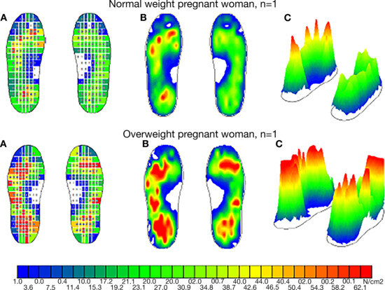
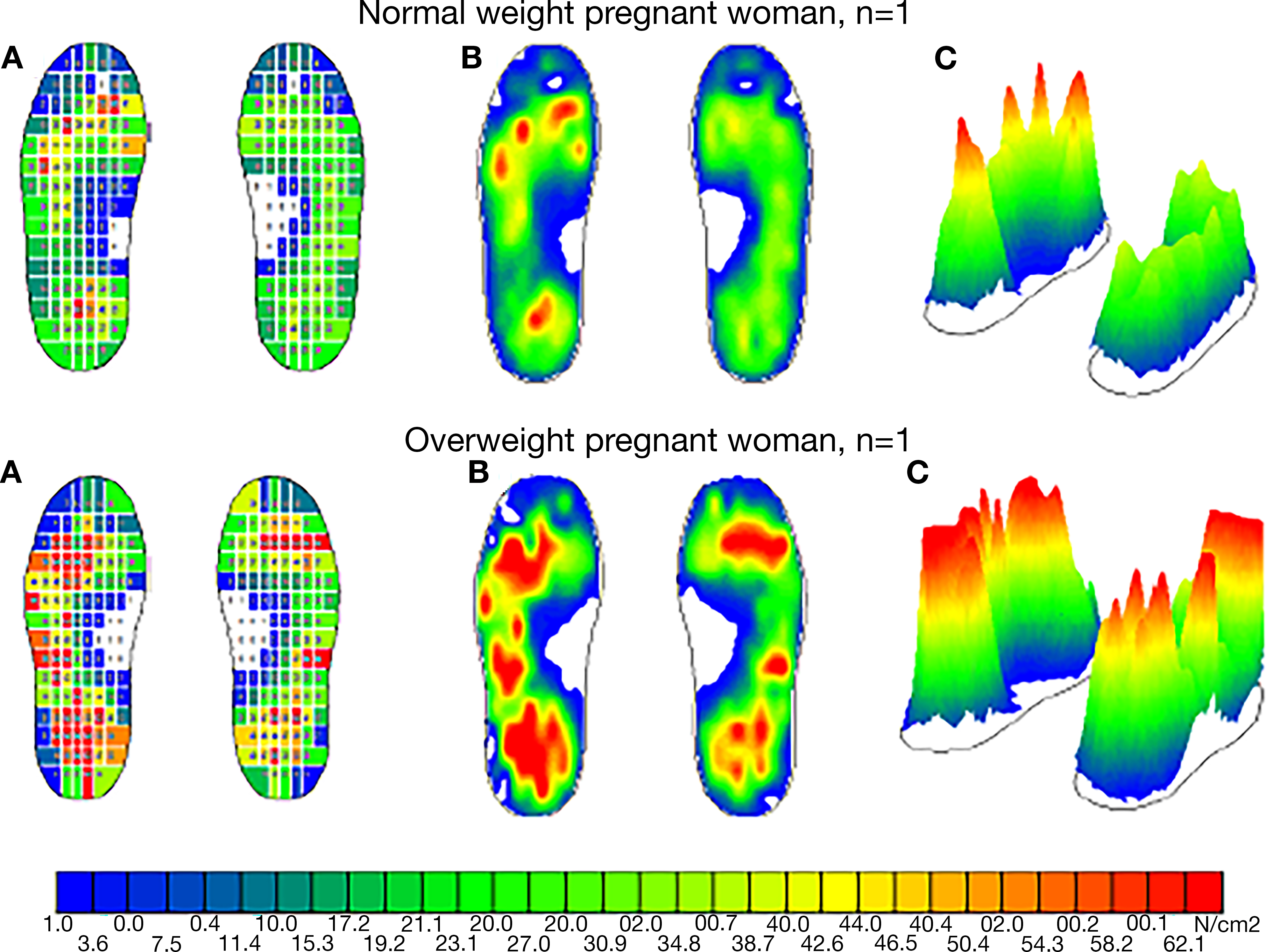
The pregnant women were asked to wear pedobarographic pressure insoles in their shoes for 3 min to allow insole acclimatization and potentially enhance the reliability of measurement. Figure 1 illustrates plantar pressure distribution for normal-weight and overweight pregnant woman during walking at their habitual speed.
Figure 1.
Foot pressure distributions for randomly selected participants from the experimental group: a normal-weight and an overweight pregnant woman both in week 25 of pregnancy: sensors on the insole (EU, 38) (A), averaged peak pressure maps (B), maximum pressure distribution from all sensors during stance (C).
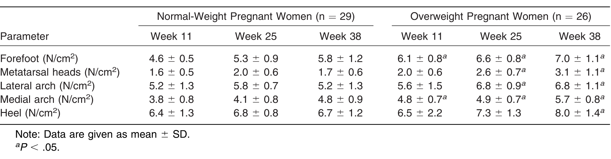
The initial contact in all of the participants was a heel strike with a visually normal heel-to-toe motion. For normal-weight pregnant women, the highest pressure amplitudes were found under the heel and the forefoot, and the lowest magnitude of pressure was recorded under the metatarsal heads. For overweight pregnant women, an increased pressure amplitude was observed under the forefoot, the metatarsal heads, the lateral arch, and the medial arch. In both groups in the third trimester, pressure under the medial arch differed for the right and left lower limbs and moved to the lateral arch. Table 3 summarizes the parameters extracted from the pedobarograph insoles during walking for normal-weight and overweight pregnant women.
Table 3.
Plantar Pressure Comparison Between 55 Normal-Weight and Overweight Pregnant Women
A significant difference in terms of pressure distribution under the forefoot and the medial arch was observed between the groups in the first trimester. The mean ± SD pressure amplitude under the forefoot was on average 32.6% higher in overweight pregnant women (4.6 ± 0.5 N/cm2 in normal-weight women versus 6.1 ± 0.8 N/cm2 in overweight women; P < .05). Similarly, the mean ± SD pressure amplitude under the medial arch was on average 26.3% higher in overweight pregnant women (3.8 ± 0.8 N/cm2 in normal-weight women versus 4.8 ± 0.7 N/cm2 in overweight women; P < .05). In the second trimester, the pressure amplitude under the forefoot, the metatarsal heads, the lateral arch, and the medial arch was on average higher by 24.5%, 30.0%, 17.2%, and 19.5%, respectively (P < .05), in overweight pregnant women compared with the normal-weight group. Moreover, in the third trimester pressure changes were observed under all of the anatomical parts of the foot (P < .05).
Differences Between Groups in GRFs During Walking
The GRF results suggest that Fz1 in overweight pregnant women in the third trimester occurred significantly later than the peak force observed in the normal-weight group, by an average of 14.2% (380 msec from the onset of stance initiation in normal-weight women versus 540 msec in the overweight group; P < .05).
The results show that normal-weight and overweight pregnant women use the same GRFs to increase the safety of movement during pregnancy (Table 4).
Table 4.
Vertical Ground Reaction Force Comparison Between 55 Normal-Weight and Overweight Pregnant Women
However, in the third trimester of pregnancy the mean ± SD maximum amplitude of force Fz1 in overweight pregnant women was significantly higher than the peak force observed in the normal-weight group by an average of 14.7% (1.16 ± 0.14 N/kg in normal-weight women versus 1.33 ± 0.13 N/kg in overweight women; P < .05). No significant difference was observed for the first peak (Fz1) in the first and the second trimesters (P > .05).
Discussion
The purpose of this study was to analyze the mechanisms of gait adaptation in normal-weight and overweight pregnant women. In this study, the evolution of spatiotemporal gait parameters during the three trimesters of pregnancy was analyzed and compared with observations in normal-weight and overweight pregnant women. Most papers showed changes in spatiotemporal gait parameters in late pregnancy, specifically, a significant increase in double support time. [4,7,18] This is in agreement with the present study, which shows that double-step duration in the third trimester of pregnancy in normal-weight and overweight pregnant women is significantly higher compared with that in the first trimester (P < .05). As a matter of fact, the prolonged double-support duration acts as a “safe strategy” to ensure the woman's equilibrium to the highest possible degree. van Zwieten et al [32] described the same phenomenon, compensating for muscle weakness in nonpregnant women. A longer stance time in the last trimester was observed by Karadag-Saygi et al. [22] Similar results were presented by Forczek and Staszkiewicz, [12] who found an increased base of support. They examined a single group of women in three different periods (before pregnancy, during the last trimester of pregnancy, and 6 mo after delivery). Statistically significant differences in body weight occurred in the three studied periods. According to Branco et al, [17] the temporal gait parameters (cycle time, step time, stance, and swing time) are influenced by pregnancy, but there is no significant difference between the second and third trimesters of pregnancy, ie, most parameters remain unchanged.
Numerous studies in the area of plantar pressure distribution in pregnant women have been performed. However, few data are available on plantar pressure distribution in overweight pregnant women. The obtained results suggest that the pattern of plantar distribution during normal walking is higher in normal-weight and overweight pregnant women for different plantar anatomical regions during pregnancy. This is in agreement with Karadag-Saygi et al, [22] who measured 35 overweight pregnant women in the last trimester. They found that pregnant women had higher forefoot pressure on the right side when standing and walking. The most significant difference in pressure amplitude in pregnant women in the first trimester was observed under the forefoot and the medial arch, on average 32.6% and 26.3% higher, respectively, in overweight pregnant women compared with normal-weight women. This increase suggests that in the case of overweight women in the first trimester, more pressure was localized under the medial arch and the forefoot than in normal-weight pregnant women. In the second trimester, the pressure amplitude was higher by 30% in overweight pregnant women compared with the normal-weight group under all of the anatomical parts of the foot, with the highest values for the metatarsal heads. In the third trimester, pressure changes were observed under all of the anatomical parts of the foot (P < .05). This adjustment may help maintain the dynamic stability of overweight pregnant women during locomotion, [20] which is consistent with the results reported by De Oliveira et al. [33] In the study, differences in plantar pressure distribution for 20 pregnant volunteers aged 18 to 35 were analyzed. As far as plantar pressure is concerned, it was found that the hind foot showed higher pressure values compared with the forefoot, which is in agreement with the present research.
The purpose of this study was to analyze changes in gait patterns in vertical GRF caused by advancing pregnancy. The results show that normal-weight and overweight pregnant women use the same GRFs to increase the safety of movement during pregnancy. However, in the third trimester of pregnancy, the maximum amplitude of force Fz1 in overweight pregnant women was significantly higher than the peak force observed in the normal-weight group by an average of 14.7% (P < .05). No significant difference was observed for the first peak (Fz1) in both the first and the second trimesters (P > .05). Similar observations were made in the study by McCrory et al, [19] where no differences in vertical GRF between trimesters were reported.
One limitation of the study was the small sample size of pregnant women. For a more detailed analysis and corroboration of the obtained results, more women should be involved in future research. Another limitation of the study was the kinematic measurement because it was not analyzed in this study. A weakness of the study design was the lack of shoe standardization, which may affect in-shoe pressure measurements. We believe that this may bias the results to a small degree because all of the participants used sport shoes with almost identical characteristics.
Conclusions
The results of this study suggest that both normal-weight and overweight pregnant women use different mechanisms of gait adaptation throughout pregnancy. During gait, the plantar loads under all of the anatomical foot parts were redistributed, and the peak force was higher throughout pregnancy, especially in overweight pregnant women. These adjustments help maintain the dynamic stability of the pregnant women during locomotion. For normal-weight and overweight pregnant women, the highest change in pressure amplitude and GRF was observed in the third trimester of pregnancy. In practice, understanding the biomechanical changes in a woman's gait can protect the musculoskeletal system during gestation. The specific clinical consideration should be made for overweight pregnant women. Based on the results obtained in this study, we suggest using foot insoles that may relieve feet pressure during gait and the discomfort related to changes in the distribution of plantar pressure in overweight pregnant women. Moreover, further research should be provided with the aim of assessing comfort during gait using different types of insoles.
Financial Disclosure: This study was supported by Bialystok University of Technology (W/WM/17/2017).
Conflict of Interest: None reported.
References
- OgambaMILoverroKLLaudicinaNM: Changes in gait with anteriorly added mass: a pregnancy simulation study. J Appl Biomech32: 379, 2016.
- JensenRKDoucetSTreitzT:Changes in segment mass and mass distribution during pregnancy. J Biomech29: 251, 1996.
- BraceroLAByrneDW:Optimal maternal weight gain during singleton pregnancy. Gynecol Obstet Invest46: 9, 1998.
- CarpesFGriebelerDKleinpaulJ: Women able-bodied gait kinematics during and post pregnancy period. Braz J Biomech9: 33, 2008.
- HaganLWongCK:Gait in pregnant women: spinal and lower extremity changes from pre- to postpartum. J Womens Health Phys Therap34: 46, 2010.
- PaukJSwinarskaD:The impact of body mass on spine alterations in pregnant women: a preliminary study. Technol Health Care26: 665, 2018.
- FotiTDavidsJRBagleyA:A biomechanical analysis of gait during pregnancy. J Bone Joint Surg Am82: 625, 2000.
- FalolaJMGouthonPKoussihouedeFE: Gait coordination in pregnancy: a study in a rural population in Africa. Sci Sports24: 49, 2009.
- KrkeljasZ:Changes in gait and posture as factors of dynamic stability during walking in pregnancy. Hum Mov Sci58: 315, 2018.
- ForczekWIvanenkoYPBielatowiczJ: Gait assessment of the expectant mothers: systematic review. Gait Posture62: 7, 2018.
- SegalNABoyerERTeran-YengleP: Pregnancy leads to lasting changes in foot structure. Am J Phys Rehabil92: 232, 2013.
- ForczekWStaszkiewiczR:Changes of kinematic gait parameters due to pregnancy. Acta Bioeng Biomech14: 113, 2012.
- BrancoMSantos-RochaRVieira1F:Biomechanics of gait during pregnancy. ScientificWorldJournal2014: 527940, 2014.
- GilleardGI:Trunk motion and gait characteristics of pregnant women when walking: report of a longitudinal study with a control group. BMC Pregnancy Childbirth13: 71, 2013.
- GottschallJSSheehanRCDownsDS:Pregnant women exaggerate cautious gait patterns during the transition between level and hill surfaces. J Electromyogr Kinesiol23: 1237, 2013.
- LymberyJKGilleardW:The stance phase of walking during late pregnancy: temporospatial and ground reaction force variables. JAPMA95: 247, 2005.
- BrancoMSantos-RochaRVieiraF: Influence of body composition on gait kinetics throughout pregnancy and postpartum period. Scientifica (Cairo)2016: 3921536, 2016.
- BrancoMSantos-RochaRAguiarL: Kinematic analysis of gait in the second and third trimesters of pregnancy. J Pregnancy2013: 718095, 2013.
- McCroryJLChambersAJDaftaryA: Ground reaction forces during gait in pregnant fallers and non-fallers. Gait Posture34: 524, 2011.
- RibeiroAPTrombini-SouzaFde Camargo Neves SaccoI: Changes in the plantar pressure distribution during gait throughout gestation. JAPMA101: 415, 2011.
- NyskaMSoferDPoratA: Plantar foot pressures in pregnant women. Isr J Med Sci33: 139, 1997.
- Karadag-SaygiEUnlu-OzkanFBasgulA:Plantar pressure and foot pain in the last trimester of pregnancy. Foot Ankle Int31: 153, 2010.
- GoldbergJBesserMPSelby-SilversteinL:Changes in foot function throughout pregnancy. Obst Gynecol97: S39, 2001.
- RasmussenKMYaktineALeds: Weight Gain During Pregnancy: Reexamining the Guidelines. Washington, DC: The National Academies Press;2009.
- DavisEMStangeKCHorowitzRI:Childbearing, stress and obesity disparities in women: a public health perspective. Matern Child Health J16: 109, 2012.
- MelzerKSchutzY:Pre-pregnancy and pregnancy predictors of obesity. Int J Obes (Lond)34(suppl 2): S44,2010.
- AtkinsonRLPietrobelliAUauyR: Are we attacking the wrong targets in the fight against obesity? the importance of intervention in women of childbearing age. Int J Obes36: 1259, 2012.
- Tenenbaum-GavishKHodM:Impact of maternal obesity on fetal health. Fetal Diagn Ther34: 1, 2013.
- LeddyMAPowerMLSchulkinJ:The impact of maternal obesity on maternal and fetal health. Rev Obstet Gynecol1: 170, 2008.
- AguiarLSantos-RochaRVieiraF: Comparison between overweight due to pregnancy and due to added weight. Gait Posture42: 511, 2015.
- PaukJIhnatouskiMNajafiB:Assessing plantar pressure distribution in children with flatfoot Arch: application of the Clarke angle. JAPMA104: 622, 2014.
- van ZwietenKJNarainFHMSchmidtKP:Paradoxical increase of pacing frequency (Hz) in early multiple sclerosis (MS) patients is unraveled by clinical observation, focusing on gait analysis. Int Sound Vibr Digest12: 9, 2011.
- De OliveiraISFigueiraJFigueiraH: Measuring the association between the biomechanics of plantar pressure and musculoskeletal changes throughout pregnancy and postpartum. Divers Equal Health Care13: 384, 2016.

© 2020 American Podiatric Medical Association