


Abstract
Distal to its origination from the sciatic nerve, the common fibular (peroneal) nerve divides into the superficial and deep fibular (peroneal) nerves. Whereas the deep fibular nerve continues its course into the anterior compartment, the superficial fibular nerve (SFN) usually arises near the fibular neck and projects distally within the lateral crural compartment before entering the superficial fascia proximal to the ankle. In this report, we describe a unilateral case where the SFN arises within the anterior crural compartment and remains there for the remainder of its course deep to the deep fascia of the leg. Surgeons should be aware of anomalies such as this, for example, when performing fasciotomies to avoid inadvertently damaging an anomalously placed SFN.
The common fibular (peroneal) nerve (CFN) arises from the sciatic nerve in the popliteal fossa and, after passing laterally around the neck of the fibula, divides into the deep fibular (peroneal) nerve (DFN) and the superficial fibular (peroneal) nerve (SFN). The DFN and SFN provide motor innervation to the anterior and lateral muscular compartments of the leg, respectively. The DFN enters the anterior crural compartment by passing through the anterior intermuscular septum, courses alongside the anterior tibial vasculature in the space between the tibialis anterior muscle and the long extensors of the pedal digits and provides motor innervation to these muscles as it proceeds distally. The DFN continues onto the dorsum of the foot to innervate the intrinsic extensors and provide cutaneous innervation to the first digital web space. The SFN remains in the lateral crural compartment and provides innervation to the fibularis longus and brevis muscles while coursing between the fibula and the muscles of the lateral compartment. The SFN pierces the crural fascia of the dorsal leg at a variable height above the talocrural joint to provide cutaneous innervation to the dorsum of the foot through the intermediate and medial dorsal cutaneous nerves of the foot.
Several works have described variations in the course of the SFN. [1,2,3,4,5,6,7,8] Adkinson et al [1] documented 85 legs in which the SFN could be found in the lateral compartment (74.1% of limbs), in the anterior compartment (14.1%), or with branches in both compartments (11.8%). The nerve is also known to course within the anterior intermuscular septum of the leg. [4,5] The DFN shows some variability in the presence of an accessory DFN and in the cutaneous field of the foot. [9] The CFN is commonly injured in up to one-third of knee dislocations [11,12,13]; of this nerve’s two branches, the DFN appears to be injured simultaneously with the CFN, [12] perhaps because the SFN branches from the CFN tend to stay in the lateral crural compartment in most cases. [13] In contrast, entrapment injuries to the SFN appear to be partially attributable to the location at which this nerve pierces the crural fascia to enter the superficial fascia of the leg. [6,7,8] Thus, patterns of branching for the CFN should be of concern to the clinician. Here, we describe the case of an unreported anomaly of the CFN entering the anterior compartment before dividing into the SFN and DFN, with further anomalies in the distal course of the SFN and the distribution of motor and cutaneous components.
Case Report
An anomalous CFN was observed in the left leg of a human cadaver (male, 67 years old) as part of a routine course in the anatomy of the lower extremity at the Kent State University College of Podiatric Medicine. The contralateral leg showed a normal CFN branching morphology and downstream distribution of the DFN and SFN.
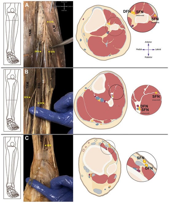
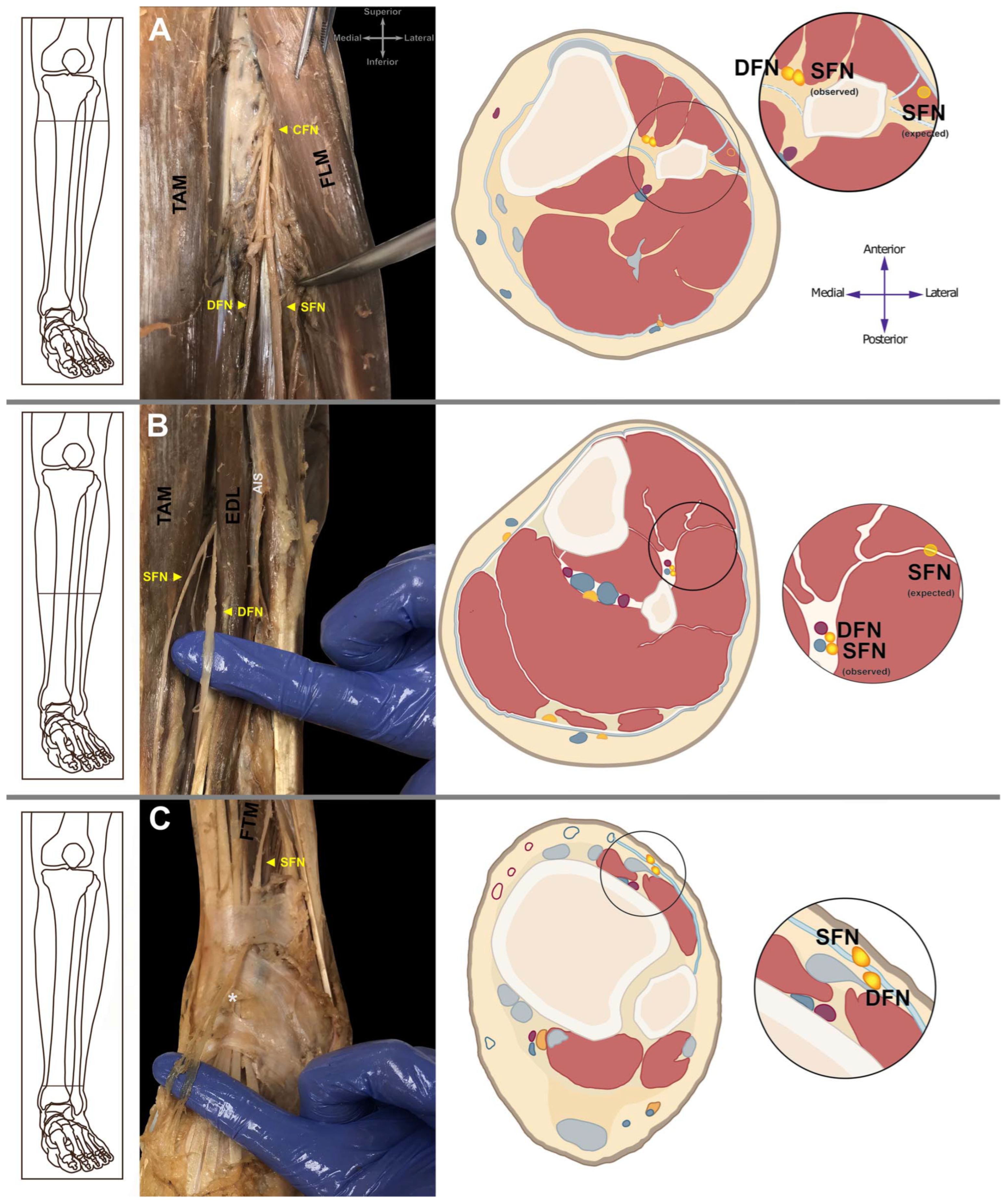
The proximal portion of the CFN in the left leg exited the popliteal fossa normally, coursing laterally around the fibular neck and toward the anterior compartment of the leg (Figure 1A). As the nerve passed deep to the fibularis longus muscle, it provided nerve fibers innervating that same muscle and the fibularis brevis muscle. The CFN then continued into the anterior crural compartment, where it branched into the SFN (now in the anterior compartment) and the DFN (Figure 1 A and B). Thus, the proximal portion of the CFN rather than the SFN provided direct branches to innervate the lateral compartment musculature.
Figure 1.
Photographic and cross-sectional demonstrations of anomalous superficial fibular nerve (SFN). All photographs are viewed from an anterolateral position. (A) Whereas the common fibular nerve (CFN) provided motor innervation to the lateral crural compartment, the SFN only became apparent in the anterior crural compartment along with the deep fibular (peroneal) nerve. In the photograph of the dissection, the CFN is observed to enter the anterior crural compartment before branching. (B) The SFN remained within the anterior compartment, evident in its placement medial to, and completely outside of, the anterior intermuscular septum. (C) After piercing the fibularis tertius muscle, the SFN pierced the crural fascia between the superior and inferior extensor retinacula. Distal to this point, the nerve divided into the medial and intermediate dorsal cutaneous nerves as expected, but the intermediate dorsal cutaneous nerve was removed from this foot at the location indicated by the light gray asterisk. DFN, deep fibular (peroneal) nerve; EDL, extensor digitorum longus muscle; FLM, fibularis longus muscle; FTM, fibularis tertius muscle; TAM, tibialis anterior muscle; AIS, anterior intermuscular septum.
Whereas the DFN appeared normal in distribution, the distal aspect of the anomalous SFN emerged from the anterior compartment by piercing the crural fascia between the superior and inferior extensor retinacula after moving through the fibularis tertius muscle (Figure 1C). As it traversed the talocrural joint, the anomalous SFN divided into the intermediate and medial dorsal cutaneous nerves of the foot, as expected.
Discussion
To our knowledge, the course and branching pattern of the CFN described in this study are unreported in the literature. Several authors have documented variation in the placement of the SFN that is similar to that observed in the present study. [1,14,15,16] Adkinson et al [1] described a similar course of the SFN as observed in our case in 12 of 85 cadaveric limbs (14.1%), but the CFN near the fibular neck branched normally in all of those limbs, with the SFN forming in the lateral compartment and then piercing the anterior intermuscular septum to enter the anterior compartment. Similarly, Ducic et al [16] found that nine of 111 limbs (8.1%) in their study showed an SFN in the anterior crural compartment, as did Bas et al [15] in six of 40 cadaveric fetal limbs (15%). However, none of these studies reported any cases where either the CFN persists into the anterior compartment or where the SFN emerges between the two crural extensor retinacula. Thus, the present case demonstrates an unreported course for the CFN and for one of its branches, the SFN. Furthermore, the pattern described here is not documented in anatomical atlases for the rhesus macaque (Macaca mulatta [17]), the baboon (Papio cynocephalus [18]), the chimpanzee (Pan troglodytes [18]), or the lowland gorilla (Gorilla gorilla [19]), suggesting that the most commonly observed pattern of the CFN, DFN, and SFN in humans is similar to that of our closest evolutionary relatives. The normal course and patterning of the CFN and its branches provide motor and cutaneous innervation to the lateral and anterior compartments of the leg, with subsequent cutaneous innervation of the dorsum of the foot derived in the most part from terminal branches of the SFN. In our case, the CFN provided small motor branches to the lateral compartment musculature, but the SFN did not form until the CFN entered the anterior compartment. This observed pattern suggests that the SFN fibers in the anterior compartment in our case may be cutaneous fibers exclusively.
A complex array of axon guidance regulation occurs during the development of peripheral nerve pathways in the limb. [20] Fidelity of the patterning of these pathways during development is important for proper motor and sensory function. In the present case, the CFN provided motor innervation to muscles of the lateral crural compartment. However, the SFN that branched from the CFN in the anterior compartment may carry only sensory neural fibers. Thus, a subtle unilateral anomaly in axon guidance cues may have allowed the motor fibers to defasciculate from the CFN in the lateral crural compartment, whereas the cutaneous sensory nerve fibers destined to innervate the dorsum of the foot remained with the CFN as it entered the anterior compartment. Although we were unable to determine whether our case demonstrated deficits in either motor or sensory function in the left leg, the final distribution of nerves suggests that muscle and cutaneous innervation was complete.
Conclusions
Our results reveal an anomaly of the branching of the CFN that may have significant clinical implications in the symptomatology and treatment of compartment syndromes of the leg, nerve impingement, and entrapment or traction injuries, even in association with ankle sprains, reduction and repair of fractures, and surgical outcomes. Although a surgeon entering the anterior compartment of the leg is concerned with protection of the DFN, damage to an anomalous SFN, even if cutaneous fibers only, may lead to undesirable outcomes. Thus, the surgeon should be aware of the possible courses of the SFN and DFN, and the possible differences in branching patterns from the CFN. This knowledge may help to explain some differences noted in the physical examination, and the anomaly may be detectable on musculoskeletal ultrasound or magnetic resonance imaging. This knowledge may alter the pattern for fasciotomy with compartment syndromes and suggests that the surgeon should locate the nerves before fracture reduction or cast placement postoperatively.
Acknowledgments
We would like to thank those who donated their bodies to medical science and education. Joani Lannoch was essential in creating the cross-sectional panels in Figure 1. Morgan Chaney thanks Caylee Heiremans, MA, MLIS for support throughout this project.
Financial Disclosure
This work was supported by the College of Podiatric Medicine and School of Biomedical Sciences at Kent State University.
Conflicts of Interest
None reported.
References
- ADKINSON, D.P.; BOSSE, M.J.; GACCIONE, D.R.; et al. Anatomical variations in the course of the superficial peroneal nerve. J Bone Jt Surg 1991, 73, 112. [Google Scholar] [CrossRef] [Scilit]
- ARORA, A.K.; VERMA, P.; ABROL, S. An anomalous variation in the division pattern of the common peroneal nerve. Int J Appl Basic Med Res 2011, 1, 118. [Google Scholar] [CrossRef] [Scilit] [PubMed]
- BROWNE, J.A.; MORRIS, M.J. Variant superficial fibular (peroneal) nerve anatomy in the middle third of the lateral leg. Clin Anat 2007, 20, 996. [Google Scholar] [CrossRef] [Scilit] [PubMed]
- Nagabhooshana, S.; Vollala, V.R.; Rodrigues, V. Anomalous superficial peroneal nerve and variant cutaneous innervation of the sural nerve on the dorsum of the foot: a case report. Cases J 2009, 2, 1. [Google Scholar] [CrossRef] [Scilit] [PubMed]
- WILLIAMS, E.H.; DELLON, L. Intraseptal superficial peroneal nerve. Microsurgery 2007, 27, 477. [Google Scholar] [CrossRef] [Scilit] [PubMed]
- TZIKA, M.; PARASKEVAS, G.; NATSIS, K. Entrapment of the superficial peroneal nerve: an anatomical insight. JAPMA 2014, 105, 150. [Google Scholar] [CrossRef] [Scilit] [PubMed]
- LEMONT, H.; CULLEN, R.W. Compression of the superficial peroneal nerve secondary to sleeping and sitting positions. JAPMA 1984, 74, 450. [Google Scholar] [CrossRef] [Scilit] [PubMed]
- KELIKIAN, A.S. Sarrafian’s Anatomy of the Foot and Ankle, 3rd Ed ed; Wolters Kluwer/Lippincott Williams & Wilkins: Philadelphia, 2011. [Google Scholar]
- SINANOVIC, O.; ZUKIC, S.; SAKIC, A.; et al. The accessory deep peroneal nerve and anterior tarsal tunnel syndrome: case report. Acta Myol 2013, 32, 110. [Google Scholar] [PubMed]
- TZIKA, M.; PARASKEVAS, G.K.; KITSOULIS, P. The accessory deep peroneal nerve: a review of the literature. Foot (Edinb) 2012, 22, 232. [Google Scholar] [CrossRef] [Scilit] [PubMed]
- NIALL, D.M.; NUTTON, R.W. KEATING JF: Palsy of the common peroneal nerve after traumatic dislocation of the knee. Bone Joint J Br 2005, 87, 664. [Google Scholar] [CrossRef] [Scilit] [PubMed]
- MEDINA, O.; AROM, G.A.; YERANOSIAN, M.G.; et al. Vascular and nerve injury after knee dislocation: a systematic review. Clin Orthop Relat Res 2014, 472, 2621. [Google Scholar] [CrossRef] [Scilit] [PubMed]
- KRIVICKAS, L.S.; WILBOURN, A.J. Peripheral nerve injuries in athletes: a case series of over 200 injuries. Semin Neurol 2000, 20, 225. [Google Scholar] [CrossRef] [Scilit] [PubMed]
- BARRETT, S.L.; DELLON, A.L.; ROSSON, G.D.; et al. Superficial peroneal nerve (superficial fibularis nerve): the clinical implications of anatomic variability. J Foot Ankle Surg 2006, 45, 174. [Google Scholar] [CrossRef] [Scilit] [PubMed]
- BAS, O.; BILGIC, S.; SALBACAK, A.; et al. Variations of the superficial peroneal nerve and its terminal branches in the Turkish newborn fetuses. Turk Neurosurg 2012, 22, 62. [Google Scholar] [CrossRef] [Scilit] [PubMed]
- DUCIC, I.; DELLON, A.L.; GRAW, K.S. The clinical importance of variations in the surgical anatomy of the superficial peroneal nerve in the mid-third of the lateral leg. Ann Plast Surg 2006, 56, 635. [Google Scholar] [CrossRef] [Scilit] [PubMed]
- HARTMAN, C.G. STRAUS WL: The Anatomy of the Rhesus Monkey; Hafner Publishing Co: New York, 1969. [Google Scholar]
- SWINDLER, D.R. WOOD CD: An Atlas of Primate Gross Anatomy; University of Washington Press: Seattle, 1973. [Google Scholar]
- Gregory, W.K.; Raven, H.C. American Museum of Natural History, et al: The Anatomy of the Gorilla; the studies of ffenry Cushier Raven, and contributions by William B. Atkinson [and others]. A collaborative work of the American Museum of Natural ffistory and Columbia University; Columbia University Press: New York, 1950. [Google Scholar]
- BONANOMI, D. PFAFF SL: Motor axon pathfinding. Cold Spring Harb Perspect Biol 2010, 2, a001735. [Google Scholar] [CrossRef] [Scilit] [PubMed]
© 2020 American Podiatric Medical Association