


Abstract
Limb salvage for Charcot's neuroarthropathy has been shown to have high complication and failure rates. The aim of our report of two cases it to present a unique complication encountered with staged limb salvage for Charcot's neuroarthropathy. In two cases, patients developed delayed tibial shaft fracture associated with previous wire placement despite insertion of locked intramedullary nail fixation that spanned the delayed fracture. Both patients experienced fractures following advancement of weight after definitive fixation. In both patients, there was noted complication with the sites of the pins and revision of external fixation before fracture. In each case, the fracture was within the construct of the intramedullary fixation and successfully treated with an extended course of nonweightbearing. Complications of external fixation and intramedullary fixation are well reported within the literature; however, tibia fracture is rare. Based on these cases, it would seem prudent to recognize the risk of delayed pin-site complications and ensure adequate length of intramedullary fixation to span the potential areas of stress.
Charcot's neuroarthropathy (CN), a syndrome associated with neuropathic changes, is becoming more prevalent, with diabetes being the most common cause.[1] It is identified by bony destruction, resorption, and eventual deformity of the foot and/or ankle and imparts a significant negative influence on quality of life.[2-5] Treatment of CN usually begins with conservative modalities and often requires surgical management.
Surgical intervention for CN is favored in many instances for its ability to improve quality of life through ambulation in the community and limiting the need for cumbersome orthoses and braces as well as reduction in the risk of developing ulcers and infection overlying bony deformities.[6,7] The primary goal of surgical intervention is to create and maintain a stable, plantigrade foot for ambulation, and several constructs including plates, screws, intramedullary nails (IMN), and external fixation have been used with varying degrees of success.
Use of circular ring fixation and IMN constructs has become more common for the reconstruction of various deformities of the lower extremities, and is particularly useful for management of diabetic neuropathic limbs.[8-16] External ring fixation offers the advantages of being less invasive than traditional internal fixation and allows monitoring of soft tissues and gradual correction of deformity. As such, it has become an integral part of the treatment algorithm in the presence of ulceration and infection. However, complications associated with external fixation are well described in the literature. The most common complication is pin-tract infection, with rates reported as high as 60%.[10,16] Other complications reported include broken wires or pins, broken foot plates, deep venous thrombosis, tibia fracture, major amputation, and even death. Intramedullary nail constructs have gained popularity in part because of their rigidity and superior biomechanical properties, but they also have high complication rates.[12-18] The overall complication rate was reported to be 45.2% by Wukich et al,[15] with nonunion being among the most commonly reported complication with IMN fixation. Despite the number of reported complications for both external and intramedullary nail fixation, tibia fracture is relatively rare among them, ranging from 1.8% to 16.7%.[10,11,19-21] The aim of this article is to illustrate two cases of tibia fracture, a rarely reported complication of both external fixation and IMN fixation, which occurred in unique and previously unreported ways.
Case Reports
Case 1
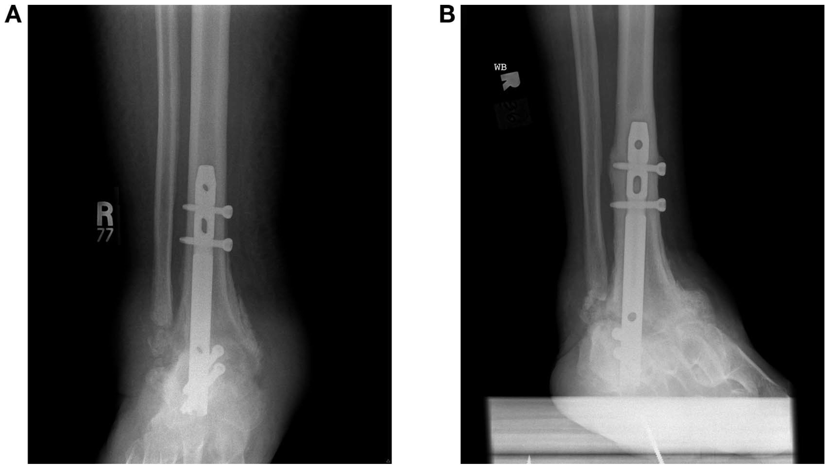
A 52-year-old woman presented with an extensive medical history, which included diabetes mellitus, peripheral neuropathy, peripheral vascular disease, and end-stage renal disease, after stepping on a piece of metal on her right heel. She had a history of poorly controlled diabetes, with a hemoglobin A1c level of greater than 10.0% at initial presentation. She did not have a retained foreign body, but developed infection and abscess, which was treated with operative debridement. Deep tissue cultures were obtained and demonstrated group B Streptococcus, which was successfully treated with oral antibiotics. No osseous involvement was noted at the time; however, the patient continued to have a chronic wound for 11 months before successful closure with a bilobed rotational flap. She was subsequently lost to follow-up and presented 2 years later with an infected ulceration to the right fifth metatarsal base. This was treated with staged debridements, negative-pressure wound therapy, and culture-guided intravenous (IV) antibiotics. Over the course of the next 6 months, she continued to have recurrent infections and ulceration of the fifth metatarsal base. She was treated with a second course of IV antibiotics and developed peripherally inserted central catheter line–associated deep venous thrombosis. She eventually underwent a complete fifth metatarsal resection and was treated with external fixation to allow for continued wound care and to maintain foot alignment (Fig. 1). One month after application of external fixation, the wound was completely healed, with resolution of all local signs of infection; however, she did have some erythema and drainage associated with one of the distal tibial pin sites. This was treated with removal of the pin and oral antibiotics. The remainder of the fixator was removed after 6 weeks and the patient was allowed a 2-week pin holiday before definitive plantar fixation with intramedullary hindfoot nail. She progressed through the early postoperative period without complication except some continued drainage from her distal tibial pin site. At 2.5 months postoperatively she was allowed touchdown weight on the operative extremity but, unfortunately, experienced a fall while showering and developed a distal tibia fracture (Fig. 2A) within the construct of the intramedullary device. This fracture coincided with the distal pin sites of previous external fixation. She underwent irrigation and debridement with placement of antibiotic beads, and operative cultures were consistent with methicillin-sensitive Staphylococcus aureus. She was treated again with IV antibiotics and then continued with suppressive therapy. The fracture went on to heal well, and over the course of 2 years, the tibia fracture stabilized, with significant hypertrophic bone formation (Fig. 2B).
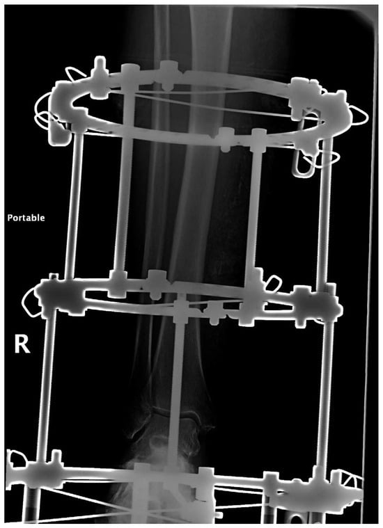
Figure 1.
Initial procedure with circular external fixator.
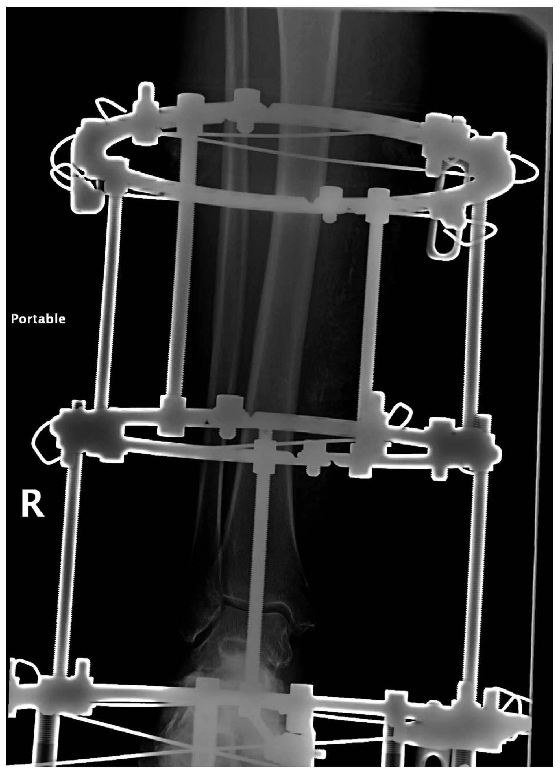
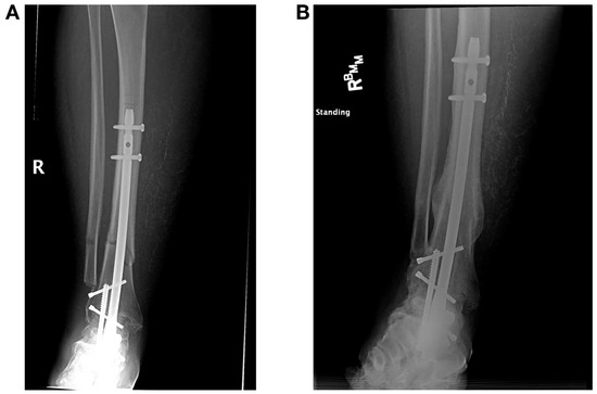
Figure 2.
A, Tibia fracture within a construct of locked intramedullary nail. B, Secondary healing with callus formation.
Case 2
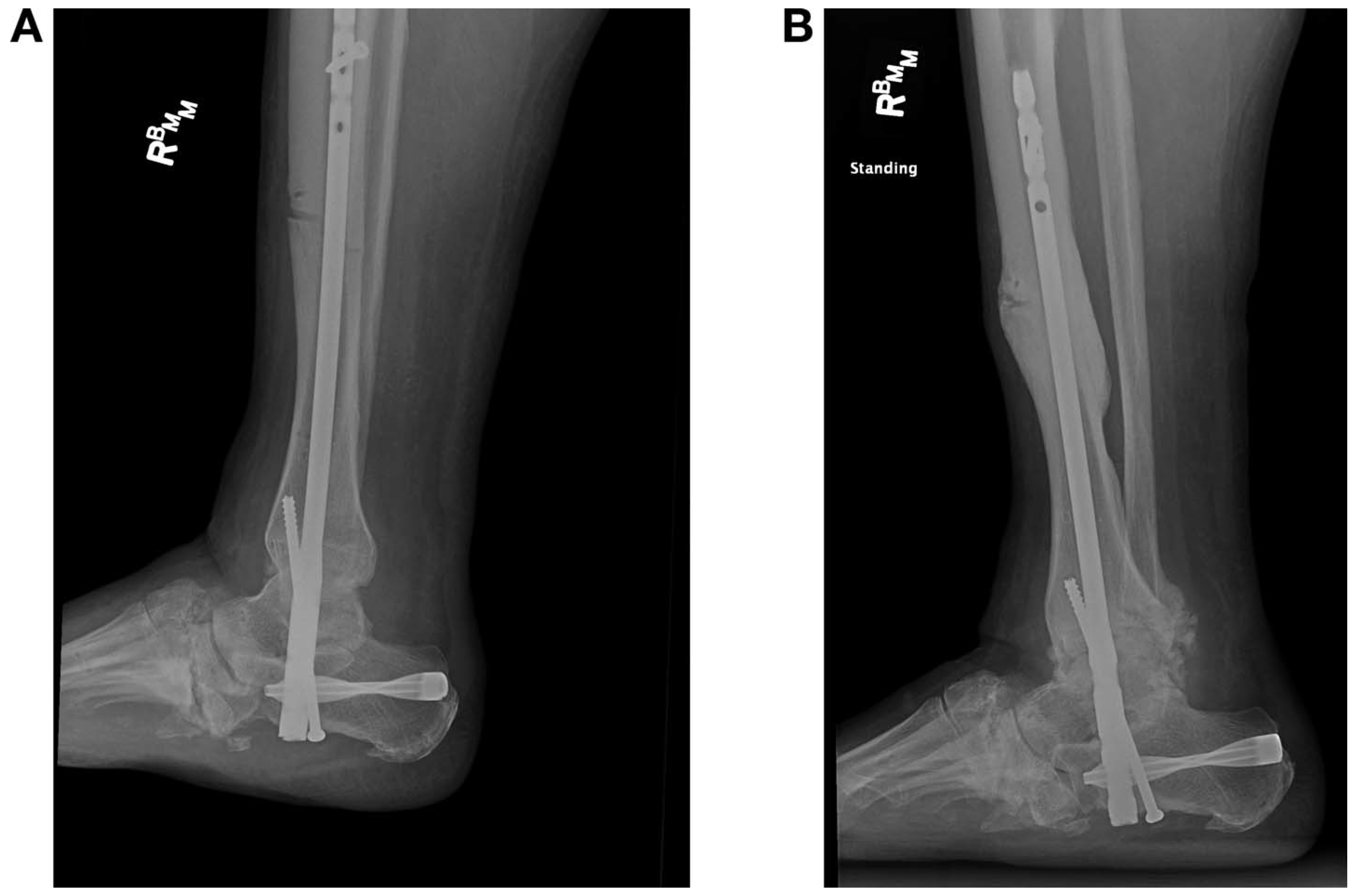
A 56-year-old man presented with a medical history of idiopathic peripheral neuropathy, venous insufficiency, and CN of both feet. He initially underwent staged reconstruction of his left midfoot and hindfoot, which developed infection of hardware approximately 3 years after surgery and which was successfully treated with removal of hardware and local wound care. He then underwent reconstruction on the right side for management of CN with midfoot collapse and plantar ulceration that had not been infected previously. Limb salvage was achieved with staged management using circular external fixator (Fig. 3A) followed by tibiotalocalcaneal arthrodesis with IMN fixation. He maintained his external fixator for 6 weeks, which was complicated with a broken distal tibia skinny wire and irritation of a proximal tibia skinny wire. Both wires were removed in the office without complication. After removal of external fixation, he underwent a 2-week pin holiday and his plantar midfoot ulceration progressed to a superficial wound before final internal fixation. He then progressed through early IMN postoperative care without complication, but unfortunately developed fracture at the proximal tibia pin sites as his weightbearing progressed 2 months postoperatively (Fig. 4A). On careful review of radiographs, the proximal pins were noted to have had unicortical fixation in the anterior tibial crest (Fig. 3B). He was treated with an additional 6-week period of nonweightbearing because the fracture was within the intramedullary fixation construct. The fracture site has healed with significant callus formation (Fig. 4B). He has since progressed to full weightbearing with bracing and healed all ulcerations without any signs of infection.
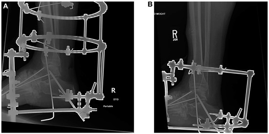
Figure 3.
A, Initial procedure with circular external fixator. B, Revision of external fixator with removal of proximal ring for proximal pin-site complications.
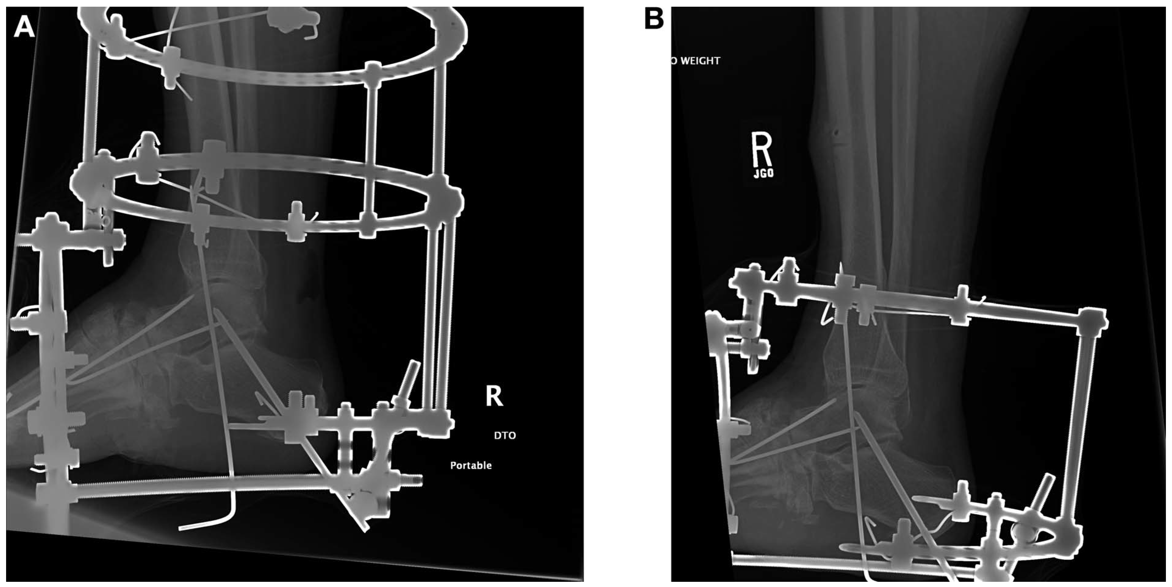
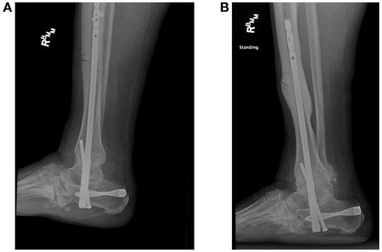
Figure 4.
A, Tibia fracture through proximal tibial pin sites within the construct of the locked intramedullary nail. B, Callus formation at fracture site.
Discussion
Although complications of circular external fixators are widely reported in the literature, tibial fracture is rare among them. Wukich et al[10] reported 58 complications in 56 patients undergoing circular ring fixation and found that diabetic patients averaged significantly more complications per person (1.3) compared with the nondiabetic group (0.6) (P = .0027), but they reported only one incident of tibia fracture. In a retrospective review of 254 patients undergoing surgical correction for Charcot deformity with static circular external fixator, Jones et al[21] reported 10 cases of tibial fracture. Seven fractures occurred among 42 patients with half pins (16.7%), whereas three occurred in 202 patients with fine wires only (1.5%). Zarutsky et al[11] used circular ring external fixation for salvage ankle arthrodesis in 43 patients and reported two (4.7%) tibia fractures. In both of our patients reported here, the tibia fracture was associated with skinny wires, and unicortical placement of the wire was noted as well. Our fractures occurred after removal of the external fixator, similar to seven of 10 fractures reported by Jones et al.[21]
Tibia fractures with hindfoot and ankle arthrodesis are also reported in the literature. Lidor et al[19] reported on 12 tibial stress fractures of the middle or distal portions of the tibia following ankle arthrodesis. Wukich et al[15] reported four tibia fractures in their diabetic cohort, with an overall fracture rate of 3.4%. Thordarson and Chang[20] reported on 12 patients undergoing tibiotalocalcaneal arthrodesis with locked IMN fixation and found two patients to have stress fracture at the level of the proximal interlocking screw, whereas seven of 12 had cortical hypertrophy at this level. The two reported fractures in our patients are unique in that they were not associated with the proximal extent of our fixation.
Tibia fractures are reported complications of both ring external fixation and tibiotalocalcaneal arthrodesis.[10,11,19-21] In the case of external fixation, it is thought of as a direct result of diminished cortical integrity and more common with half-pin fixation, whereas in tibiotalocalcaneal arthrodesis, it is likely a resultant stress riser at the proximal extent of the fixation. In our cases, we experienced tibial fractures associated with staged reconstruction following the advancement of weightbearing with definitive fixation by means of intramedullary nailing. In both cases, the fracture sites were associated with prior external fixation skinny wire pin sites as opposed to half-pins and were not consistent with stress risers at the proximal extent of definitive IMN fixation.
Conclusions
In our experiences with staged reconstruction and diabetic limb salvage, we have learned that complications can and will occur. In such a difficult, complex patient population, managing the variety of complications is key. With regard to staged tibiotalocalcaneal arthrodesis with IMNs, we prefer a longer nail for a couple of reasons. First, with shorter nails, there may be a noticeable difference in motion, with an increase in cortical hypertrophy or even a window-wiper effect at the proximal aspect of the nail in the relatively soft tibial metaphysis (Fig. 5). This may contribute to significant valgus deformity as the nail loosens in the metaphyseal bone and the cortical bone responds over time. Second, to minimize stress risers, particularly at the level of the previous pin sites, we use longer intramedullary fixation, typically between 260 mm and 340 mm in length. This longer fixation is secured in solid tibial diaphysis and avoids the complications of a short nail while bypassing the pin sites and reducing the stress through an already weakened area. Despite this, these two cases suggest that even with adequate fixation, the combination of mechanical insult, osseous deficits associated with diabetes, and extended periods of nonweightbearing may still cause significant pathology. Fortunately, longer fixation provided relative stability at the tibial fracture sites to allow secondary bone healing and helped both of these patients avoid additional surgery for their fractures.
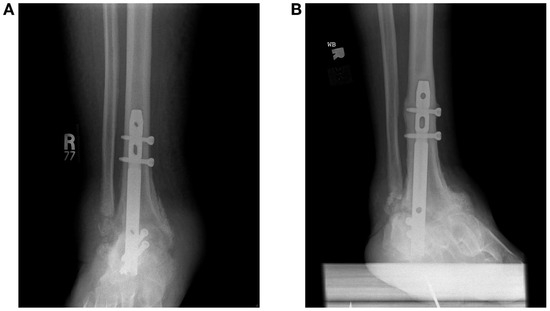
Figure 5.
A, Short (150-mm) intramedullary nail. B, Subsequent cortical hypertrophy and window-wiper effect at the proximal extent of the nail.
Funding
None reported.
Conflicts of Interest
None reported.
References
- ArmstrongDGPetersEJ:Charcot's arthropathy of the foot. JAPMA92: 390, 2002.
- PinzurMSEvansA:Health-related quality of life in patients with Charcot foot. Am J Orthop32: 492, 2003.
- PakarinenTKLainHJMaenpaaH: Long-term outcome and quality of life in patients with Charcot foot. Foot Ankle Surg15: 187, 2009.
- RaspovicKRWukichDK:Self-reported quality of life in patients with diabetes: a comparison of patients with and without Charcot neuroarthropathy. Foot Ankle Int35: 195, 2014.
- KroinESchiffAPinzurMS: Functional impairment of patients undergoing surgical correction for Charcot foot arthroplasty. Foot Ankle Int38: 705, 2017.
- SaltzmanCLHagyMLZimmermanB: How effective is intensive nonoperative initial treatment of patients with diabetes and Charcot arthropathy of the feet?Clin Orthop Relat Res435: 185, 2005.
- PinzurMSSchiffAP:Deformity and clinical outcomes following operative correction of Charcot foot: a new classification with implications for treatment. Foot Ankle Int39: 265, 2018.
- EasleyMLooneyCWellmanS: Ankle arthrodesis using ring external fixation. Tech Foot Ankle Surg5: 150, 2006.
- SaynerLRosenblumB:External fixation for Charcot foot reconstruction. Curr Surg62: 618, 2005.
- WukichDKBelczykRJBurnsPR: Complications encountered with circular ring fixation in persons with diabetes mellitus. Foot Ankle Int29: 994, 2008.
- ZarutskyERushSSchuberthJ:The use of circular wire external fixation in the treatment of salvage ankle arthrodesis. J Foot Ankle Surg44: 22, 2005.
- CaravaggiCCimminoMCarusoS: Intramedullary compressive nail fixation for the treatment of severe Charcot deformity of the ankle and rearfoot. J Foot Ankle Surg45: 20, 2006.
- Dalla PaolaLVolpeAVarottoD: Use of retrograde nail for ankle arthrodesis in Charcot neuroarthopathy: a limb salvage procedure. Foot Ankle Int28: 971, 2007.
- MendicinoRWCatanzaritiARSaltrickKR: Tibiotalocalcaneal arthrodesis with retrograde intramedullary nailing. J Foot Ankle Surg43: 83, 2004.
- WukichDKMalloryBRSuderNC: Tibiotalocalcaneal arthrodesis using retrograde intramedullary nail fixation: comparison of patients with and without diabetes mellitus. J Foot Ankle Surg54: 876, 2015.
- ElAlfyBAliAMFawzySI:Ilizarov external fixator versus retrograde intramedullary nailing for ankle joint arthrodesis in diabetic Charcot neuroarthropathy. J Foot Ankle Surg56: 309, 2017.
- BerendMEGlissonRRNunleyJA:A biomechanical comparison of intramedullary nail and crossed lag screw fixation for tibiotalocalcaneal arthrodesis. Foot Ankle Int18: 639, 1997.
- FlemingSSMooreTJHuttonWC:Biomechanical analysis of hindfoot fixation using an intramedullary rod. J South Orthop Assoc7: 19, 1998.
- LidorCFerrisLRHallR: Stress fracture of the tibia after arthrodesis of the ankle of the hindfoot. J Bone Joint Surg Am79: 558, 1997.
- ThordarsonDBChangD:Stress fractures and tibial cortical hypertrophy and tibiotalocalcaneal arthrodesis with an intramedullary nail. Foot Ankle Int20: 497, 1999.
- JonesCPYoungbloodSAWaldropN: Tibial stress fracture secondary to half-pins in circular ring external fixation for Charcot foot. Foot Ankle Int35: 572, 2014.
© 2025 American Podiatric Medical Association