


Abstract
A case describing an O-to-Z double-advancement flap used to treat a 62-year-old woman with a slowly enlarging exophytic mass in the plantar aspect of the right foot is presented. Clinical details, surgical technique, and histologic photographs are described. This case report highlights the rare exophytic presentation of a pedal angioleiomyoma, which has not been described in the literature before.
Angioleiomyoma is a rare, benign, soft-tissue mass that originates in the tunica media of blood vessels. [1] It is also known as vascular leiomyoma. Vascular leiomyomas can occur anywhere in the body, but the most common presentation is subcutaneously on the lower extremity. [2] Angioleiomyomas most commonly affect middle-aged women between the ages of 40 and 60 years. The ratio of men to women affected has been estimated to be anywhere from 1:1.6 to 2.3. [3] Although the most common location for an angioleiomyoma is the foot, their overall incidence is not very high. Angioleiomyomas constitute only approximately 0.2% of benign foot masses. [4]
Its exact cause is not known, but angioleiomyoma is associated with many factors, including pregnancy, traumatic venous stasis, infection, and hormonal changes. [5] There are three subcategories of vascular leiomyomas: capillary or solid, venous, and cavernous, the most common being the capillary type. [3] Histologically, capillary leiomyomas show smooth muscle cells surrounding small vascular channels, lacking abnormal cells or mitotic activity. [6,7]
Angioleiomyoma has most commonly been described as a painful or tender subcutaneous mass most often in the lower extremity; however, it can occur on any location of the body. [3] It has also been described as nodular, nonpainful, and occasionally with osseous consistency. [8] Multiple case studies have shown calcifications within the mass on microscopic visualization, and it presented with pain or tenderness in most cases. [9] A literature review shows multiple publications of case studies consistent with diverse presentation, location, and comorbidities. Multiple masses have been described as slow growing, well-circumscribed, and nonencapsulated, with locations on the posterior heel, the plantar arch, and within the tarsal tunnel. [10-14]
Angioleiomyoma should be distinguished from all nodular and exophytic soft-tissue lesions within the lower extremity based on histologic appearance. This diagnosis is confirmative and final with full excision of the lesion but can also be used to help rule out malignancy and other premalignant lesions. Although angioleiomyomas are benign and slow growing, some may present rapidly. Surgical excision is the treatment of choice for all tender or painful soft-tissue masses.
When vascular leiomyomas occur in the foot, patients will present to the clinic with the most common complaint of pain to the plantar aspect associated with walking on a firm mass beneath the skin. This study describes the case of an excision of an exophytic subcutaneous plantar mass and primary closure using an O-to-Z double-advancement flap. On histologic examination, the mass was found to be an angioleiomyoma.
Case Report
A 62-year-old woman with a medical history significant for hyperlipidemia, cardiac murmur, and osteopenia presented to the clinic with a painful and enlarging soft-tissue mass to the plantar aspect of her right midfoot. The patient reported that the lesion had been present for approximately 4 years and had increased in size during this period to the point that weightbearing was moderately uncomfortable. She denied clinical signs of aggressive tumor features such as rapid rate of growth and change, ulceration, bleeding at the lesion site, local swelling, or tender regional lymph nodes. Her family history was unknown because of being an Asian immigrant. Her social history was significant for prior cigarette use.
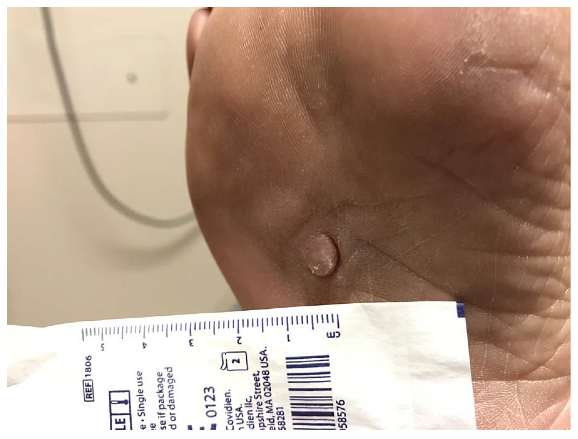
Visual examination of the soft-tissue mass revealed an approximately 7 × 7 × 10-mm exophytic tumor (Fig. 1). Palpation of the mass demonstrated a soft but solid texture without fluctuance. It was appreciated to be located in the subcutaneous tissue without deep fascia involvement. The mass was nonpulsatile and did not transilluminate. There were no localized swelling or pigment changes noted. Gross sensation and vascular status were intact to bilateral feet.
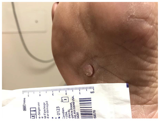
Figure 1.
Exophytic tumor on the plantar aspect of a 62-year-old patient's right foot.
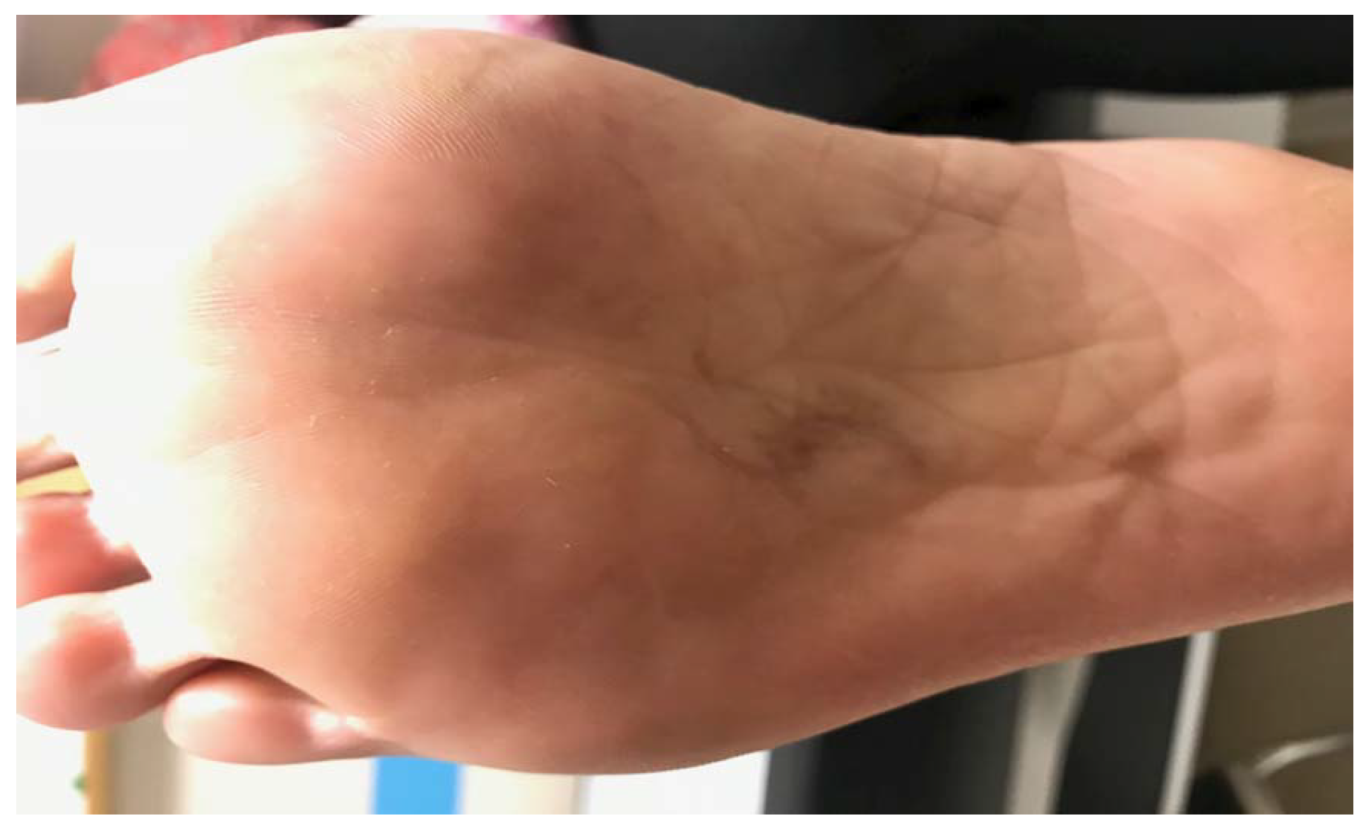
An excisional biopsy and double-advancement flap (O-to-Z) was subsequently performed under local anesthesia (Figs. 2–5). Following primary closure, the patient was permitted to bear weight as tolerated in a surgical shoe with a felt offloading pad for 2 weeks. The surgical specimen was sent for pathologic evaluation and was diagnosed as an angioleiomyoma (Figs. 6–9). The patient returned to the clinic at 2 weeks for suture removal. At 1 year postoperatively, the scar was noted to be soft and supple (Fig. 10). The patient had no complaints and her pain was alleviated.
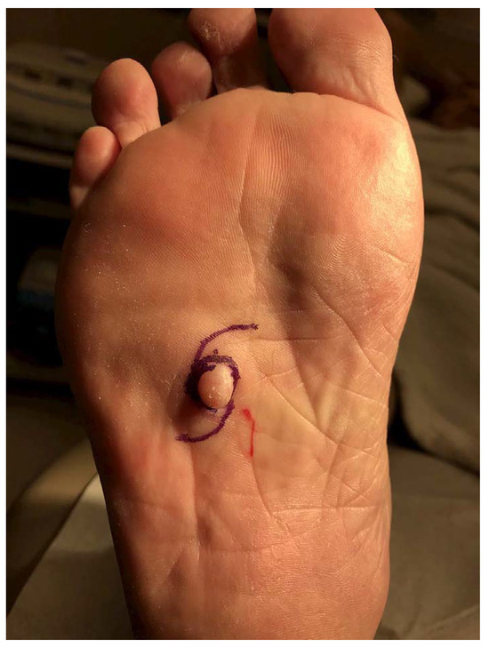
Figure 2.
Proposed plan for excision of primary tumor and closure with O-to-Z double-rotation flap.
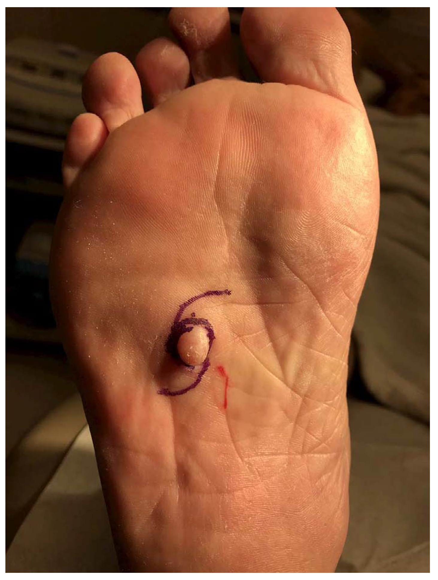
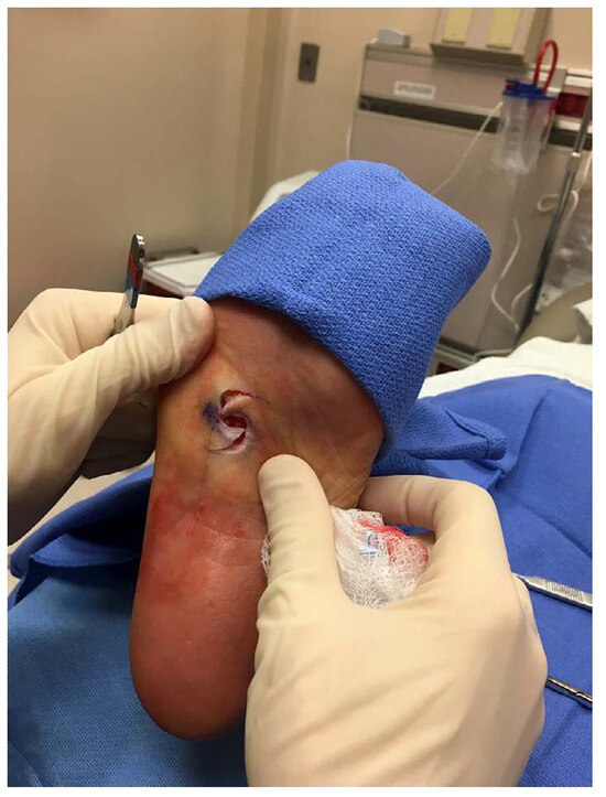
Figure 3.
After excision and before flap advancement.
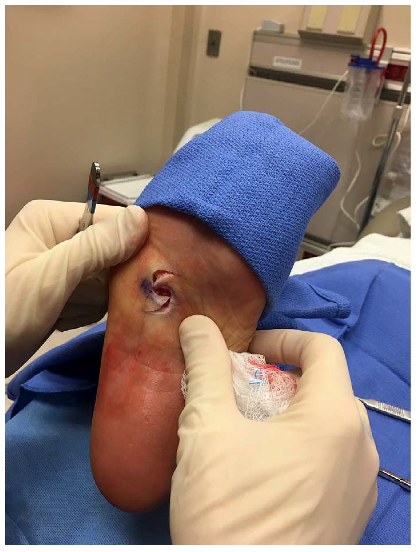
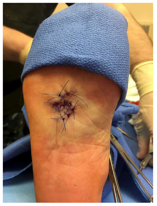
Figure 4.
Final closure showing Z-shaped appearance.
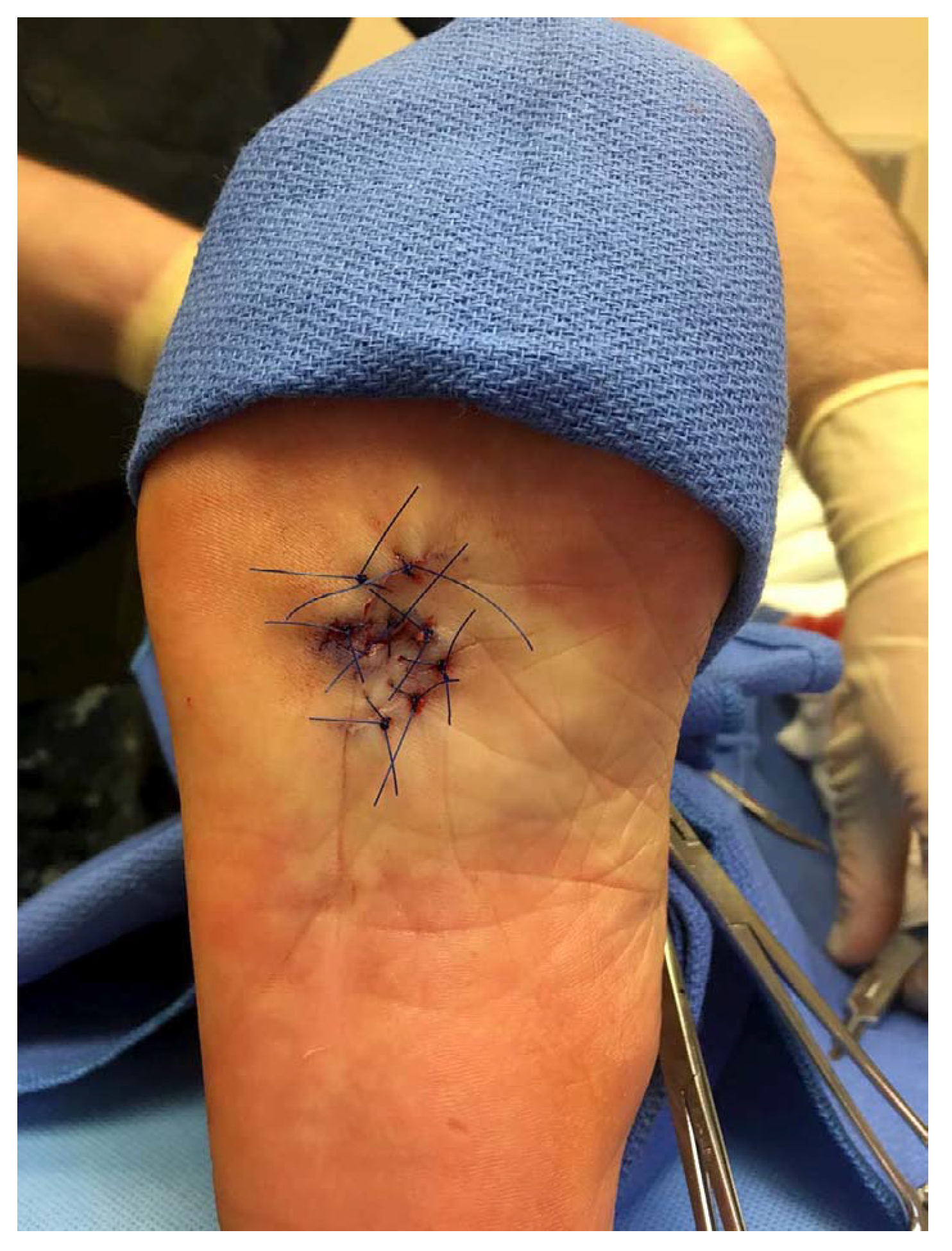
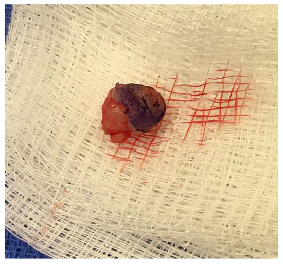
Figure 5.
Resection of lesion en toto.
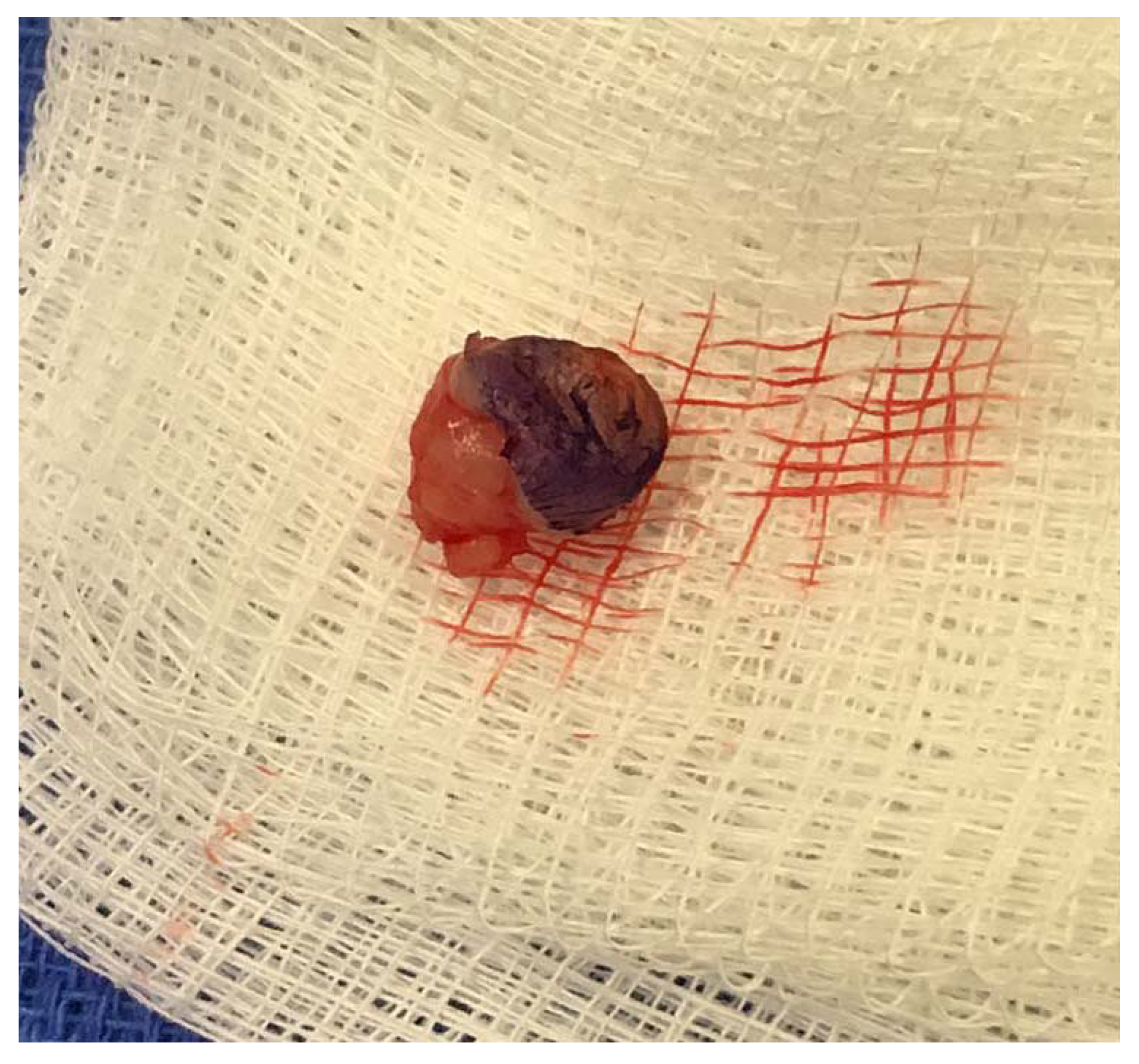
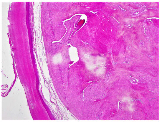
Figure 6.
At low power, in the left side of the image, there is normal overlying acral skin characterized by a thick layer of compact keratin overlying thin epidermis with normal appearing keratinocytes. Underlying this acral skin is a very well-circumscribed, rounded nodule composed of alternating fascicles of bland appearing eosinophilic cells. Even at low power, we appreciate that within the alternating fascicles are blood vessels with fairly thick vessel walls.
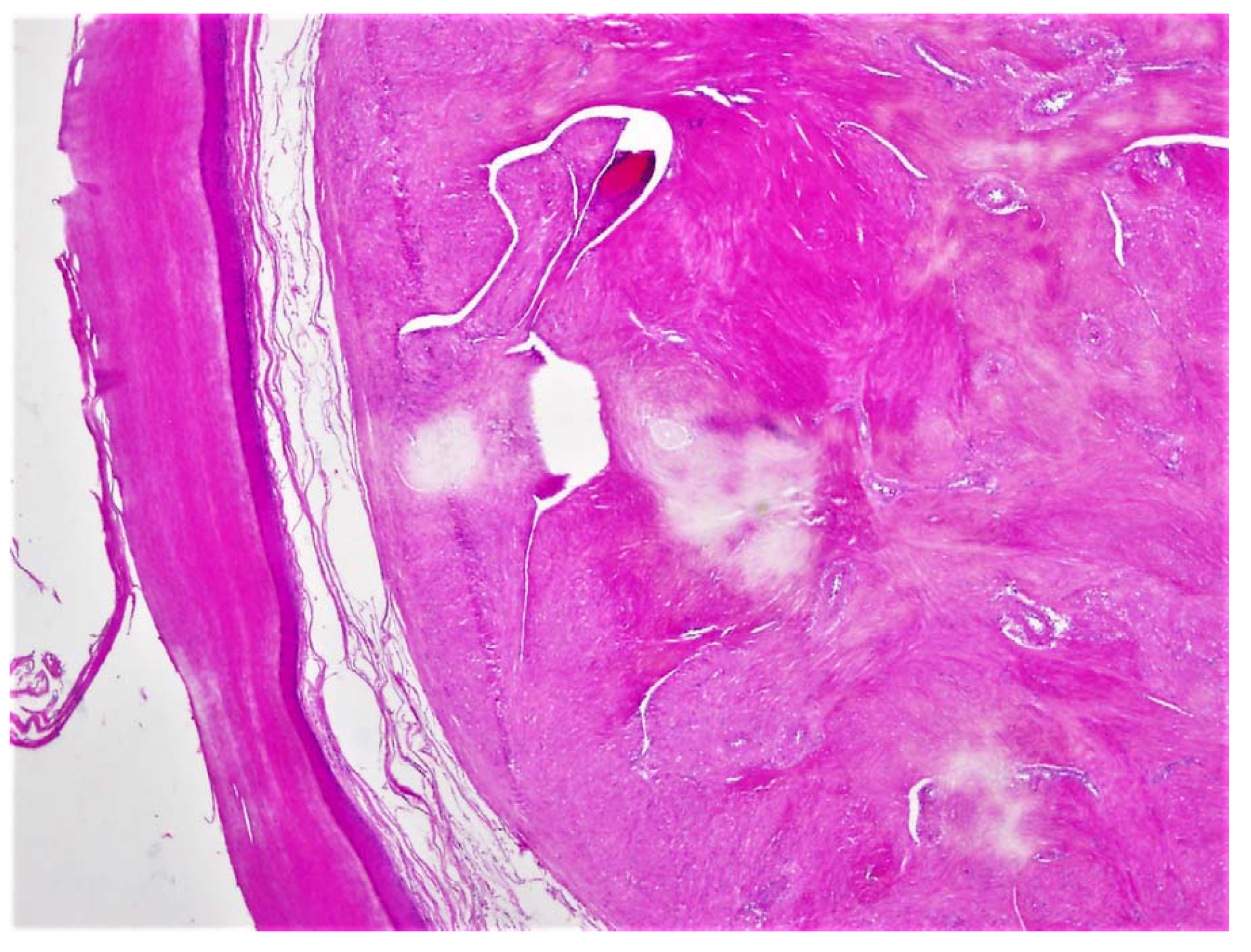
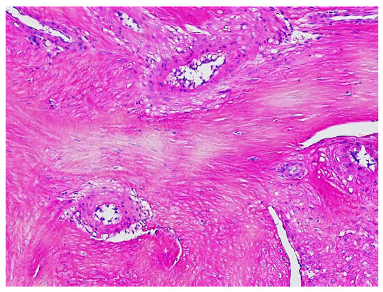
Figure 7.
At higher power, the alternating fascicles become more visible and the bland spindled cell nuclei that compose the fascicles become more prominent. The vessels are lined by normal appearing endothelium; however, they lack the normal elastic lamina. The overall appearance of bland spindled cells with abundant eosinophilic cytoplasm in alternating fascicles with intervening vessels without elastic lamina is consistent with a diagnosis of angioleiomyoma.
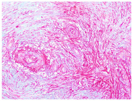
Figure 8.
A Masson-Trichrome stain is shown in which smooth muscle fibers are red. The stain demonstrates smooth muscle within the vessel walls, as well as fascicles of smooth muscle surrounding the vessel highlighting both the vascular and smooth muscle component of the angioleiomyoma. Between the smooth muscle fibers collage fibers, stained blue by the Masson-Trichrome may also be identified.
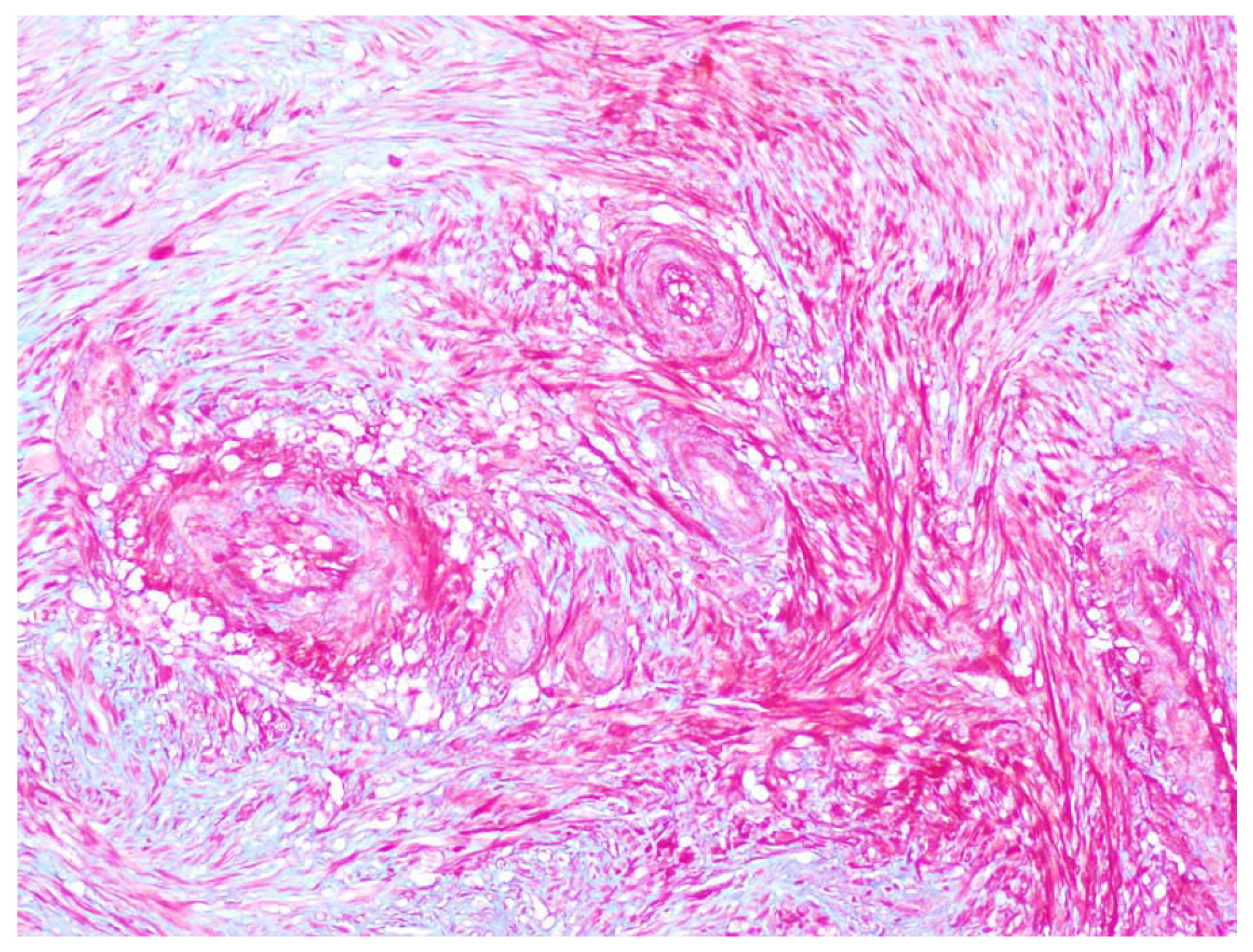
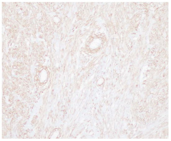
Figure 9.
An immunohistochemical stain for smooth muscle actin (SMA) is also shown. This antibody binds to smooth muscle fibers and again demonstrates smooth muscle within the walls of the vessels as well as fascicles of smooth muscle surrounding the vessels, consistent with the diagnosis of angioleiomyoma.
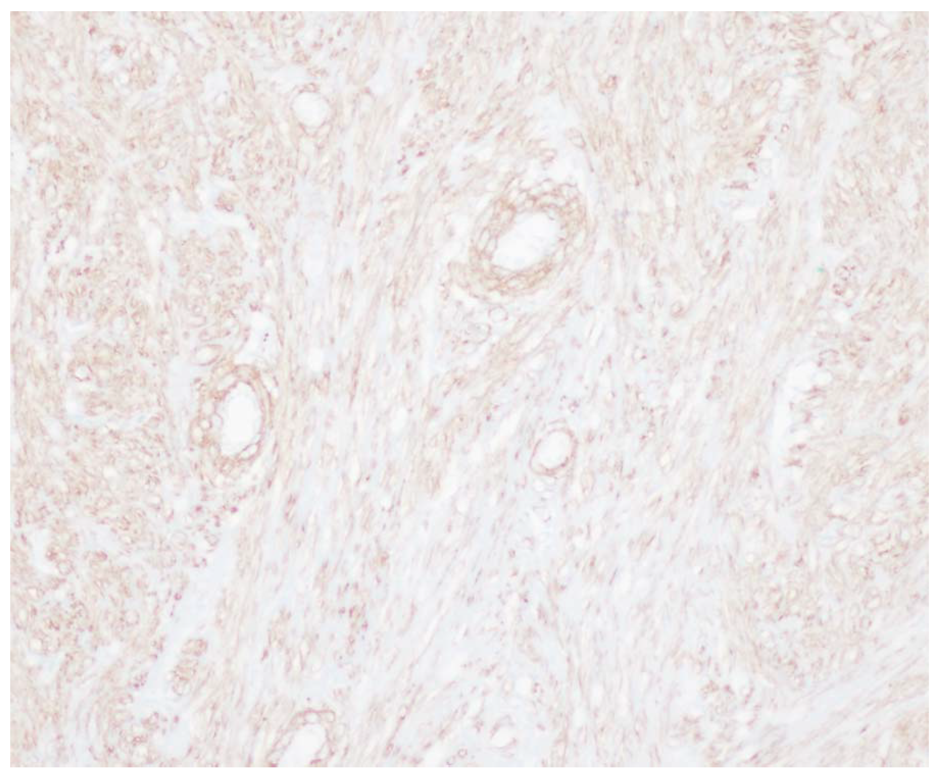
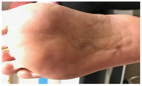
Figure 10.
One year after initial excision.
Discussion
Although it has been well documented for angioleiomyomas to occur in the foot, to our knowledge, this is the only case of histopathologically confirmed angioleiomyoma in a patient who presented with an exophytically growing soft-tissue mass. The presentation is rare compared with the presentation of many other case studies described within the literature, which are typically subfascia masses.
Surgical excision is beneficial and can be performed through a single linear incision for most subcutaneous nodular masses; however, because of the exophytic nature of the angioleiomyoma in our patient, there would be a soft-tissue deficit postoperatively, leading to tense tissues and possible dehiscence on weightbearing. It was for this reason that an O-to-Z double-advancement skin flap was used. This technique was chosen in place of a standard 3:1 excision to decrease the amount of tissue waste.
The optimal design of the O-to-Z flap has been studied by Buckingham et al. [15] The basic design begins with a circle-shaped excision of the lesion. Two curvilinear incisions at opposite sides of the primary circular defect are then created. The flaps are undermined, rotated, and advanced, resulting in a final Z closure.
Conclusions
Angioleiomyoma should be within the differential diagnosis for soft-tissue masses in situations in which the mass is either subcutaneous or exophytic in nature, as this article has described. Surgical excision is still the treatment of choice and can include a flap excision technique when the soft-tissue mass displays exophytic growth to decrease the soft-tissue void during excision and to reduce the waste of tissue.
Acknowledgment
Jeptha T. Johnson III, MD, Department of Pathology and Laboratory Services at Walter Reed National Military Medical Center for providing the pathologic images and description.
Financial Disclosure:
None reported.
Conflict of Interest:
None reported. The views expressed are solely those of the authors and do not reflect the official policy or position of the US Navy, the Department of Defense, or the US Government.
References
- WooKS.KimSH.KimHS.: Clinical experience with treatment of angioleiomyoma. Arch Plast Surg41: 374, 2014.
- SantucciA.AlbiniM.VenturaA.: Clinical and histological features of vascular leiomyoma of the foot: case report and literature review. Foot Ankle Surg6: 267, 2000.
- HachisugaT.HashimotoH.EnjojiM.Angioleiomyoma: a clinico-pathologic reappraisal of 562 cases. Cancer54: 126, 1984.
- BerlinSJ.A review of 2720 lesions of the foot. JAPMA70: 318, 1980.
- SweeneyJ.KeatingsSE.Angioleiomyoma. J Foot Surg22: 21, 1983.
- HanftJR.CarbonellJA.DoHQ.Angioleiomyoma of the lower extremity. JAPMA87: 388, 1997.
- SprinkleR.SanguezaO.WellsA.Angioleiomyoma of the lower leg. JAPMA107: 244, 2017.
- MaheshwariAV.TempleHT.MurocachoCA.Calcified angiomyomas of the foot: a case report. Foot Ankle Int29: 449, 2008.
- KacerovskaD.MichalM.KreuzbergB.: Acral calcified vascular leiomyoma of the skin: a rare clinicopathological variant of cutaneous vascular leiomyomas: report of 3 cases. J Am Acad Dermatol59: 1000, 2008.
- YatesBJ.Angioleiomyoma: clinical presentation and surgical management. Foot Ankle Int22: 670, 2001.
- HamouiM.LargeyA.AliM.: Angioleiomyoma in the ankle mimicking tarsal tunnel syndrome: a case report and review of the literature. J Foot Ankle Surg49: 398, 2010.
- LepoffA.MakarovV.WilliamsM.Angioleiomyoma of the plantar foot. JAPMA108: 262, 2018.
- MurataH.MatsuiT.HorieN.: Angioleiomyoma with calcification of the heel: report of two cases. Foot Ankle Int28: 1021, 2007.
- SprinkleRLBIII,SanguezaOP.WellsAE.Angioleiomyoma of the lower leg. JAPMA107: 244, 2017.
- BuckinghamED.QuinnFB.CalhounKH.Optimal design of O-to-Z flaps for closure of facial skin defects. Arch Facial Plast Surg5: 92, 2003.
© 2020 American Podiatric Medical Association