


Abstract
Tillaux fracture is known to occur in adolescents once it happens during the transition period when the medial and central physis has finished closure, but the lateral physis is still opened. The trauma mechanism is typically external rotation ankle injury resulting in an avulsion fracture of the anterolateral tibial plafond. This fracture has rarely been reported in adults, especially associated with other injuries. We report a case of Tillaux fracture in an adult, associated with a Volkmann fracture and a Maisonneuve fracture, that were surgically treated with open reduction and internal fixation and had an excellent outcome. Recognizing and appropriately treating these injuries is key in the prevention of further degenerative arthritis and instability.
Tillaux fracture is an avulsion fracture of the anterolateral aspect of the tibial plafond owing to the pull of a taut anteroinferior tibiofibular ligament. In adults, different from children, the ligament generally gives away before avulsion of its attachment to the anterolateral tibial plafond occurs, leading to relative rarity of this avulsion pattern. [1-4] The mechanism of this fracture is typically supination with external rotation. [1,5] In children, the size of the fracture depends on the extent of the fusion medially because distal tibial epiphyseal closure has an asymmetrical pattern. Physeal closure starts from the center and then progresses toward the medial side. Finally, fusion of the lateral aspect of the distal tibial occurs. The medial closure occurs at approximately 13 to 14 years of age, with lateral closure beginning at 14.5 to 16 years of age. The interval (∼18 months) during which the distal tibial physis completes its closure and gains maturity is referred to as the transition period. Adolescent Tillaux fracture occurs during the transition period, when the medial and central physes have finished closure but the lateral physis is still open. [2,5,6]
Volkmann fracture generally occurs in ankle fracture due to either mechanism of pronation or supination with external rotation. External rotation forces make the talus rotate further laterally and the posteroinferior tibiofibular ligament stretch, thus either the posteroinferior tibiofibular ligament will rupture or an avulsion fracture from the posterolateral tibia will occur. [2]
Maisonneuve fracture is an injury caused by a pronation–external rotation force at the ankle that results in rupture along the syndesmosis with its diastasis, lateral talar shift, and spiral fracture of the proximal half of the fibula. [7]
Tillaux fractures have rarely been reported in adults. [1,2] Of the six published case reports in patients older than 18 years, only one had Tillaux fracture associated with Volkmann fracture, [2] and none had association with Maisonneuve fracture.
In the present report, we describe the case of a Tillaux fracture in an adult associated with Volkmann and Maisonneuve fractures. To our knowledge, no data are available regarding the association of all of these lesions.
Case Report
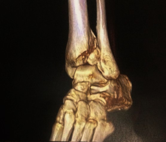
A 74-year-old woman presented to the emergency department after being hit by a car. The patient was unable to bear weight on her left lower extremity. On clinical examination, swelling and significant tenderness were present over the proximal extremity of her leg to her ankle. The neurovascular examination findings were normal. Radiographs showed a spiral fracture of the proximal half of the fibula (Maisonneuve fracture) (Fig. 1A) and a posterior malleolus fracture (Volkmann fragment) (Fig. 1B). Computed tomography revealed a fracture of the anterolateral aspect of the tibial plafond (Tillaux-Chaput fracture) (Fig. 2). The patient also had a pelvic fracture that was not displaced.
Figure 1.
Preoperative radiographs showing (A) anteroposterior view of the leg showing a spiral fracture of the proximal half of the fibula (Maisonneuve fracture) and (B) lateral view of the ankle showing a displaced posterior malleolus fracture (Volkmann fragment).
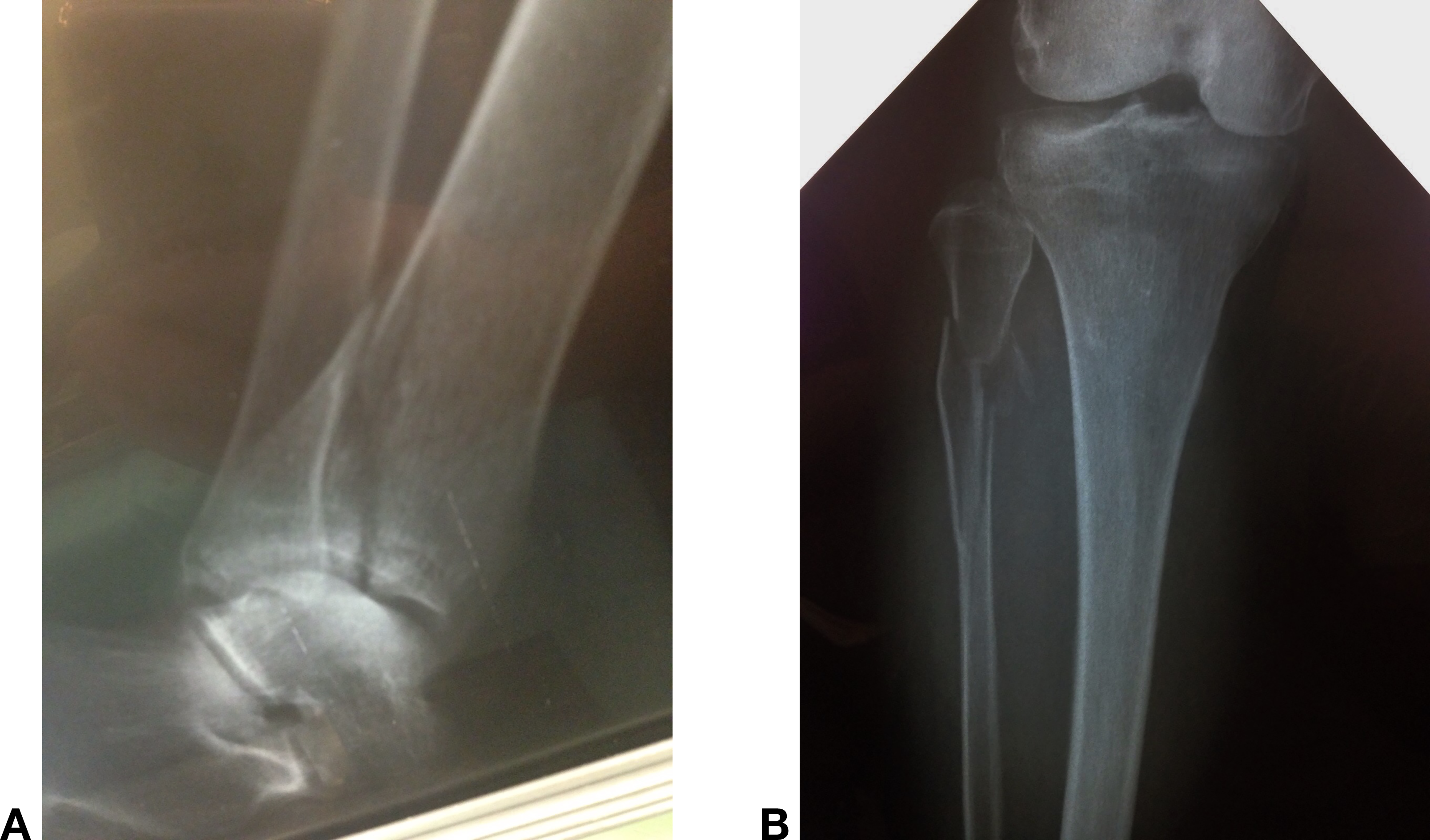
Figure 2.
Three-dimensional computer tomography scan showing a fracture of the anterolateral aspect of the tibial plafond (Tillaux-Chaput fracture).
Surgical Technique
The patient underwent surgery 14 days after the initial injury. With the patient under spinal anesthesia, under tourniquet control, and in the right lateral decubitus position, a posterolateral incision was made. The Volkmann fragment was reduced and fixed with a third tubular buttress plate. Thus, the decubitus was changed to dorsal position and the syndesmosis was stabilized using a single 3.5-mm fully threaded cortical screw engaging three cortices. Through an anterolateral approach, the Tillaux fracture was reduced and fixed with two cannulated lag screws (Fig. 3). The entire procedure was performed under fluoroscopic guidance (Figs. 4–6). Postoperatively, the patient was placed in a below-the-knee nonweightbearing plaster cast for 6 weeks. Within 12 weeks, clinical and radiographic assessments revealed the fracture to be well consolidated without any complications (Figs. 7 and 8). At that time, full weightbearing was allowed. Twelve months after surgery, the patient returned with no symptoms or pain and complete range of motion of the ankle. The American Orthopaedic Foot & Ankle Society ankle and hindfoot scale score was 100.
Figure 3.
Intraoperative photograph showing the Tillaux fracture that was reduced and fixed with two cannulated lag screws through the ankle’s anterolateral approach.
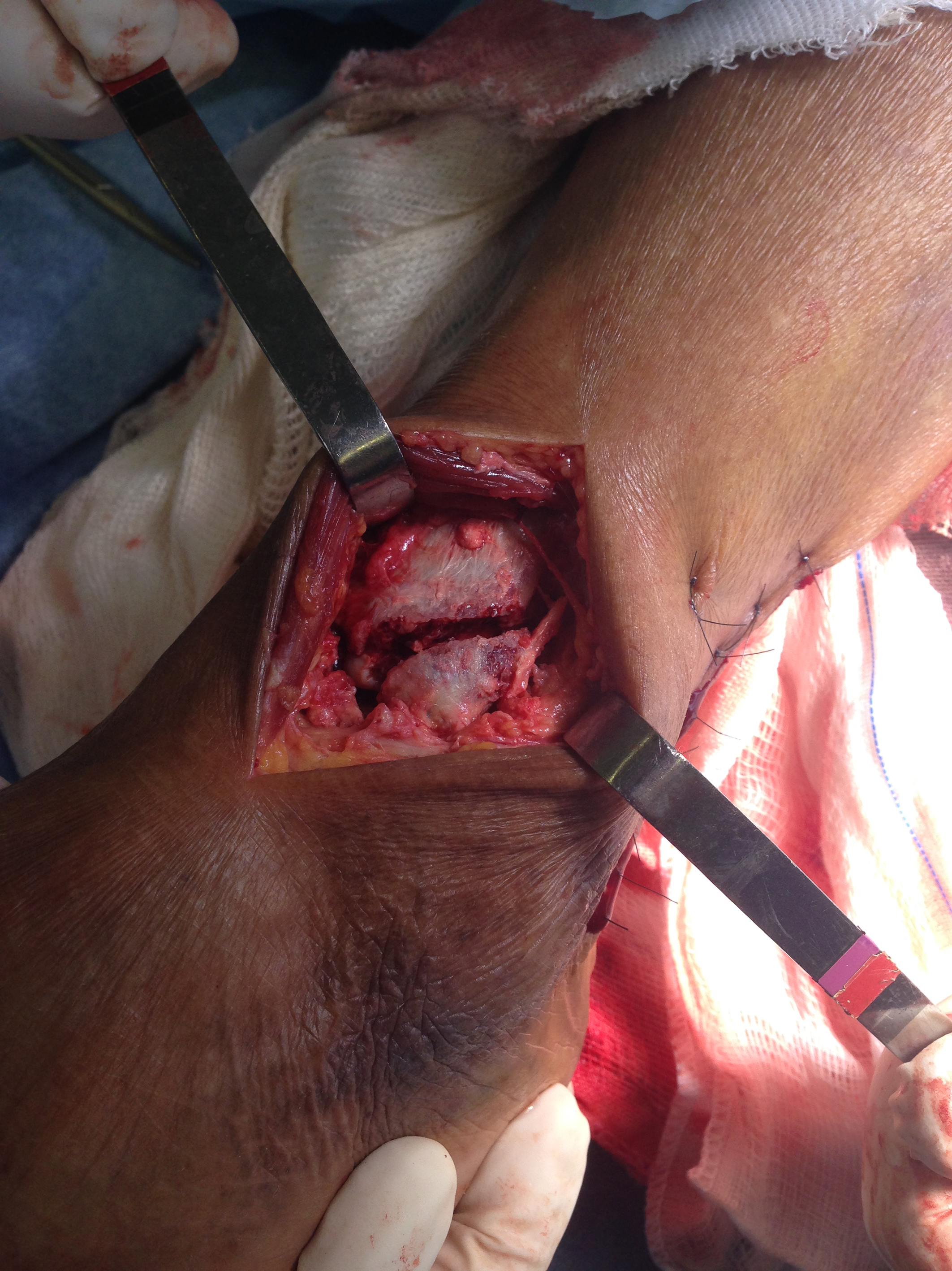
Figure 4.
Anteroposterior mortise intraoperative radiographs after reduction and fixation of all fractures.
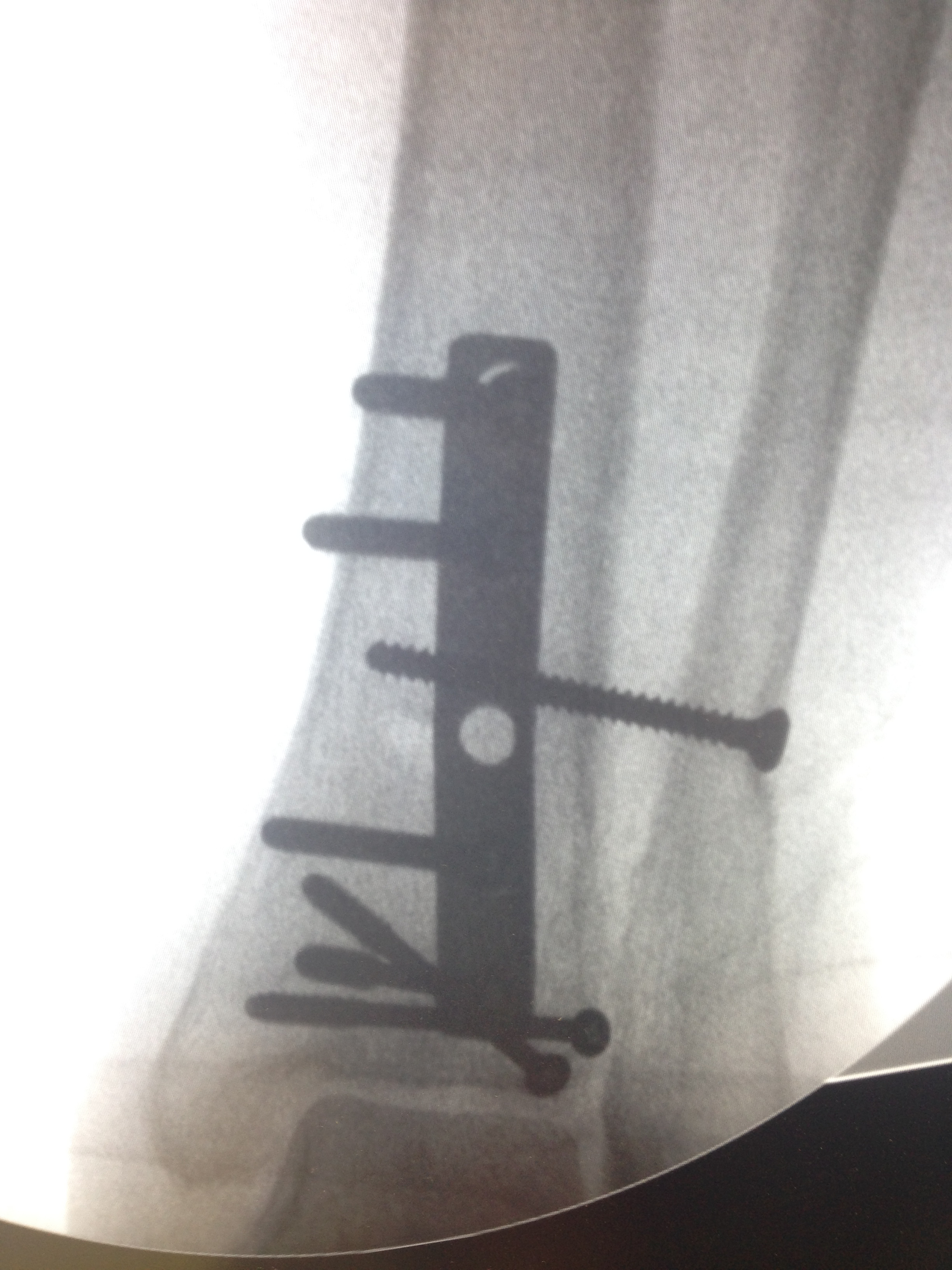
Figure 5.
Anteroposterior intraoperative radiographs after reduction and fixation of all fractures.
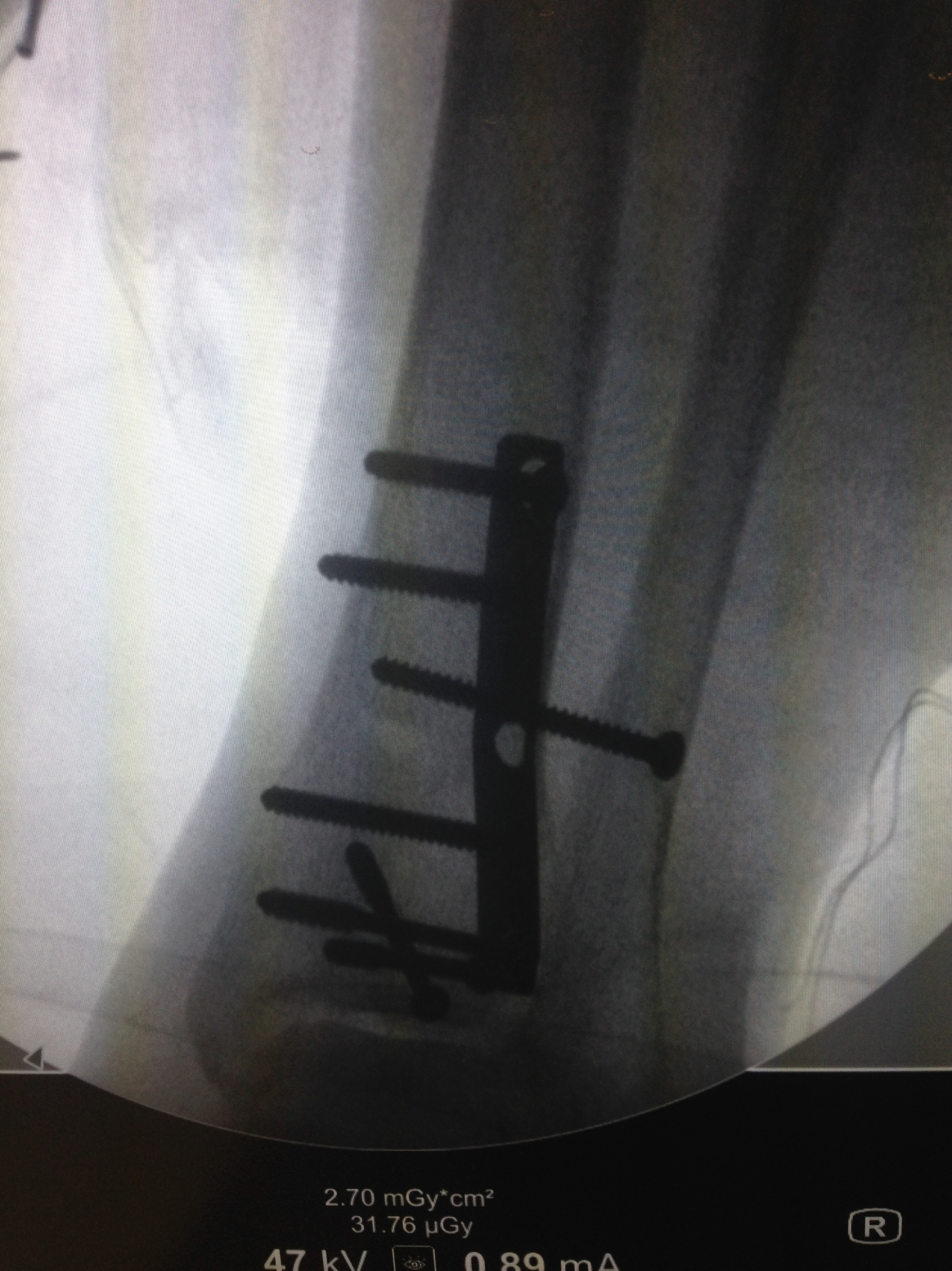
Figure 6.
Lateral intraoperative radiographs after reduction and fixation of all fractures.
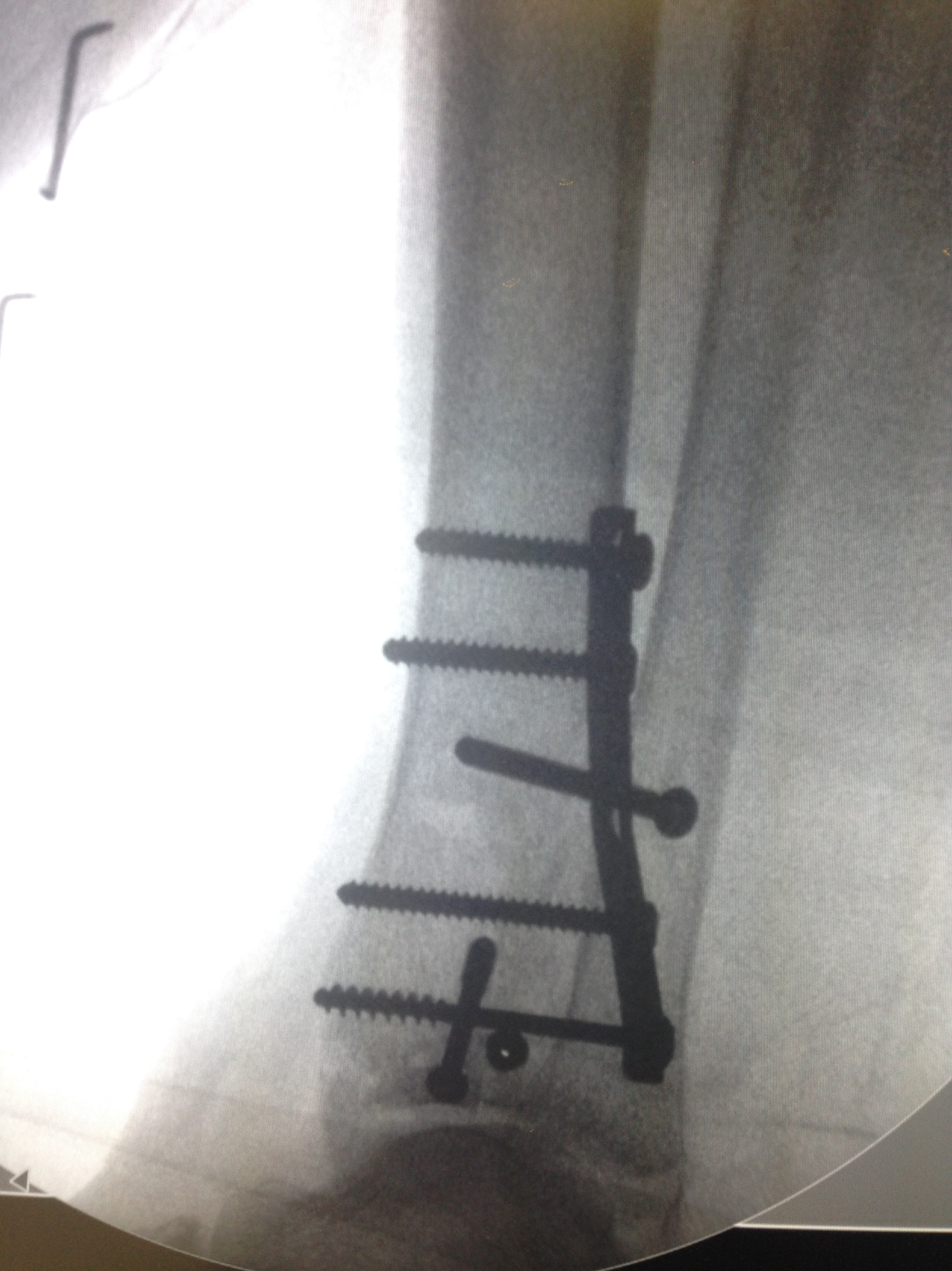
Figures 7.
Anteroposterior postoperative photograph showing complete bone healing and joint congruency after 12 months.
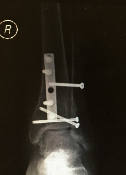
Figure 8.
Lateral postoperative photograph showing complete bone healing and joint congruency after 12 months.
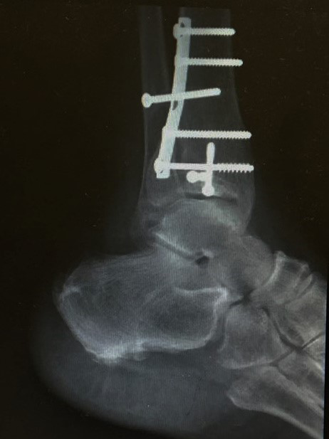
Discussion
Tillaux fracture has rarely been reported in adults. A literature review presented only six other reports with eight cases. [2] This lesion is known to occur in adolescents and is defined as an avulsion fracture of the anterolateral distal tibial epiphysis. It occurs during the transition period, at age 12 to 14 years in girls and 15 to 18 years in boys, when the medial and central physes have finished closure but the lateral physis is still open, making it weaker. [8,9] The mechanism of this injury is external rotation of the ankle that leads to an avulsion fracture owing to the pull of the anteroinferior tibiofibular ligament. [1,5] Volkmann fracture corresponds to an avulsion fracture of the posteroinferior tibiofibular ligament from its tibial attachment. Simultaneous Tillaux and Volkmann fractures in the adult patient is extremely rare, with only two cases previously reported. [2]
Tillaux fracture has rarely been associated with medial malleolus fracture or deltoid injury, [10] and the syndesmosis should be carefully evaluated. The present case shows a unique association: Tillaux and Volkmann fractures associated with a high fibula fracture or Maisonneuve fracture.
The possible mechanism of injury in the present case was pronation–external rotation, according to Lauge-Hansen classification, characterized by, progressively, deltoid ligament rupture or fracture of the medial malleolus; disruption of the anterior syndesmosis and rupture of the interosseous ligament and membrane; spiral fracture of the fibula in its proximal half; and either rupture of the posterior syndesmosis or fracture of the posterior tibia. [7]
Tillaux fracture associated with Volkmann fracture is also caused by supination–external rotation. However, in that mechanism the fibula fracture occurs at the level of syndesmosis, and in the case reported herein there was a Maisonneuve avulsion fracture instead.
Diagnosis is challenging in cases such as this. Tillaux and Volkmann fractures can be missed in direct radiographs with anteroposterior and lateral radiographic views of the ankle. Furthermore, Maisonneuve fracture can be neglected if leg radiography is not performed. Oblique ankle radiographic views can help in the diagnosis, and additional computed tomography is recommended if these lesions are suspected to help with diagnosis and surgical treatment.
Maisonneuve fracture treatment is surgical, with anatomical reduction and syndesmosis stabilization. Isolated Tillaux and Volkmann fractures can be treated nonsurgically if there is no dislocation. When displacement is greater than 2 mm, reduction and fragment fixation is advised to prevent further degenerative arthritis and instability.
In the present patient, there was a unique association of lesions, thus the syndesmosis was disrupted at two points, with sequent ankle instability requiring fixation.
Conclusions
The Tillaux fracture is an uncommon lesion in adults, and its association with Volkmann and Maisonneuve fractures is even rarer. These lesions may be neglected when careful physical examination and imaging are not performed. Due to marked instability caused in the ankle, its surgical treatment is indispensable, avoiding early osteoarthritis.
Financial Disclosure
None reported.
Conflict of Interest
None reported.
References
- Kumar N,Prasad M:Tillaux fracture of the ankle in an adult: a rare injuryJ Foot Ankle Surg53: 757, 2014.
- Kose O,Yuksel HY,Guler F,et al: Isolated adult Tillaux fracture associated with Volkmann fracture: a unique combination of injuries: report of two cases and review of the literature. J Foot Ankle Surg55: 1057, 2016.
- Miller MD:Arthroscopically assisted reduction and fixation of an adult Tillaux fracture of the ankle. Arthroscopy13: 117, 1997.
- Duchesneau S,Fallat LM:The Tillaux fracture. J Foot Ankle Surg35: 127, 1996.
- Patel A,Shur V:Juvenile Tillaux ankle fracture pattern in a skeletally mature adult. The Foot16: 54, 2006.
- Jennings MM,Lagaay P,Schuberth JM:Arthroscopic assisted fixation of juvenile intra-articular epiphyseal ankle fractures. J Foot Ankle Surg46: 376, 2007.
- Smith MG,Ferguson E,Kurdy NM:Persistent diastasis in a Maisonneuve fracture: interposition of a tibial osteochondral fragment: a case report. J Foot Ankle Surg44: 225, 2005.
- Simon WH,Floros R,Schoenhaus H,et al: Juvenile fracture of Tillaux: a distal tibial epiphyseal fracture. JAPMA79: 295, 1989.
- Sharma B,Reddy IS,Meanock C:The adult Tillaux fracture: one not to miss. BMJ Case Rep2013:bcr2013200105, 2013.
- Marti CB,Kolker DM,Gautier E:Isolated adult Tillaux fracture: a case report. Am J Orthop34: 337, 2005.
© 2019 American Podiatric Medical Association