


Abstract
A 36-month follow-up of the management of bilateral adolescent unicameral bone cysts in a high school gymnast treated with a calcium sulfate/calcium phosphate (CSCP) bone void filler (BVF) is presented. The more developed left calcaneal cyst was managed with a traditional, open approach consisting of allogenic bone graft, CSCP BVF mixed with platelet-rich plasma. The less developed right calcaneal cyst was managed with a less used approach, a percutaneous bone cortex incision with only the CSCP BVF. The rationale for the selection between the open and percutaneous approaches, long-term BVF incorporation, and positive patient outcome allowing a quick return to athletics are presented.
Virchow first described juvenile bone cysts in 1876.[1] A unicameral bone cyst can occur in any bone but occurs more frequently in long bones. A unicameral bone cyst can occur at any age, but occurs often in the first two decades of life, most frequently occurring in children between the ages of 5 and 15 years.[2] This bone cyst accounts for 3% of all cyst-like bone conditions.[3,4] The most common body location of unicameral bone cyst is the proximal humerus and proximal femur; however, every bone in the human skeleton can be affected. The calcaneus is the most common tarsal bone affected by unicameral bone cyst[5,6]; other foot bones can be involved.[7,8,9]
The unicameral bone cyst is a benign, radiolucent, fluid-filled lesion of bone. The cysts can weaken the bone cortex, predisposing that bone to a pathologic fracture. The surgical management of the unicameral bone cyst is diverse. Unicameral bone cysts located in the feet are managed with debridement of the cyst lining followed by bone marrow aspirate concentrate incorporation,[10,11,12] injection of a calcium sulfate/calcium phosphate composite,[13,14] injection with allogenic bone graft,[15,16,17] and less frequently by filling with cortisone.[18,19,20,21] Surgical long bone unicameral bone cyst management includes stabilization with an intramedullary nail[22,23] or cyst decompression with cannulated screws.[24] The intramedullary nailing can also be combined with a synthetic bone substitute[22,23] or with platelet-rich plasma (PRP).[25]
Kadhim et al[26] selected 62 articles from a total of 463 articles for meta-analysis. The selected articles needed to have a minimum sample size of 15 patients and have provided data on radiographic healing outcome. The cumulative sample size was 3,211 patients with 3,217 unicameral bone cysts. The study summary or pool estimate of methylprednisolone acetate injection resulted in a healing rate (77.4%) comparable to that for bone marrow injection (77.9%). A higher healing rate was observed with methylprednisolone acetate injection when inner wall disruption was performed. The pool estimate of bone marrow with demineralized bone matrix injection was high (98.7%). The unicameral bone cyst healing rate after surgical curettage was comparable whether autograft or allograft was used (90%). Unicameral bone cyst treatment with flexible intramedullary nails without curettage provided an almost 100% healing rate, whereas continuous decompression with cannulated screws provided an 89% healing rate. Conservative treatment resulted in a healing rate of 64.2% (95% confidence interval, 26.7%–101.8%).[26] Levy et al[5] reviewed 10 studies with 171 patients with 181 calcaneal unicameral bone cysts with a minimum of 1 year of follow-up and level I to IV evidence. The review by Levy et al. demonstrated that all of the patients treated with open curettage and bone augmentation had significant improvement in heel pain (P < .001). Only 1.1% ± 1.0% of the cysts treated conservatively had healed on radiography compared with 93.0% ± 13.0% of the cysts treated surgically (P < .001). Autografting had a significantly greater percentage of radiographic cyst consolidation than did allografting (97.4% ± 11.1% versus 85.1% ± 15.8%, P < .001, Z = 3.5).[5] Pedzisz et al[27] presented a group of nine patients (four male patients and five female patients) with solitary bone cysts managed with cyst wall soft-tissue lining removal followed by cavity filling with allogenic bone grafts mixed with PRP. This authors stated that all cysts were completely filled with new bone and all of the patients were without pain at 12 months.
Adolescent unicameral pedal bone cysts that require surgical intervention generally involve open curettage with grafting. This surgical intervention involving open curettage with grafting will yield satisfactory results consisting of graft incorporation, pain resolution, and resumption of weightbearing activities in 8 to 12 weeks.[2,12] There are always situations where the patient wishes or needs to return to full weightbearing activities sooner than 8 to 12 weeks. This case study illustrates an example of a patient involved in competitive sport with successful return to play in less than 12 weeks following treatment for an unicameral bone cyst.
Case Report
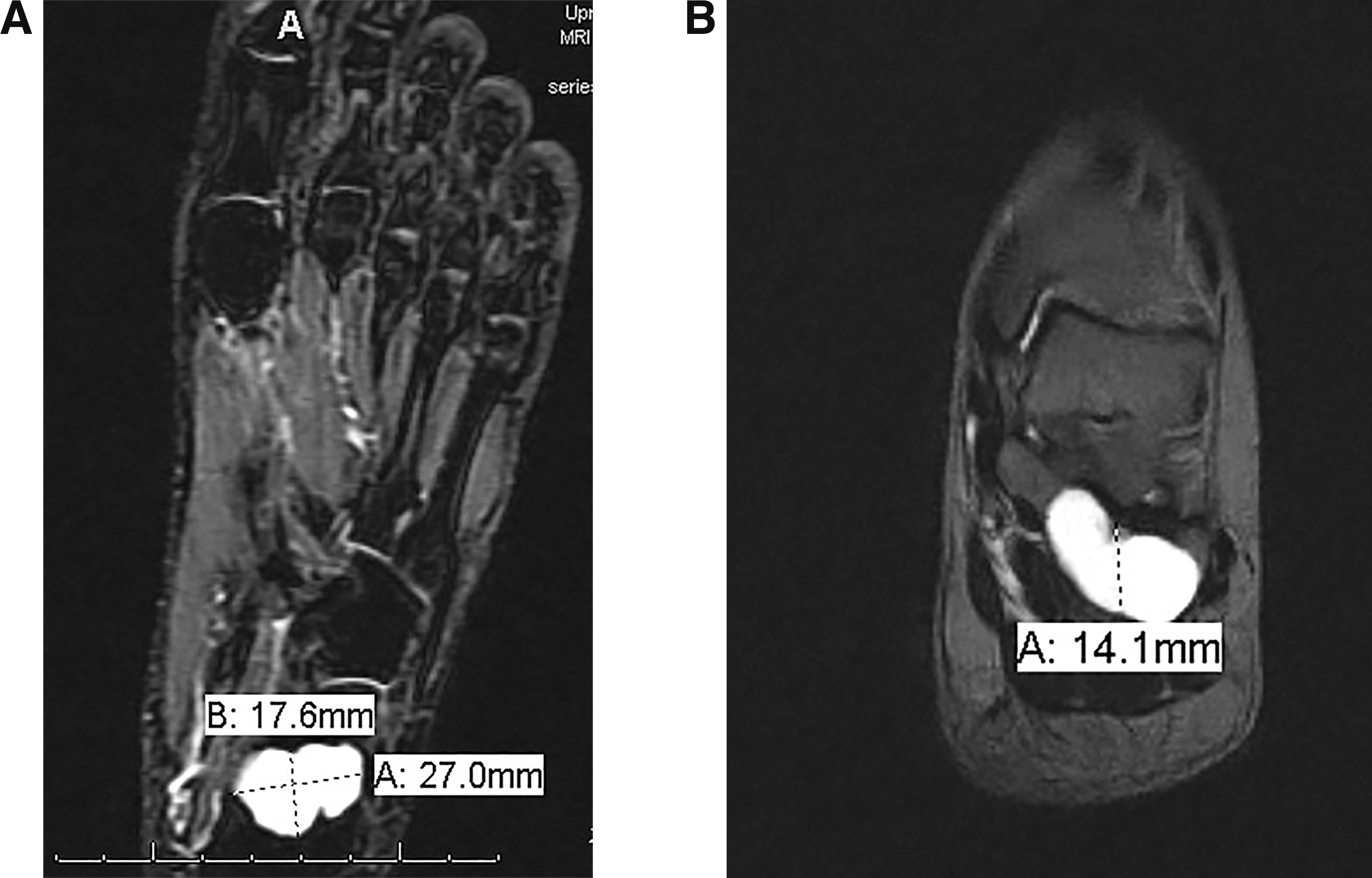
A 14-year-old girl presented with her parents for a second opinion regarding significant pain at her left rear foot. This patient was on track for a college gymnastics scholarship. This patient had dropped a large can on her left foot 9 months previously and had had no improvement in her pain. Previous treatment by a different physician included prescription nonsteroidal anti-inflammatory drugs, offloading with crutches, and pain management, without any improvement. The previous physician had not obtained any foot radiographs. The patient's physical examination was significant only for marked pain on lateral and medial manual left calcaneal body compression. The contralateral foot was unremarkable on examination. Initial radiographs demonstrated a well-circumscribed lucency with a narrow zone of transition in the body of the left calcaneus (Fig. 1). The left foot computed tomographic (CT) (Fig. 2) and magnetic resonance imaging (MRI) (Fig. 3) scans demonstrated a well-circumscribed lobulated uniform cyst in the calcaneal body without cortical breakthrough consistent with a unicameral bone cyst.
Figure 1.
Preoperative left lateral foot radiograph.
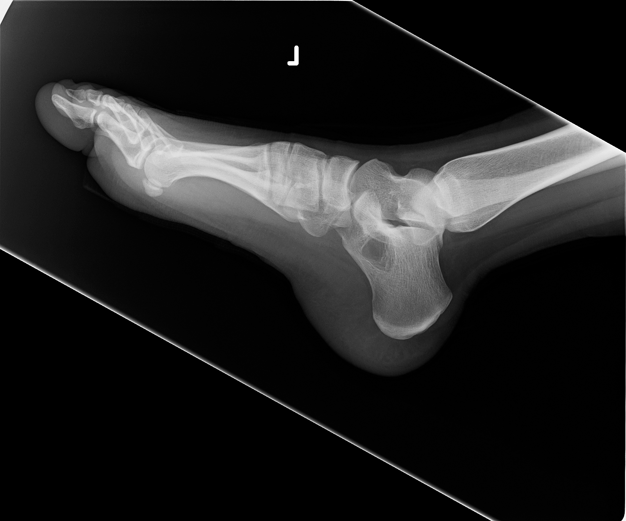
Figure 2.
A, Preoperative left lateral foot computed tomographic scan. B, Preoperative left axial foot computed tomographic scan.
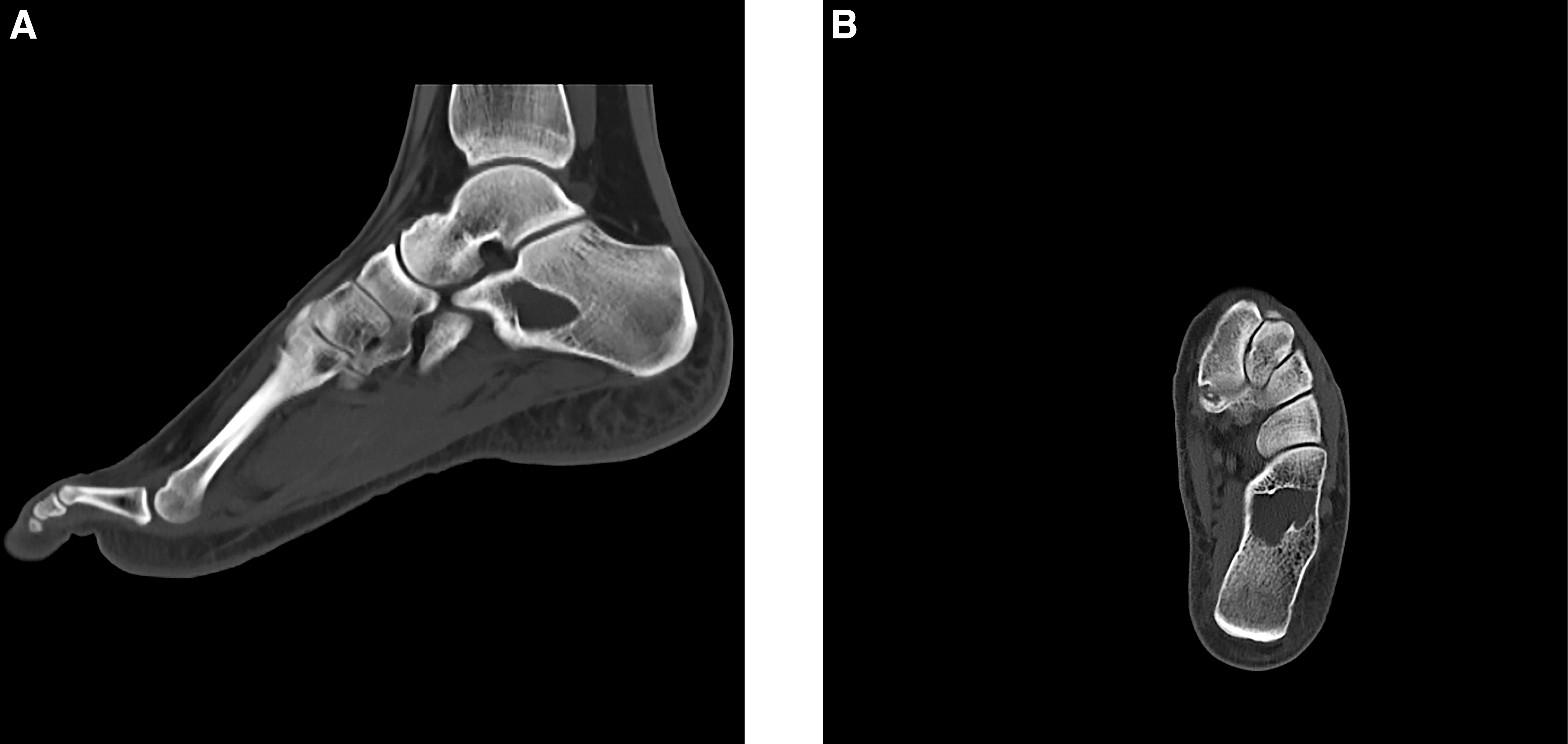
Figure 3.
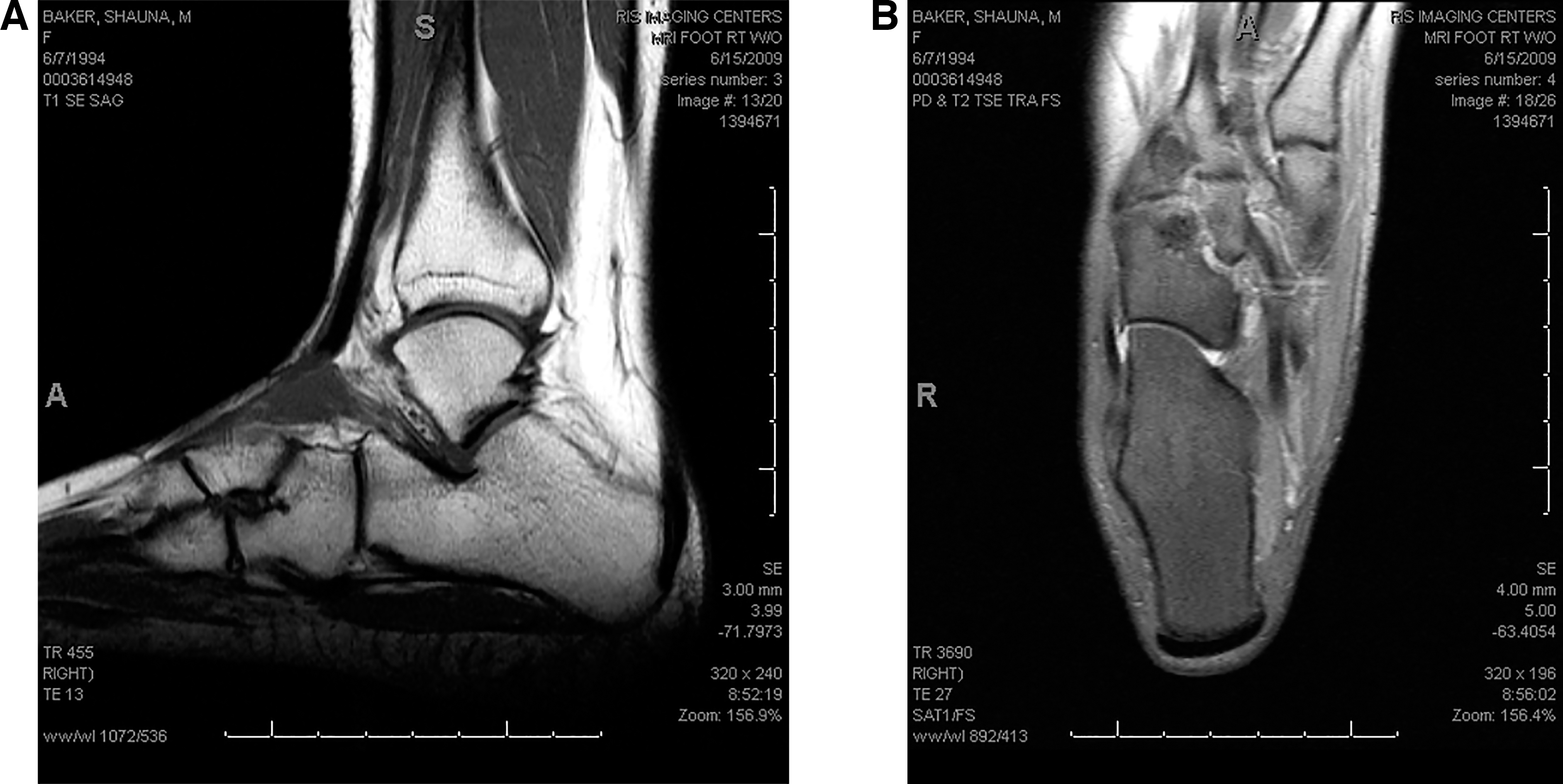
A, Preoperative left axial foot magnetic resonance imaging scan. B, Preoperative left coronal foot magnetic resonance imaging scan.
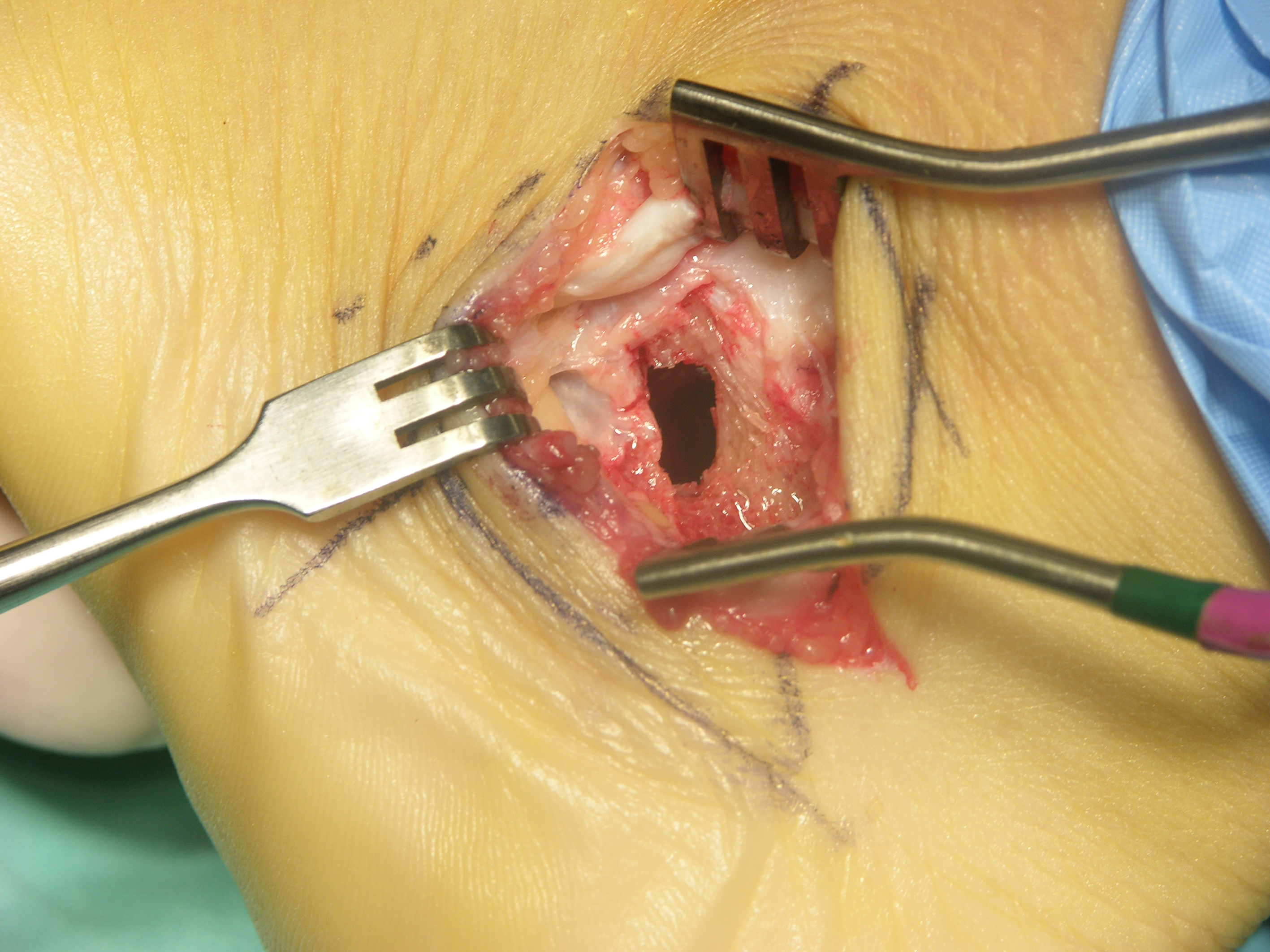
Because this patient was a high school gymnastic athlete on track for a gymnastic scholarship, the patient and her parents did not wish to proceed with autogenous bone grafting to avoid a second surgical site at the hip. The parents and the patient requested a treatment plan that would allow the quickest return to gymnastics. An allogenic bone graft supplemented with calcium sulfate/calcium phosphate (CSCP) bone void filler (BVF) and PRP was to be used. A lateral calcaneal cortical bone window was created, allowing excellent access to the bone cyst (Fig. 4). The intraoperative findings were consistent with a unicameral bone cyst, and diagnosis was confirmed by means of pathology report. The bone cyst was curettaged and evacuated. A defect of approximately 20 cc remained in the calcaneus.
Figure 4.
Lateral calcaneal cortical window.
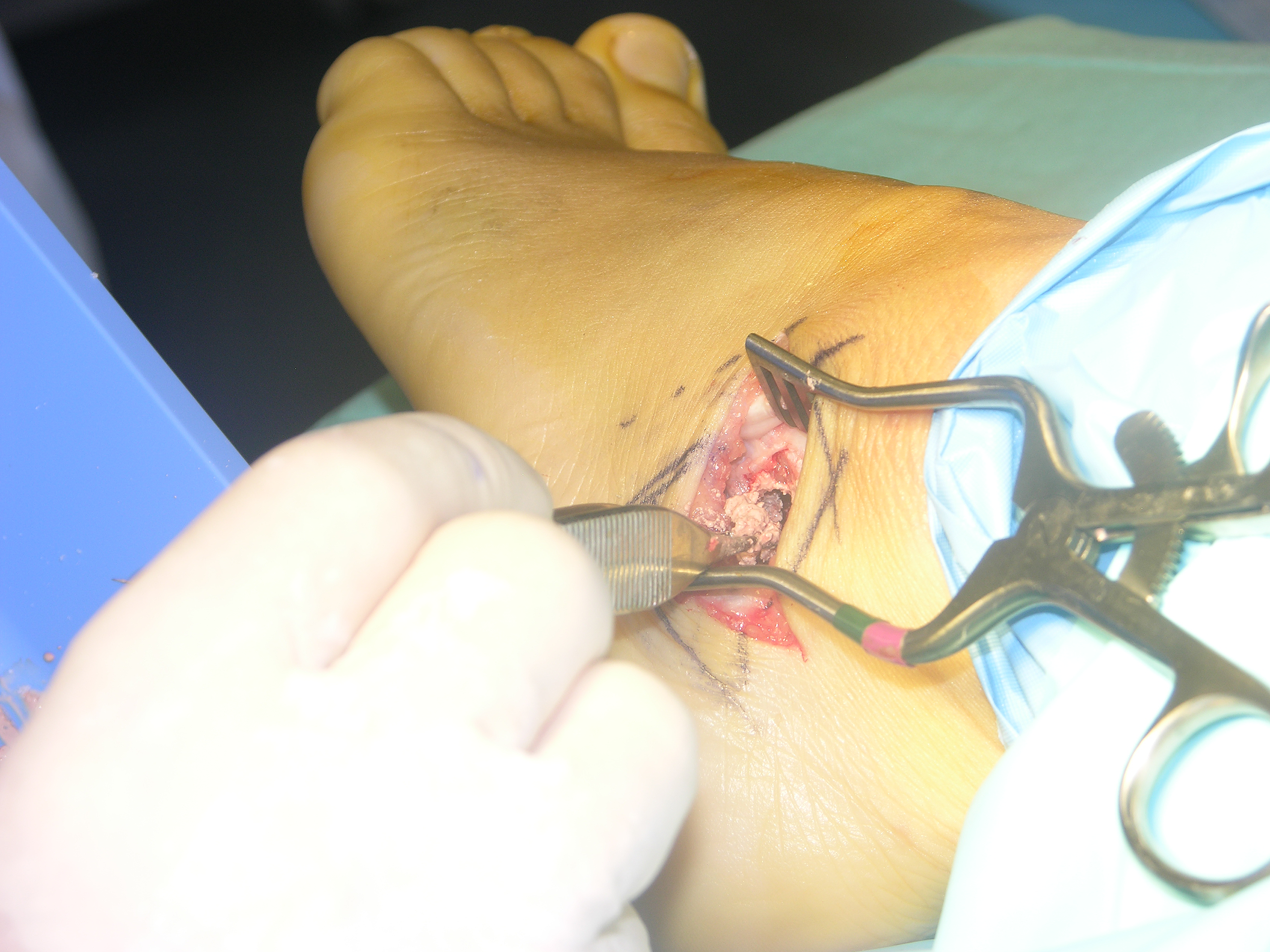
Through venous access using a 21-gauge incising cannula, 50 cc of the patient's autogenous cellular tissue (whole blood) was procured, consisting of hematopoietic tissue cells and other components, using the BioMet GPS II system (Zimmer Biomet, Warsaw, Indiana). The patient's harvested blood cells were then subjected to apheresis, and underwent a process of cellular separation, isolation, and concentration. The PRP layer was then collected, mixed with the BVF (Fig. 5). The PRP was substituted for the Omnipaque in the BVF kit. This mixed BVF and PRP was then added to the allogeneic cancellous bone chips (Fig. 6). Then, the allogeneic cancellous bone chips, PRP, and BVF combination was packed into the calcaneal bone defect (Fig. 7). The calcaneal cortical window was replaced followed by typical soft-tissue closure.
Figure 5.
Platelet-rich plasma and Cerament (BONESUPPORT Holding AB, Lund, Sweden).
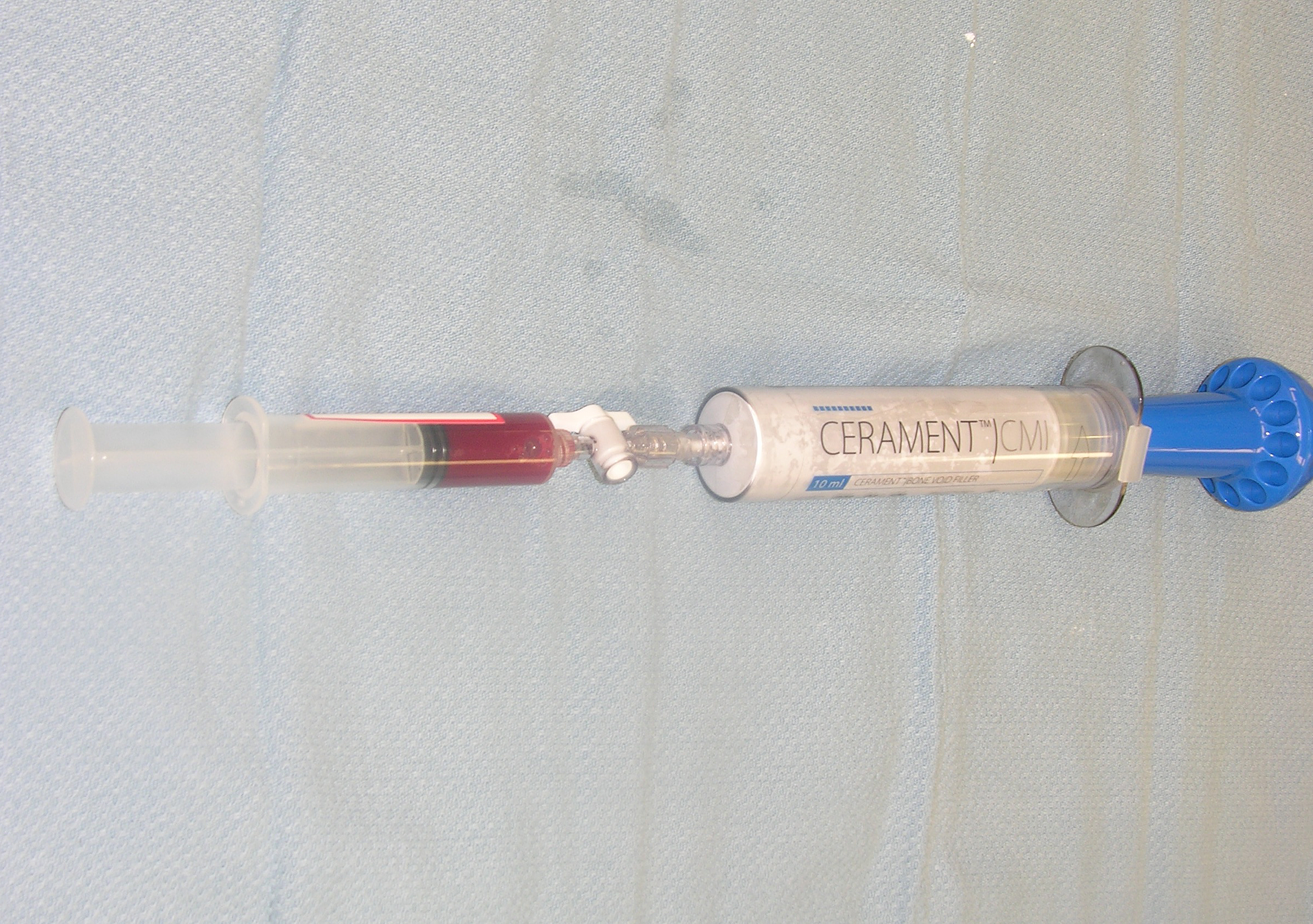
Figure 6.
Bone void filler and platelet-rich plasma combination.
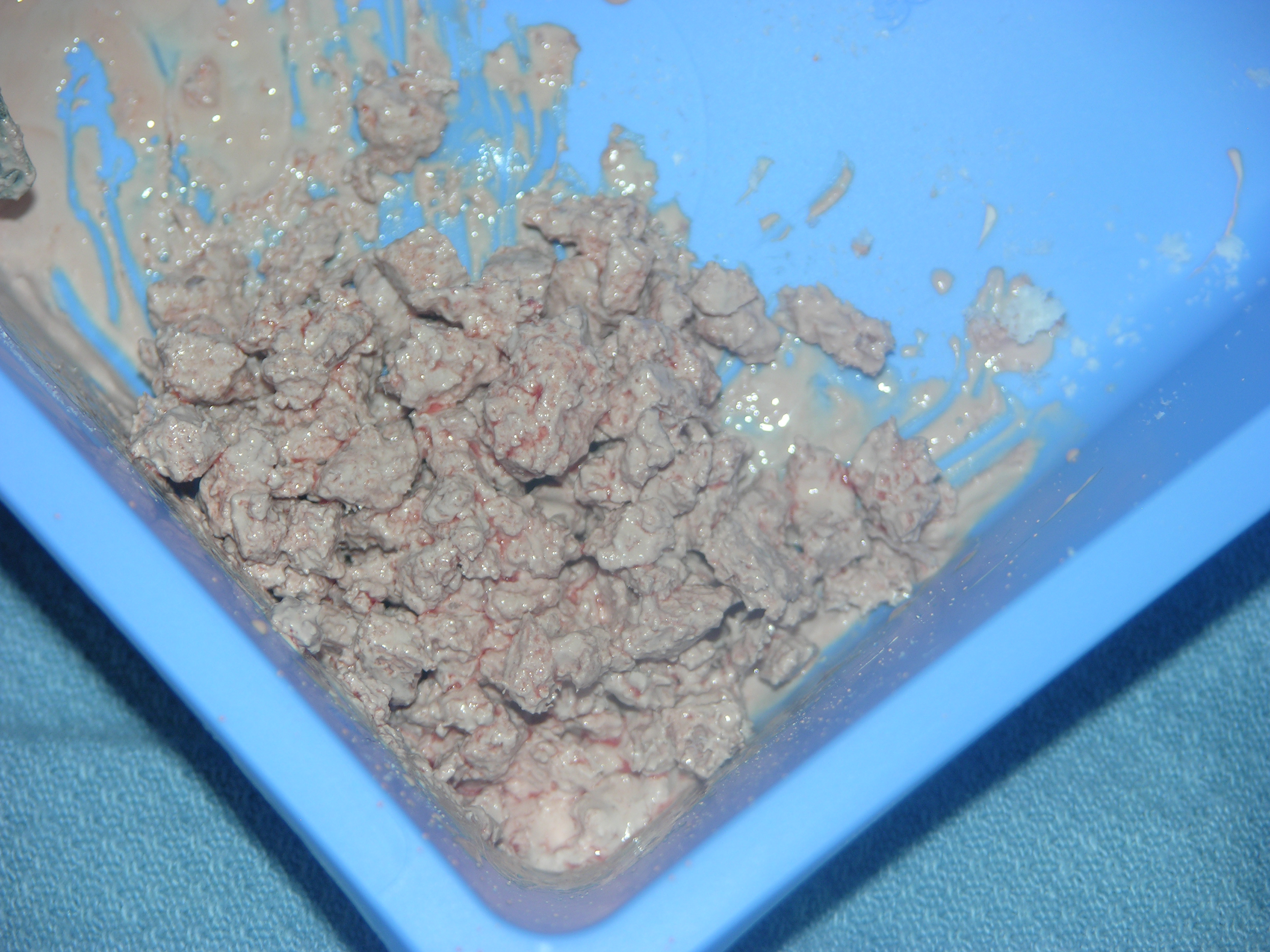
Figure 7.
Delivery of the allogenic bone graft, platelet-rich plasma, and Cerament.
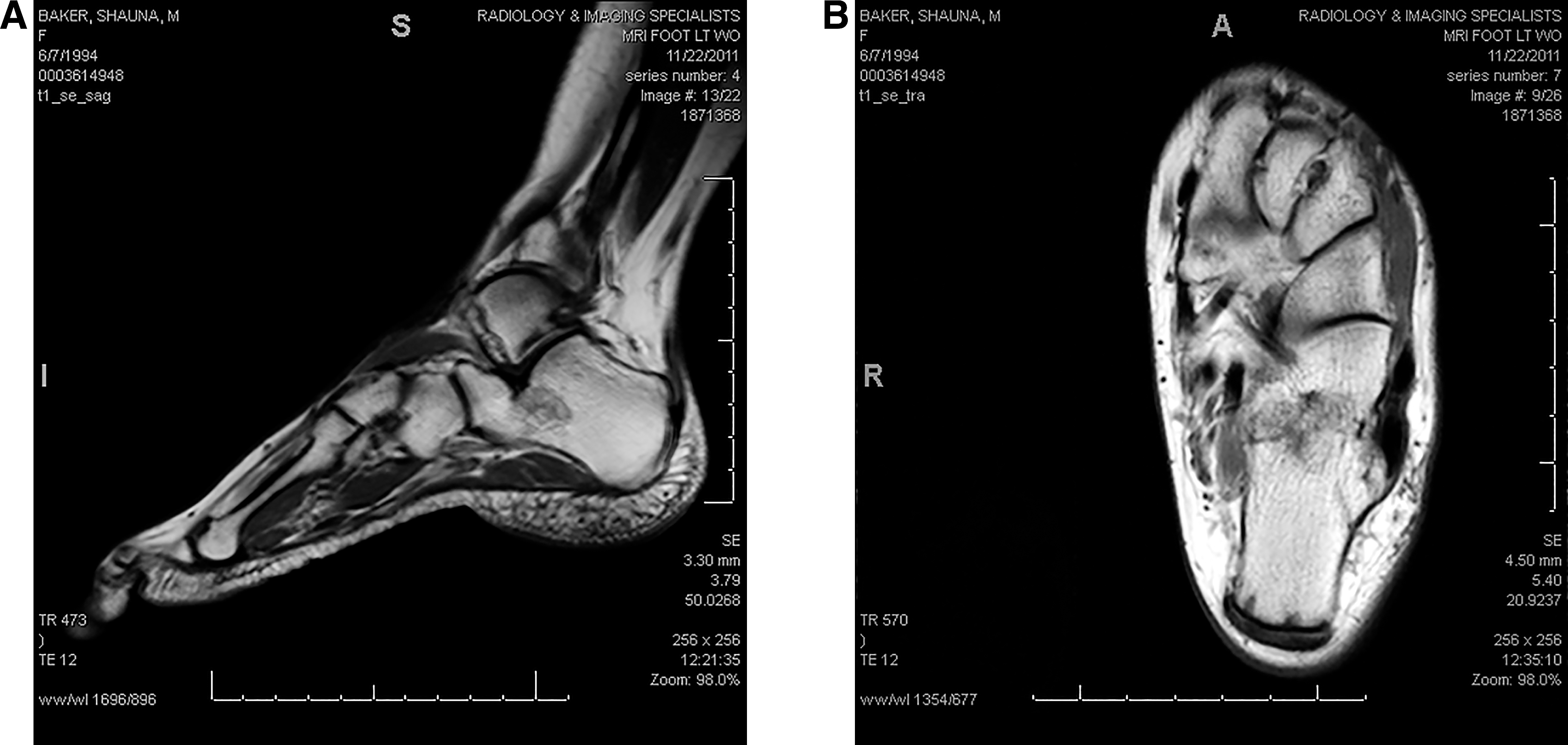
The patient progressed very well, and the patient's pain subsided very quickly 1 week after surgery. After 3 weeks of left foot nonweightbearing, the patient progressed to 50% weightbearing after week 3 and then progressed to complete weightbearing without restrictions on postoperative day 44 (postoperative week 5). Physical therapy was not needed. There was no pain on examination to the left heel, and the patient was discharged without restrictions and returned to gymnastics. Postoperative left foot radiographs, MRI scans, and CT scans were obtained 3 years after surgery. The postoperative left foot radiograph demonstrated a normal spongiosum trabecular pattern with no recurrence of the lucency in the body of the calcaneus (Fig. 8). The postoperative left foot CT scan demonstrated complete trabecular regeneration at the previous well-circumscribed lobulated uniform cyst within the calcaneal body (Fig. 9). The postoperative left foot MRI scan also demonstrated complete trabecular regeneration at the previous well-circumscribed lobulated uniform cyst within the calcaneal body (Fig. 10). This patient was followed to 4 years after surgery. At this time, there was no pain to the left calcaneus on examination and the patient exhibited a normal gait, with no activity restrictions, fully engaged in gymnastics.
Figure 8.
Left foot 3-year postoperative radiograph.
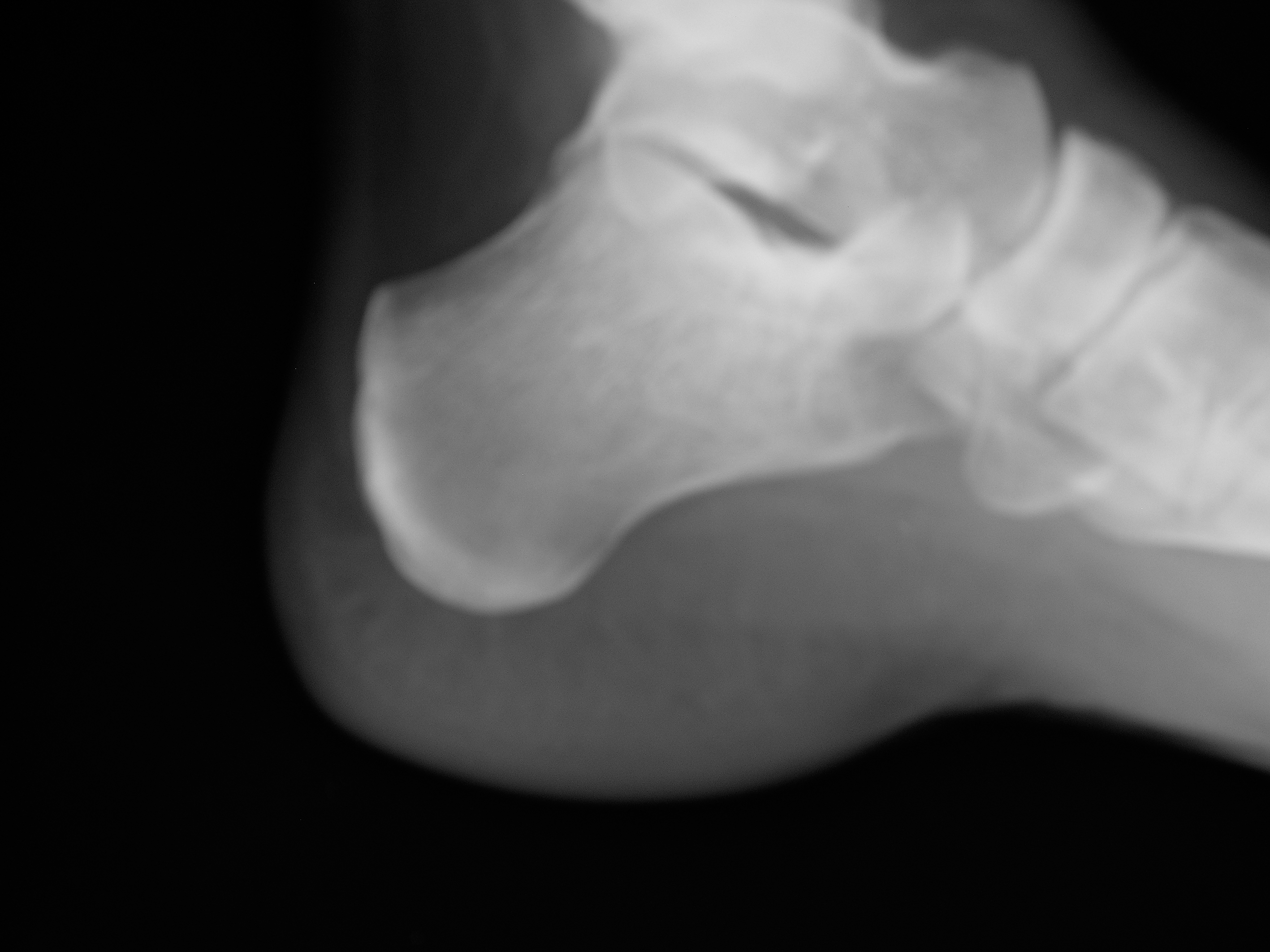
Figure 9.
A, Left foot 3-year postoperative lateral computed tomographic scan. B, Left foot 3-year postoperative axial computed tomographic scan.
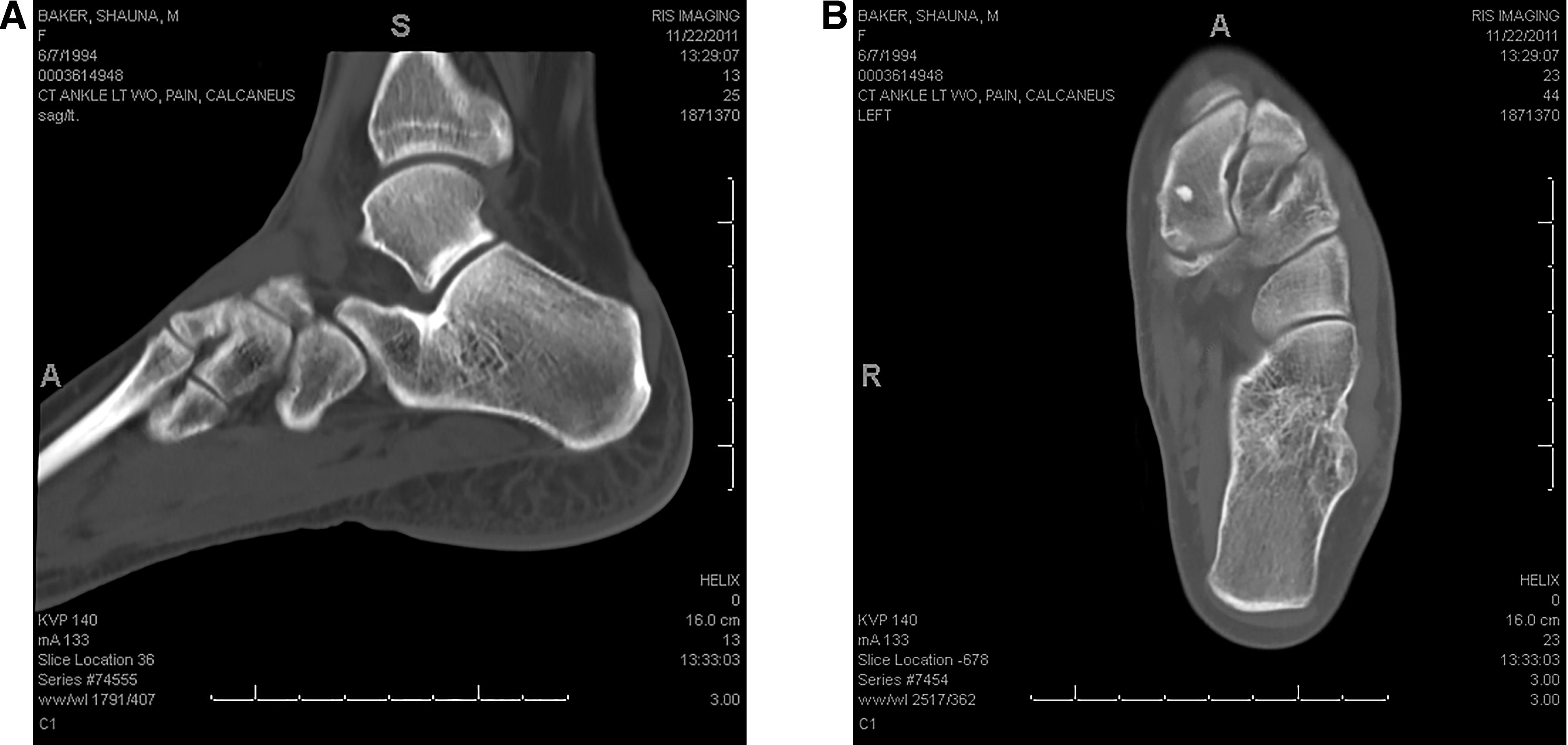
Figure 10.
A, Left foot 3-year postoperative lateral magnetic resonance imaging scan. B, Left foot 3-year postoperative axial magnetic resonance imaging scan.
Eight months after the patient's initial presentation to the author for the left heel, she presented with significant right heel pain, insidious in onset, of 2 months' duration. The patient felt that the pain in her right heel was the same as her left heel 8 months previously. Initial radiographs were negative for lucency or other pathology (Fig. 11). Initial CT films were read as normal. After the author reviewed the CT films, areas of delineated trabecular rarefaction with tiny tubular lucency were seen in the body of the right calcaneus. An additional review of the CT films by the radiologist confirmed this and the radiologist gave a formal diagnosis of early developing cyst (Fig. 12). A preoperative right foot MRI study was read as a normal examination (Fig. 13). Surgical intervention was chosen to stop the progression of a completely formed bone cyst.
Figure 11.
Preoperative right lateral foot radiograph.
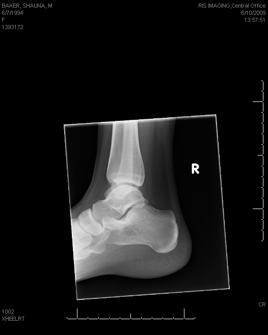
Figure 12.
A, Preoperative lateral right foot computed tomographic scan. B, Preoperative axial right foot computed tomographic scan.
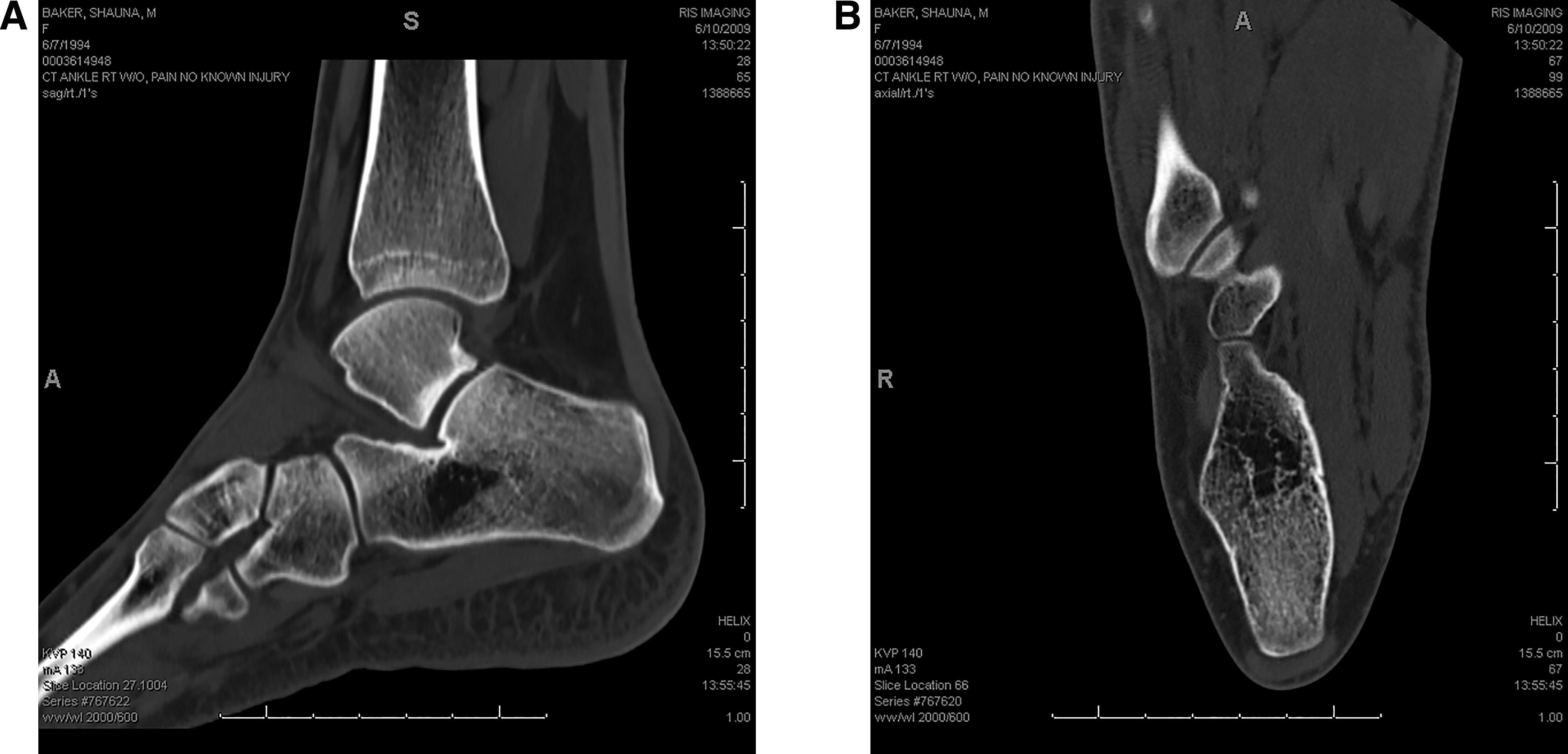
Figure 13.
A, Preoperative lateral right foot magnetic resonance imaging scan. B, Preoperative axial right foot magnetic resonance imaging scan.
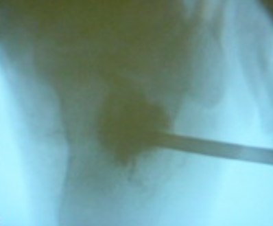
A simple lateral calcaneal bone cortex incision was completed with a smooth 9/64-inch pin into the developing cyst (Fig. 14). The BVF was mixed according to protocol without any additional material. Using a Jam Shedi needle, 10 cc of the BVF was delivered percutaneously into the developing bone cyst under intraoperative fluoroscopy (Fig. 15). There was excellent filling of the developing bone cyst. Simple soft-tissue closure was completed. The patient tolerated the procedure well and was able to bear weight the next day with crutches and progressed to full weightbearing at postoperative week 3. The patient continued to progress unremarkably through the postoperative period and was discharged without restrictions at postoperative week 6 with return to gymnastics. Physical therapy was not needed.
Figure 14.
Right foot approach for percutaneous delivery of Cerament.
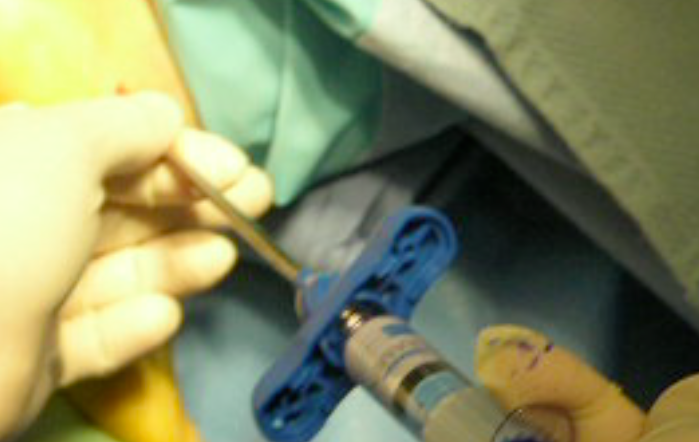
Figure 15.
Right foot percutaneous delivery of Cerament.
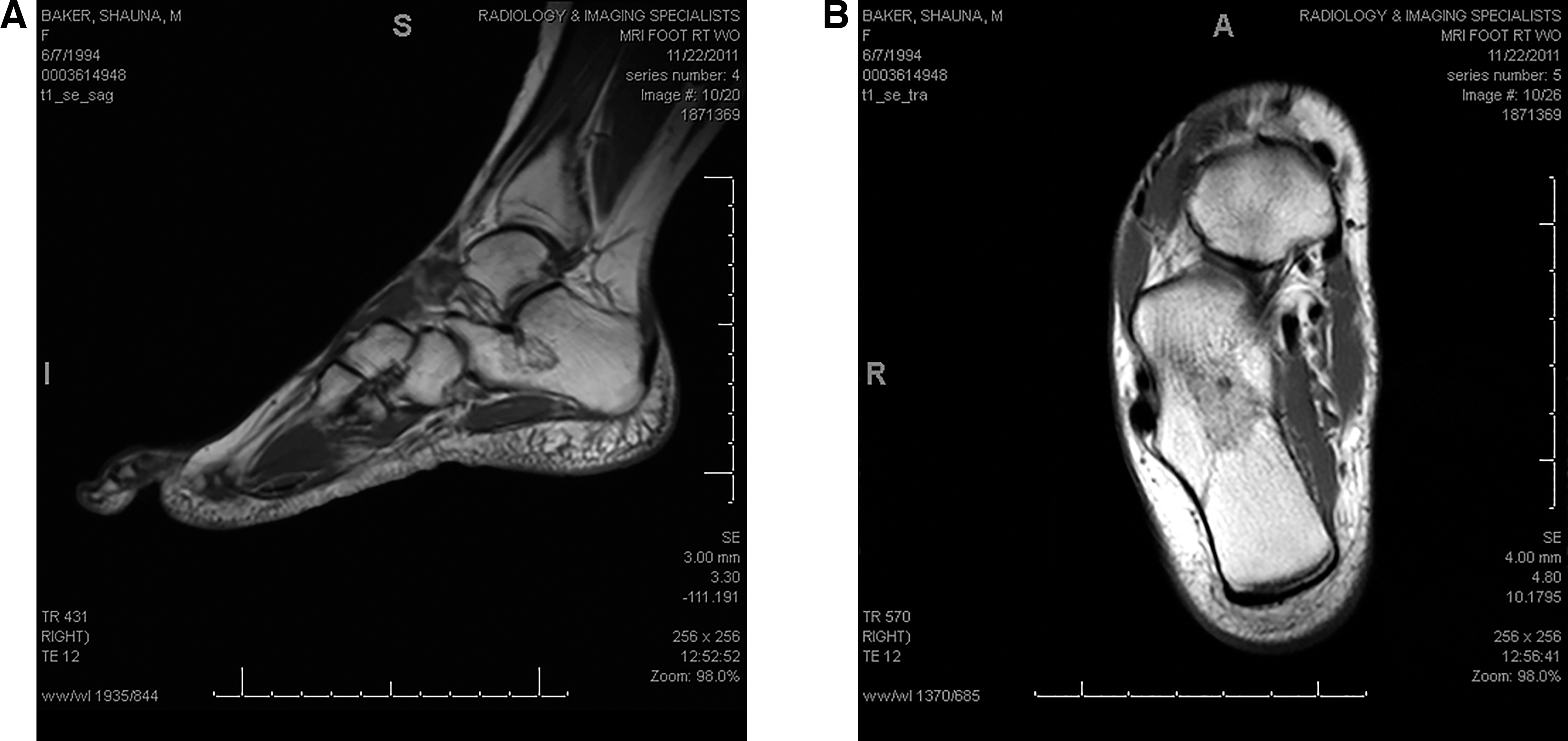
Postoperative radiographs and MRI and CT scans were obtained at 2 years 3 months after right foot surgery. The postoperative right foot radiograph demonstrated a normal spongiosum trabecular pattern, with no recurrence of the lucencies in the body of the calcaneus (Fig. 16). The postoperative right foot CT film demonstrated complete trabecular regeneration at the previous developing cyst within the calcaneal body (Fig. 17). The postoperative right foot MRI scan also demonstrated complete trabecular regeneration at the developing cyst within the calcaneal body (Fig. 18). This patient was followed to 3 years after surgery. At this time, there was no pain to the right calcaneus on examination and the patient exhibited a normal gait, with no activity restrictions and was fully engaged in gymnastics.
Figure 16.
Right foot postoperative lateral radiograph.
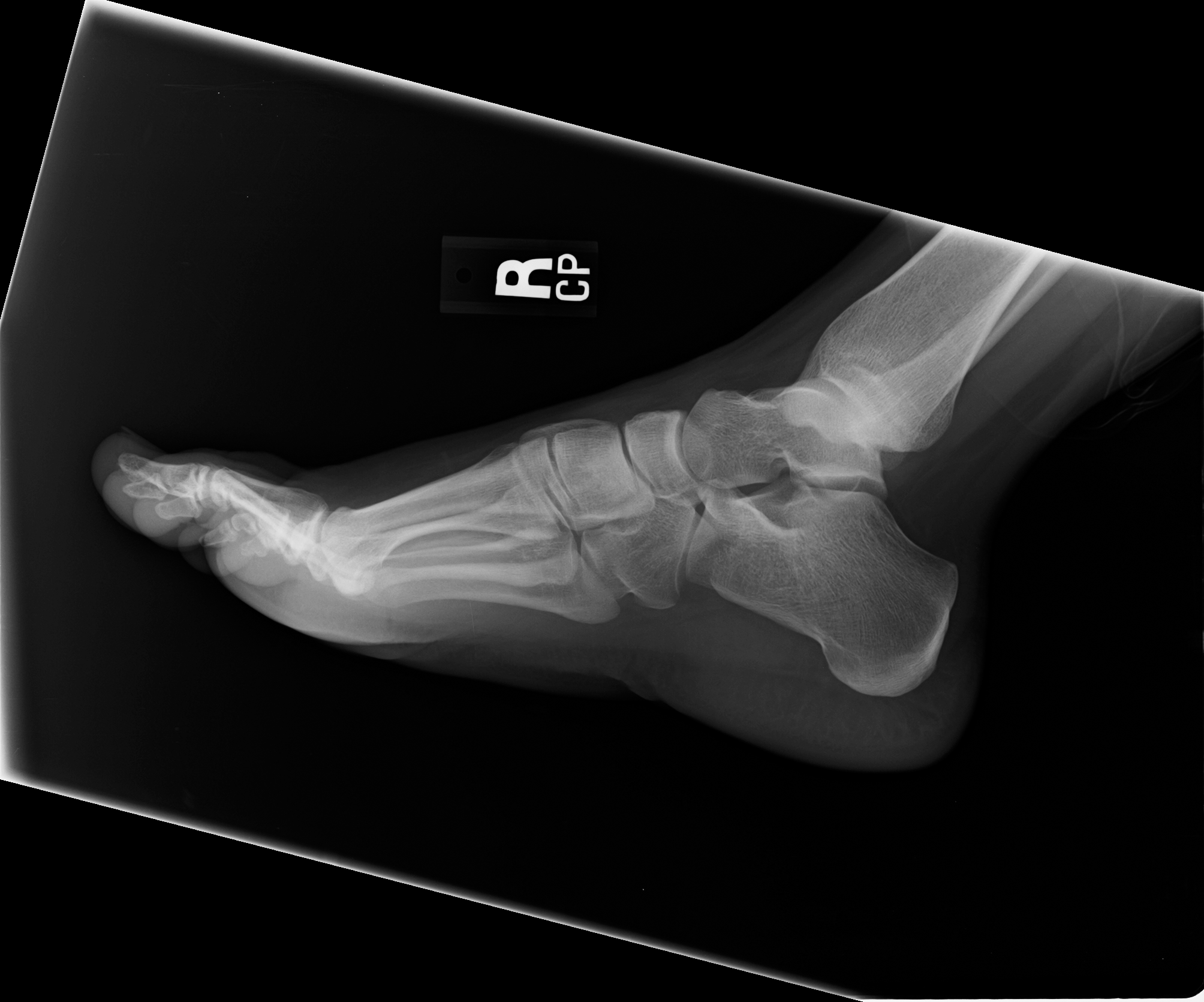
Figure 17.
A, Right foot postoperative lateral computed tomographic scan. B, Right foot postoperative axial computed tomographic scan.
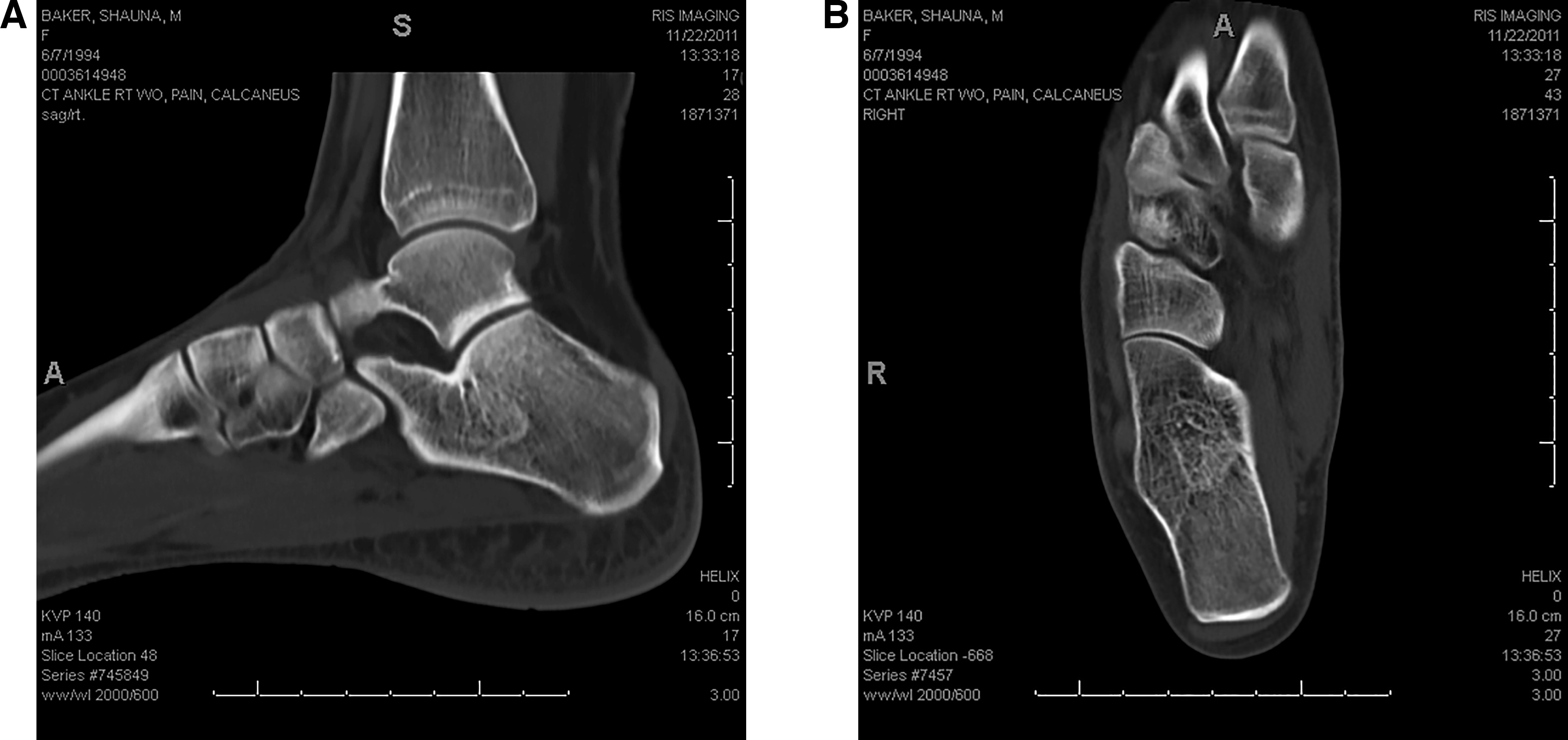
Figure 18.
A, Right foot postoperative lateral magnetic resonance imaging scan. B, Right foot postoperative axial magnetic resonance imaging scan.
Discussion
The indication for treatment of a unicameral bone is to manage symptoms—primarily pain—and to prevent a pathologic fracture. The left heel treatment goal was to return this patient to gymnastics as soon as possible. The BVF was selected because of the partial content of hydroxyapatite (calcium phosphate), which attracts osteoblast cells (thus, the increasing osteoconductive properties of the graft to supplement the allogenic bone graft). The CSCP BVF was mixed with the activated PRP to establish an osteoinductive property to the allogenic bone graft.[27,28,29,30,31,32,33] This combination nicely penetrated the allogeneic spongiosum bone graft, holding the bone chips together. This final mixture made handling the graft much easier and allowed 100% contact with the unicameral bone cyst wall surface. The goal for the right heel was also to return this patient to gymnastics as soon as possible. The percutaneous injection of the BVF arrested the bone cyst formation and allowed restoration of the calcaneal trabecular structure. The BVF succeeded because of the partial content of hydroxyapatite (calcium phosphate), which attracts osteoblast cells to restore the calcaneal trabecular structure. In both cases, there was excellent restoration of the calcaneal trabecular anatomy, resolution of pain, and full return to function and gymnastics without restrictions.
Conclusions
Regarding the left calcaneus containing a well-formed unicameral bone cyst, the BVF consisting of CSCP can be an effective and easy treatment modality in conjunction with allogenic bone graft and PRP. As seen in the right calcaneal treatment of the early forming bone cyst, percutaneous BVF delivery can be an effective and easy treatment. Both individual calcaneal cyst treatments allowed the patient to return to gymnastics quicker than if autogenous bone graft was incorporated into both calcanei, especially regarding the right percutaneous approach.
Financial Disclosure: None reported.
Conflict of Interest: None reported.
References
- Virchow R: “On the formation of bony cysts,” in Über die Bildung von Knochencysten, p 369, S-B Akad Wiss, Berlin, 1876.
- Cottalorda J, Kohler R, Sales de Gauzy J, et al: Epidemiology of aneurysmal bone cyst in children: a multicenter study and literature review. J Pediatr Orthop B 13: 389, 2004.
- Godette GA, Rooney RJ: Two different non-neoplastic lesions in the same long bone. Contemp Orthop 30: 395, 1995.
- Lokiec F, Ezra E, Khermosh O, et al: Simple bone cysts treated by percutaneous autologous marrow grafting. A preliminary report. J Bone Joint Surg Br 78: 934, 1995.
- Levy DM, Gross CE, Garras DN: Treatment of unicameral bone cysts of the calcaneus: a systematic review. J Foot Ankle Surg 54: 652, 2015.
- Epstein J, Wertheimer SJ: Unicameral bone cyst of the calcaneus: literature review and case studies. JAPMA 74: 76, 1984.
- Schick FA, Daniel JN, Millern JS: Unicameral bone cyst of the medial cuneiform. JAPMA 106: 357, 2016.
- Chieppa WA, Shinder M: Unicameral bone cyst of the talus. JAPMA 79: 441, 1989.
- Martin SJ, Schiller JE: Unicameral bone cyst of the second metatarsal with pathologic fracture. JAPMA 77: 143, 1987.
- Arazi M, Senaran H, Memik R, et al: Minimally invasive treatment of simple bone cysts with percutaneous autogenous bone marrow injection. Orthopedics 28: 108, 2005.
- Docquier PL, Delloye C: Autologous bone marrow injection in the management of simple bone cysts in children. Acta Orthop Belg 70: 204, 2004.
- Docquier PL, Delloye C: Treatment of aneurysmal bone cysts by introduction of demineralized bone and autogenous bone marrow. J Bone Joint Surg Am 87: 2253, 2005.
- Gentile JV, Weinert CR, Schlechter JA: Treatment of unicameral bone cyst in pediatric patients with an injectable regenerative graft: a preliminary report. J Pediatr Orthop l33: 254, 2013.
- Evaniew N, Tan V, Parasu N, et al: Use of a calcium sulfate-calcium phosphate synthetic bone graft composite in the surgical management of primary bone tumors. Orthopedics 36: e216, 2013.
- Park IH, Micic ID, Jeon IH: A study of 23 unicameral bone cysts of the calcaneus: open chip allogeneic bone graft versus percutaneous injection of bone powder with autogenous bone marrow. Foot Ankle Int 29: 164, 2008.
- Rougraff BT, Kling TJ: Treatment of active unicameral bone cysts with percutaneous injection of demineralized bone matrix and autogenous bone marrow. J Bone Joint Surg Am 84: 921, 2002.
- Glaser DL, Dormans JP, Stanton RP, et al: Surgical management of calcaneal unicameral bone cysts. Clin Orthop Relat Res 360: 231, 1999.
- Capanna R, Campanacci DA, Manfrini M: Unicameral and aneurysmal bone cysts. Orthop Clin North Am 27: 605, 1996.
- Capanna R, Dal Monte A, Gitelis S, et al: The natural history of unicameral bone cyst after steroid injection. Clin Orthop Relat Res 166: 204, 1982.
- Parsch K, Arnold P, Jani L, et al: Juvenile bone cysts: relative value and therapy results of cortisone injections [in German]. Orthopade 24: 65, 1995.
- Mylle J, Burssens A, Fabry G: Simple bone cysts: a review of 59 cases with special reference to their treatment. Arch Orthop Trauma Surg 111: 297, 1992.
- Kanellopoulos AD, Mavrogenis AF, Papagelopoulos PJ, et al: Elastic intramedullary nailing and DBM-bone marrow injection for the treatment of simple bone cysts. World J Surg Oncol 5: 111, 2007.
- Roposch A, Saraph V, Linhart WE: Flexible intramedullary nailing for the treatment of unicameral bone cysts in long bones. J Bone Joint Surg Am 82: 1447, 2000.
- Brecelj J, Suhodolcan L: Continuous decompression of unicameral bone cyst with cannulated screws: a comparative study. J Pediatr Orthop B 16: 367, 2007.
- Marion R, Daniel S, Lucas M, et al: Elastic Stable Intramedullary Nailing (ESIN), Orthoss and Gravitational Platelet Separation–System (GPS): an effective method of treatment for pathologic fractures of bone cysts in children. BMC Musculoskelet Disord 12: 45, 2011.
- Kadhim M, Thacker M, Kadhim A, et al: Treatment of unicameral bone cyst: systematic review and meta analysis. J Child Orthop 8: 171, 2014.
- Pedzisz P, Zgoda M, Kocon H, et al: Treatment of solitary bone cysts with allogenic bone graft and platelet-rich plasma: a preliminary report. Acta Orthop Belg 76: 374, 2010.
- Kalen A, Wahlstrom O, Linder CH, et al: The content of bone morphogenetic proteins in platelets varies greatly between different platelet donors. Biochem Biophys Res Commun 375: 261, 2008.
- Leitner GC, Gruber R, Neumuller J, et al: Platelet content and growth factor release in platelet-rich plasma: a comparison of four different systems. Vox Sang 91: 135, 2006.
- Weibrich G, Kleis WK, Hitzler WE, et al: Comparison of the platelet concentrate collection system with the plasma-rich-in-growth-factors kit to produce platelet-rich plasma: a technical report. Int J Oral Maxillofac Implants 20: 118, 2005.
- Buchwald D, Kaltschmidt C, Haardt H, et al: Autologous platelet gel fails to show beneficial effects on wound healing after saphenectomy in CABG patients. J Extra Corpor Technol 40: 196, 2008.
- Martinez-Zapata MJ, Marti-Carvajal A, Sola I, et al: Efficacy and safety of the use of autologous plasma rich in platelets for tissue regeneration: a systematic review. Transfusion 49: 44, 2009.
- Ekback G, Ryttberg L, Axelsson K, et al: Preoperative platelet-rich plasmapheresis and hemodilution with an autotransfusion device in total hip replacement surgery. J Clin Apher 15: 256, 2000.
© 2019 American Podiatric Medical Association