


Abstract
Osteoid osteoma is a benign tumor originating from osteoblasts, and it is mostly seen in long bones of lower limbs. The distal phalanx of the foot is an atypical location for an osteoid osteoma, and lesions occurring in this location may be a diagnostic challenge. A 22-year-old man presented with a complaint of severe pain on the second distal phalanx of his right foot. An osteoid osteoma was suspected after radiologic evaluation. The lesion was surgically excised and removed completely by curettage. Histopathologic evaluation confirmed the diagnosis of an osteoid osteoma. The patient was followed-up for a 9-month period without any symptoms or recurrence.
Osteoid osteoma is a benign tumor that originates from osteoblasts. Approximately 50% of osteoid osteomas occur in long bones of lower limbs.[1] The incidences in phalanges of the hand and foot are reported to be 7% and 1%, respectively.[2,3] This benign tumor causes severe pain, which is worse at night, and responds dramatically to nonsteroidal anti-inflammatory drugs (NSAIDs).[4] Osteoid osteoma of the distal phalanx of the foot is extremely rare. The diagnosis may be challenging, as the soft-tissue swelling, nail deformity, and atypical radiographic appearance can make distinguishing it from a chronic osteomyelitis, epidermoid cyst, enchondroma, or glomus tumor difficult. Thus, the diagnosis and treatment may be delayed.[5-7] In this article, we report the case of a 22-year-old man with an atypical location of an osteoid osteoma at the distal phalanx of the foot.
Case Report
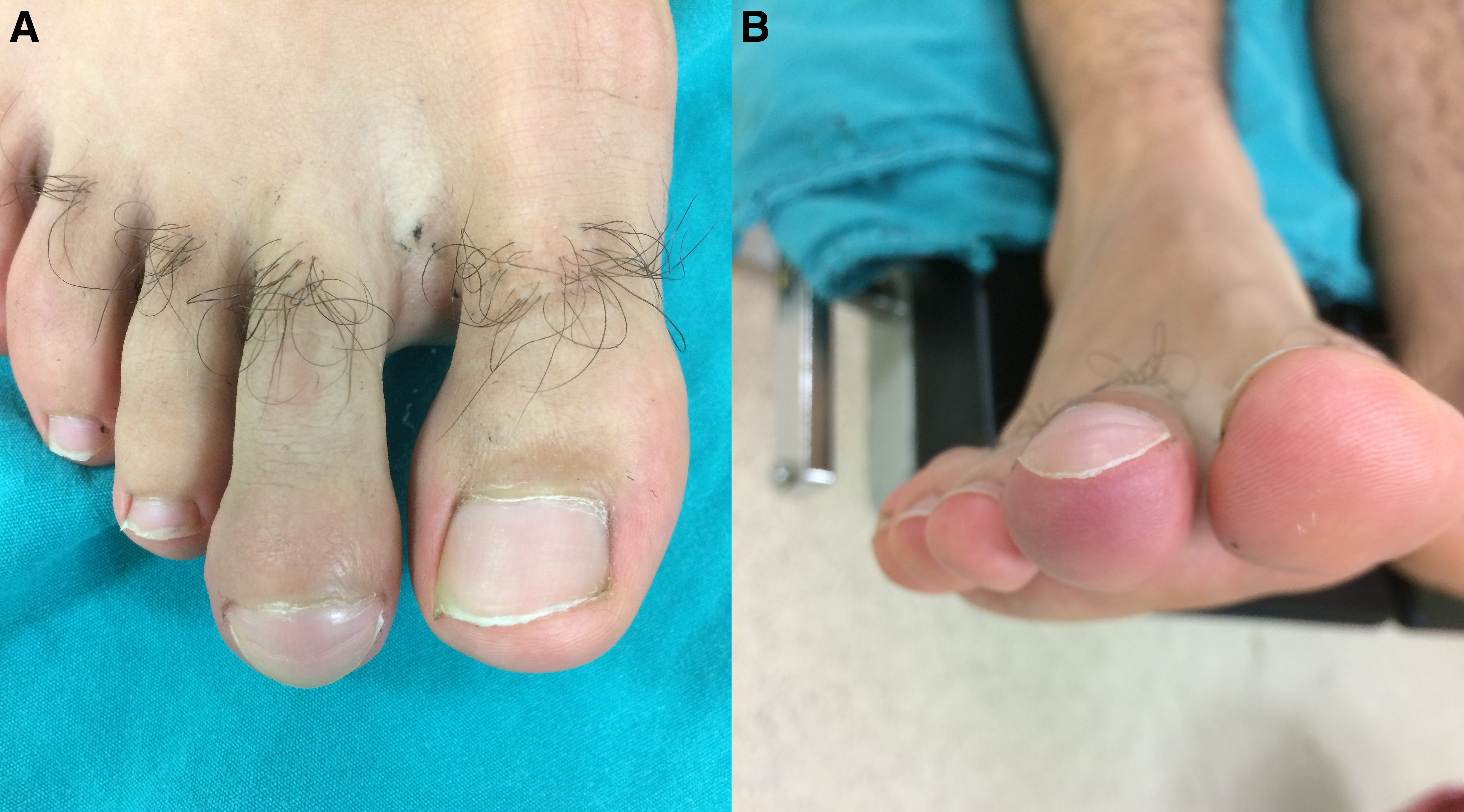
A 22-year-old man was admitted to our outpatient clinic with a complaint of severe pain at the second distal phalanx of his right foot. He declared that the pain was present for the last 4 months and responded well to NSAIDs. He denied any history of trauma to the region. Physical examination demonstrated swelling and tenderness at the second distal phalanx of the right foot without hyperemia (Fig. 1). We considered chronic osteomyelitis, epidermoid cyst, enchondroma, or glomus tumor in the differential diagnosis list.
Figure 1.
Clinical view of the right foot of the patient. Swelling and tenderness at the second distal phalanx of the right foot without hyperemia is shown.
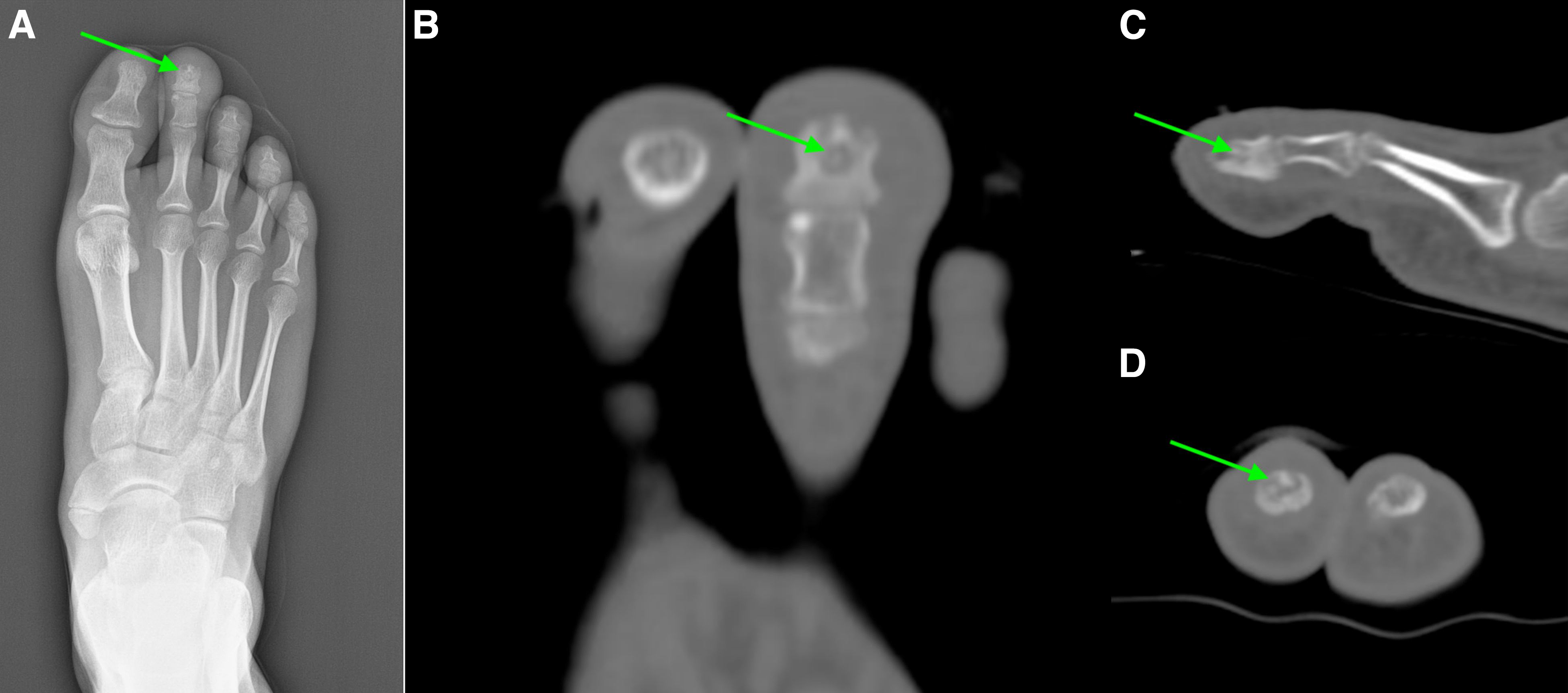
His laboratory findings, including white blood cell count, erythrocyte sedimentation rate, and serum C-reactive protein levels, were within the normal limits. A radiograph of the region demonstrated cortical sclerosis at the second distal phalanx of the right foot. The appearance was nonspecific for determining a definitive diagnosis and the patient was scheduled for a computed tomography (CT) examination. On CT, the lesion had a hypoattenuating nidus with surrounding sclerosis and cortical thickening (Fig. 2). An osteoid osteoma was at the top of the differential list.
Figure 2.
Anteroposterior radiograph of the right foot (A) shows increased density at the distal phalanx of the second digit of the right foot with a central radiolucent area (arrow). Axial (B), sagittal (C), and coronal (D) CT images of the right foot demonstrate a hypoattenuating nidus with surrounding sclerosis and cortical thickening at the affected phalanx (arrow).
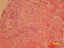
The patient was scheduled for surgical excision of the tumor. A longitudinal incision on the second distal phalanx of the foot was performed and the nidus was completely removed by curettage (Fig. 3). Histopathologic examination of the surgical specimen confirmed the diagnosis of an osteoid osteoma (Fig. 4). The patient's pain was relieved after surgery. The patient was asymptomatic and had no recurrence for a follow-up period of 9 months.
Figure 3.
Intraoperative photograph of the lesion is shown. The lesion underwent a successful surgical curettage.
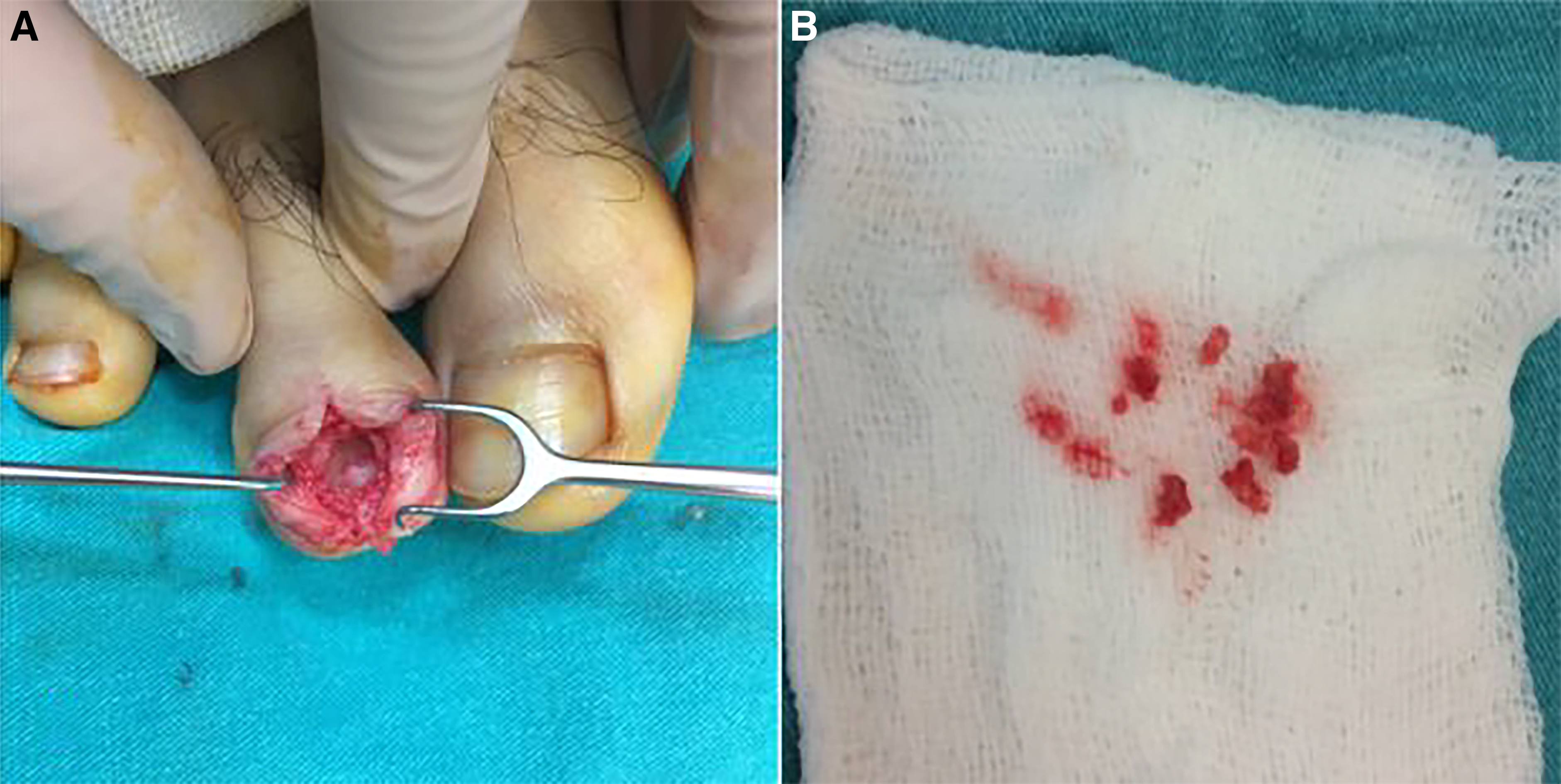
Figure 4.
Histopathologic examination demonstrated bone trabecula circumscribed with sclerotic nidus of woven bone. The final diagnosis was osteoid osteoma.
Discussion
Osteoid osteoma is one of the lesions encountered frequently in daily practice, as it constitutes 15% of all benign bone lesions. It is most commonly seen in patients aged between 5 and 25 years and is two times more frequently seen in male patients. Approximately 50% of osteoid osteomas occur in long bones of lower limbs.[1] The incidences in phalanges of the hand and foot are reported to be 7% and 1%, respectively.[2,3] The metaphysis and diaphysis of the long bones are the most common locations. More than half of the lesions are located in femur and tibia, with approximately 30% located in the proximal femur.[8] It can also be seen in the spine, carpal and tarsal bones, phalanges, ribs, and pelvis. Our patient was a 22-year-old man with an atypical location of an osteoid osteoma at the distal phalanx of the foot.
The typical clinical presentation of osteoid osteoma is the complaint of a pain that worsens at night. The pain is relieved dramatically by the administration of NSAIDs and is worse after alcohol intake. It often radiates to the neighboring insertions, which may delay the accurate diagnosis. These typical clinical signs are seen in almost 80% of the cases.[9] However, the remaining 20% of patients have an atypical clinical presentation or atypical location, which makes accurate diagnosis difficult.[10] Osteomyelitis, epidermoid cyst, enchondroma, or glomus tumor may be considered in the differential diagnosis list.[5,6,7] Osteoid osteoma of the phalanges of the feet may cause soft-tissue swelling that may mimic an infection, and patients may receive antibiotic therapy wrongly.[10]
Conclusions
Osteoid osteomas are classified according to their location in the bone. There are subperiosteal, cortical, and medullary (cancellous) types. The cortical type is the most common and usually occurs in the diaphyses and metaphyses of the long bones of the lower limbs. The typical radiographic appearance of osteoid osteoma is fusiform thickening on one side of the tubular bone with a radiolucent area in the middle of this thickened bone. This centrally located radiolucent area is called a “nidus” and is usually smaller than 2 cm.[11] On CT, the nidus is seen as a well-defined, round, or oval area with low attenuation.[12] Disuse because of pain may cause a generalized decrease in bone density of the affected bone.[13] Although magnetic resonance imaging (MRI) has a limited role in demonstrating the nidus, it has low to intermediate signal on T1-weighted images and variable signal intensity on T2-weighted images.[12] Adjacent bone marrow or soft-tissue edema may accompany severe edema accompanying the lesion and the sclerosing nature of the nidus may decrease the diagnostic value of MRI. Bone scintigraphy demonstrates increased osteoblastic activity of the lesion.
Financial Disclosure: None reported.
Conflict of Interest: None reported.
References
- Boedelon RL, Cracco A, Book MK: Osteoid osteoma producing premature fusion of the epiphysis of the distal phalanx of the big toe: a case report. J Bone Joint Surg Am 57: 120, 1975.
- Frieberger RH, Loitman BS, Helperm M, et al: Osteoid osteoma: a report on 80 cases. AJR Am J Roentgenol 82: 194, 1959.
- Picci P, Mirra JM: “Osteoid Osteoma, ”in Bone Tumors, Vol 1, edited by JM Mirra, p 226, Lea & Febiger, Philadelphia, 1989.
- Laurence N, Epelman M, Markowitz RI, et al: Osteoid osteomas: a pain in the night diagnosis. Pediatr Radiol 42: 1490, 2012.
- Foucher G, Lemarechal P, Citron N, et al: Osteoid osteoma of the distal phalanx: a report of four cases and review of the literature. J Hand Surg Br 12: 382, 1987.
- Burger IM, McCarthy EF: Phalangeal osteoid osteoma. Br J Radiol 62: 321, 1989.
- Jones SN, Stoker DJ: Radiology at your fingertips: lesions of the terminal phalanx. Clin Radiol 39: 487, 1988.
- Campanacci M: “Osteoblastoma,”in Bone and Soft Tissue Tumors, edited by M Campanacci,WF Enneking, p 415, Springer, New York, 1999.
- Dorfman HD, Czerniak B: “Benign Osteoblastic Tumors,”in Bone Tumors, p 85, Mosby, St. Louis, MO, 1998.
- Başar H, Topkar OM, Erol B: Osteoid osteoma of distal phalanx of toe: a rare cause of foot pain. Case Rep Orthop 2014: 560892, 2014.
- Papathanassiou ZG, Megas P, Petsas T, et al: Osteoid osteoma: diagnosis and treatment. Orthopedics 31: 1118, 2008.
- Chai JW, Hong SH, Choi JY, et al: Radiologic diagnosis of osteoid osteoma: from simple to challenging findings. Radiographics 30: 737, 2010.
- Swee RG, McLeod RA, Beabout JW: Osteoid osteoma: detection, diagnosis, and localization. Radiology 130: 117, 1979.
© 2019 American Podiatric Medical Association