


Abstract
Medial talonavicular dislocation associated with cuboid fracture is rare. We report an 18-year-old man with this injury who exhibited excellent results after open reduction and stabilization of the joint with temporary Kirshner wires.
The midtarsal joint is formed by the talonavicular and calcaneocuboid joints and transects the longitudinal axis of the medial and lateral foot. Midtarsal joint injuries are rare, easily-missed injuries. Main and Jowett [1] classify midtarsal injuries into five groups as medial, lateral, longitudinal compression, plantar, and crush according to the acting force and direction of the deformity. The types of medial and lateral midtarsal injuries are related to the severity of the acting force, and ligamentous strain, fracture, fracture dislocation, and swivel dislocation could occur.
Swivel dislocation is isolated talonavicular dislocation, and it is a rare midtarsal injury. It is differentiated from subtalar and Chopart dislocations, as the talocalcaneal and subtalar joints are intact. Rotational forces along the longitudinal axis of the foot while the talocalcaneal ligaments are intact cause disruption of the talonavicular stabilizers, and this results in medial or lateral talonavicular dislocation.[1] In medial swivel dislocation, a medially directed deforming force causes medial dislocation of the talonavicular joint with medial rotation of the forefoot. In lateral swivel dislocation, the laterally directed deforming force causes lateral dislocation of the talonavicular joint with lateral rotation of the forefoot. The spring ligament complex is the most important static stabilizer of the talonavicular joint and acts as a sling for the talar head. Rupture of the spring ligament complex results with plantar and lateral rotation of the talar head.[2] Although the talocalcaneal ligament is intact, the calcaneus rotates or swivels on this ligament.[3] Swivel dislocations could be associated with navicular, calcaneus, cuboid, and metatarsal fractures.[3,4,5,6] Clinical examination along with foot and ankle radiographs are necessary to diagnose midtarsal injuries. Urgent treatment of these injuries is important; otherwise, they may cause permanent disabilities. Early anatomical reduction and stable fixation improve the clinical results in talonavicular joint dislocations. In this study, we report a rare case of medial talonavicular dislocation associated with nondisplaced horizontal cuboid fracture, with excellent results following early open reduction.
Case Report
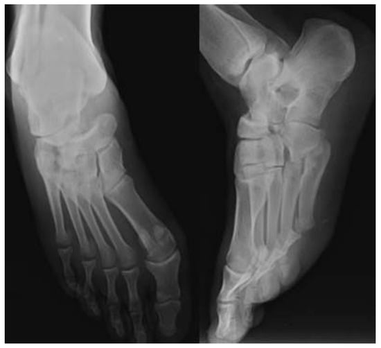
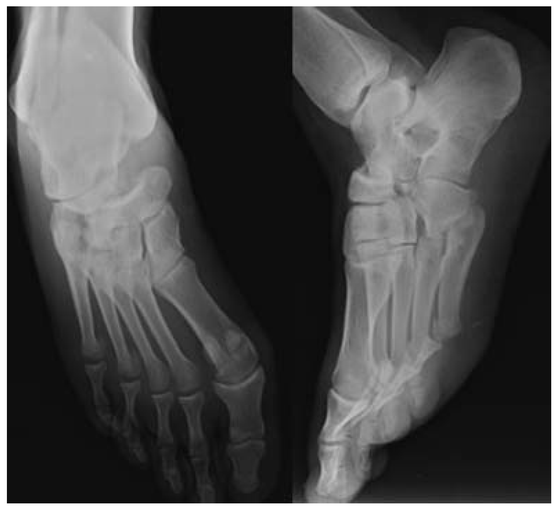
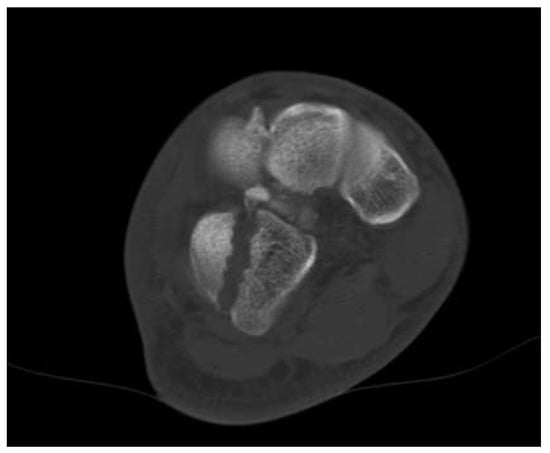
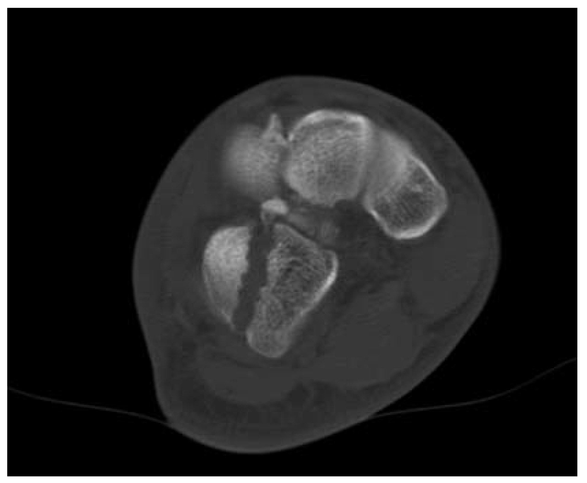
An 18-year-old construction worker presented to the emergency room after falling from a height of 3 m. Severe swelling and tenderness on the medial and dorsal side of the right foot was reported. He was unable to bear weight. Neurovascular examination of the right foot was normal. Anteroposterior oblique radiographs of the foot were obtained (Figure 1). Isolated dislocation with compression fracture of the medial talar head was noted on radiography. On computed tomography, a minimally displaced horizontal cuboid fracture was also observed (Figure 2).
Figure 1.
Right foot anteroposterior and oblique radiographs.
Figure 2.
Right foot computed tomographic axial sequence showing minimally displaced horizontal cuboid fracture.
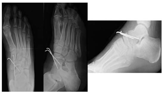
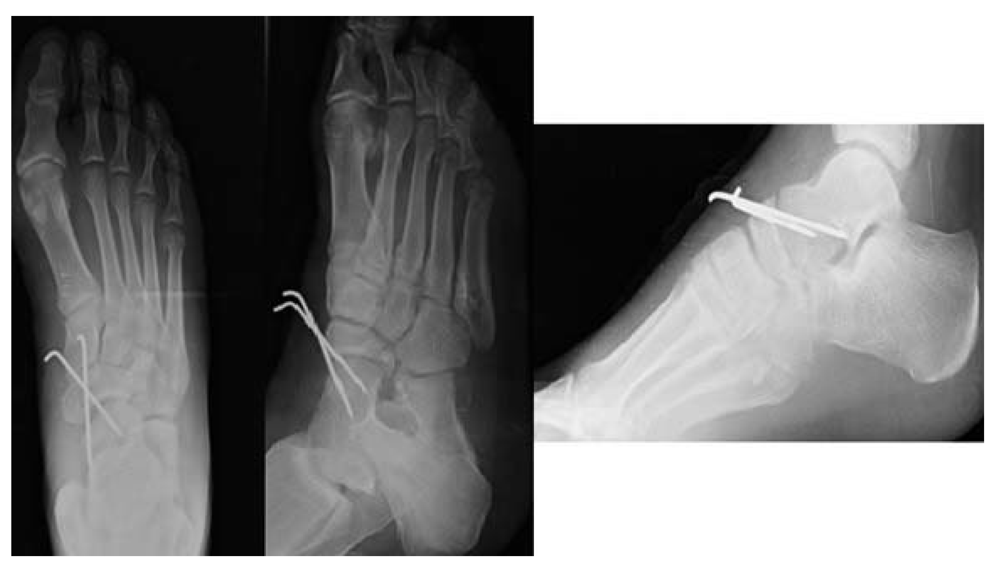
Closed reduction manipulation under sedation was not successful in the emergency room. Open reduction under spinal anesthesia was performed 12 hours after injury in the operating room. A 4-cm longitudinal incision over the talonavicular joint was used for reduction. The extensor hallucis longus tendon and dorsalis pedis artery were retracted laterally to reach the talonavicular joint. Impacted fragments of the medial talar head were excised. Reduction was performed with traction, external rotation, and abduction of the forefoot. Postreduction examination under fluoroscopic vision revealed instability of the talonavicular joint. The talonavicular joint was stabilized with two Kirschner wires placed from the navicular bone to the talus (Figure 3). The cuboid fracture was stable. A short leg cast was applied. The Kirschner wires and the cast were removed 6 weeks after surgery. The patient was allowed to bear weight, with a gradual increase of load. At 3- and 6-month follow-up, his foot was painless and stable. He returned to work in the third month after trauma. Subtalar and ankle movements were normal.
Figure 3.
Postoperative right foot anteroposterior/ oblique radiographs and ankle lateral radiograph.
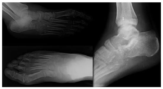
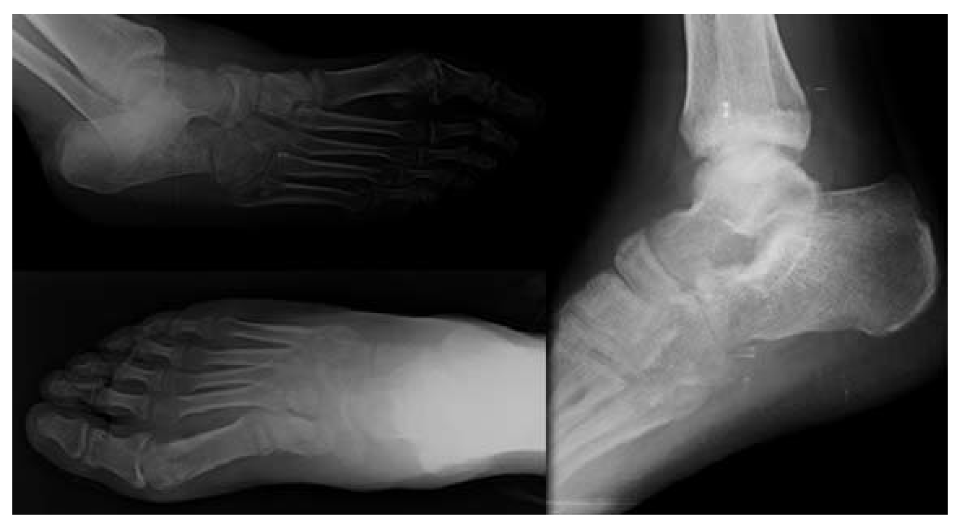
At his last follow-up, at 18 months, his foot was stable and he was complaining of only mild pain on the lateral side of his midfoot after long walks. The talonavicular joint was noted to be normal on radiographs obtained 18 months postoperatively (Figure 4).
Figure 4.
Right foot anteroposterior/oblique radiographs and ankle lateral radiograph obtained 18 months postoperatively.
Discussion
Isolated talonavicular dislocations are rare midtarsal injuries. The direction and severity of the forces acting over the midtarsal joints determine the type of injury. In this article, medial swivel dislocation associated with cuboid fracture was reported. Medial swivel dislocation is caused by a medially directed deforming force, and the talonavicular joint dislocated medially with medial rotation of the forefoot.
Swivel dislocations could be associated with tarsal and metatarsal fractures.[3,4,5,6] Isolated talonavicular dislocations associated with cuboid fractures are generally seen with lateral swivel dislocations as ‘‘nutcracker fractures,’’ because the cuboid is crushed between the talar head and the metatarsal bones.[7] Cuboid fractures associated with medial swivel dislocations are extremely rare.[5] The fracture mechanism of the cuboid bone is different in medial swivel dislocation. In medial swivel dislocation, medially directed deforming forces cause adduction and external rotation of the forefoot. The navicular bone dislocates dorsally and medially and causes impaction on the superomedial surface of the talus. The spring ligament complex ruptures with dislocation of the talonavicular joint. The sling effect of the spring ligament complex under the talar head is lost. The cuboid bone is tensioned with forefoot adduction. Plantar and lateral rotation of the talar head following rupture of the spring ligament complex causes impaction of the talar head to the tensioned cuboid bone.
Diagnosis of isolated talonavicular dislocations and associated lesions can be challenging, particularly in patients with multiple injuries. A high level of suspicion in addition to extensive clinical examination and radiographic evaluation are necessary to diagnose midtarsal injuries. Computed tomography is helpful in case of clinical suspicion and for further evaluation of the midtarsal injuries. In this case, horizontal cuboid fracture was missed on direct radiography and diagnosed with computed tomography.
Early anatomical reduction and stable fixation improve the clinical results in talonavicular dislocations. Delay in treatment will cause an increased risk of avascular necrosis and osteoarthritis. In the literature, pes equinovarus deformity development was mentioned in a missed and untreated case.[8] Closed reduction is the first step in the treatment; however, reduction in a closed manner is not always possible.[1,4,9,10] It is estimated that small fragments caused by an impaction fracture of the talar head and locking of the navicular bone over the superolateral impacted surface of the talar head blocked the reduction in this case.
Postreduction stability should be evaluated carefully for successful results. Damage to the spring ligament complex and joint capsule as well as impaction of the talar head could cause instability of the joint. In case of instability of the joint, smooth Kirschner wires are used for temporary fixation of the joint. Sometimes, primary arthrodesis of the talonavicular joint could be an alternative [11,12] if the talonavicular joint articular surface is severely damaged. In this case, we preferred Kirschner wires for temporary fixation, as the joint was observed to be unstable after reduction because of the young age of the patient and because the articular surface was intact.
Conclusions
We report this case because of the rarity of medial swivel dislocations associated with cuboid fracture, to highlight the importance of computed tomography in further evaluation of midtarsal injuries, and to explain the different mechanisms of cuboid fractures associated with medial swivel dislocation. Furthermore, excellent outcome with early appropriate treatment is emphasized in this article.
Funding
None reported.
Conflicts of Interest
None reported.
References
- MAIN BJ, JOWETT RL: Injuries of the midtarsal joint. J Bone Joint Surg 57: 89, 1975.
- KAVANAGH EC, KOULOURIS G, GOPEZ A, ET AL: MRI of rupture of the spring ligament complex with talo-cuboid impaction. Skeletal Radiol 36: 555, 2007.
- PILLAI A, CHAKRABARTI D, HADIDI M: Lateral swivel dislocation of the talo-navicular joint. Foot Ankle Surg 12: 39, 2006.
- SAMOLADAS E, FOTIADES H, CHRISTOFORIDES J, ET AL: Talonavicular dislocation and nondisplaced fracture of the navicular. Arch Orthop Trauma Surg 125: 59, 2005.
- WILLIAMS DP, HANOUN A, HAKIMI M, ET AL: Talonavicular dislocation with associated cuboid fracture following low-energy trauma. Foot Ankle Surg 15: 155, 2009.
- INAL S, INAL C: An unusual variety of simultaneous fracture dislocation pattern: medial swivel dislocation of talonavicular joint with displaced fractures of the fourth and fifth metatarsals. Foot Ankle Surg 52: 501, 2013.
- HERMEL MB, GERSHON-COHEN J: The nutcracker fracture of the cuboid by indirect violence. Radiology 60: 850, 1953.
- HAFEZ MA, BAWARISH MA, GUVVALA R: Closed talar 1 body fracture with talonavicular dislocation: a case report. Foot Ankle Int 21: 599, 2000.
- RYMASZEWSKI LA, ROBB JE: Mechanism of fracture-dislocation of the navicular: brief report. J Bone Joint Surg Br 70: 492, 1988.
- VAISHYA R, PATRICK JH: Isolated dorsal fracture-dislocation of the tarsal navicular. Injury 22: 47, 1991.
- VENTHAM NT, PHADNIS J, SUJENTHIRAN A, ET AL: Isolated transcalcaneal talonavicular dislocation: a severe injury related to a low-energy mechanism. J Foot Ankle Surg 52: 367, 2013.
- HARRIS AP, JOHNSON J, WARYASZ GR: Isolated dorsal dislocation of the talonavicular and calcaneocuboid articulations (Chopart joint) from a low-energy mechanism. Am J Emerg Med 34: 1733, 2016.
© 2019 American Podiatric Medical Association