


To the Editor:
There have been numerous reports of tears of the plantar fascia, and research has shown plantar fascial tears often occur after trauma.[1,2,3,4,5,6,7,8] These tears are commonly located at the insertion of the plantar fascia into the calcaneus.
This report presents a case review of a plantar fascia tear in the area of the midfoot that occurred without a history of trauma. In this case, the clinical presentation of the tear mimicked a plantar fibroma. Ledderhose[9] initially described these masses in 1897 as plantar fibromatosis, a distinct disease. Fibromas on the bottom of the foot usually appear as solitary or multiple small fixed subcutaneous masses, which may be painful, and grow slowly.[10,11]
A search of the literature found no instance of a midfoot plantar fascial tear in the absence of trauma, nor any instance of a tear resembling a fibroma.[12] As a result, this case is purportedly the first documented report of a midfoot tear of the plantar fascia in absence of trauma. Given this experience, awareness of midfoot tears of the plantar fascia is important to provide optimum treatment for the problem.
Case Report
A 60-year-old, 5-foot-3.5-inch female, weighing 158 pounds, presented to the author’s Crown Point, Indiana office with an 8-month history of bilateral foot pain, with the right foot being significantly more painful. The pain occurred when standing and walking. The patient had previously been treated by her family physician with anti-inflammatories, and she also had tried soaks and varying her footwear.
The patient’s medical history was significant for hypertension, hip and shoulder problems, and prior ankle sprains. Her current medications were metoprolol 50 mg daily and sertraline 100 mg daily. She denied tobacco or alcohol abuse and was allergic to aspirin and penicillin. She had undergone three Cesarean section procedures. Her family history revealed that her mother had hypertension, a maternal aunt had thyroid problems, and a deceased sibling had epilepsy.
The lower-extremity examination was within normal limits for circulation, integument, reflexes, and sensation. Musculoskeletal examination revealed a mild hallux valgus deformity and mild digital contractures. There was pain on palpation of the tibial sesamoid bone on the right foot and on palpation of the plantar fascia. Radiographs confirmed a tibial sesamoid fracture. Her primary problems were a sesamoid bone fracture on the right and plantar fasciitis bilaterally. The patient was treated conservatively with a walking boot and anti-inflammatories.
The patient’s sesamoid problems resolved routinely, but during the following 7 months, her plantar fasciitis symptoms continued. The plantar fasciitis was significantly more symptomatic on the right side. Treatment included custom-made orthotics, night splints, anti-inflammatories, two injections of cortisone into the right heel, and— toward the end of this 7-month period—physical therapy.
During the physical therapy treatments, the patient presented to the author’s office complaining primarily of plantar fasciitis-related symptoms on her right foot. She noted that her physical therapist had noticed a small ‘‘nodule’’ on the bottom of the left foot. The patient indicated that the nodule pain was mild and denied any history of trauma. Physical examination revealed a small palpable mass in the left foot that was only mildly painful with palpation. The surrounding skin color and temperature were normal, and there was no edema. The circulation and muscle strength were normal. Radiographs of the area of the foot where the mass was located were negative, and a diagnostic ultrasound revealed that the mass was located in the plantar fascia. The working diagnosis was plantar fibroma. Magnetic resonance imaging (MRI) was recommended to confirm the diagnosis. The patient deferred, and she was instructed to return if the problem worsened.
The patient did return 3 months later relating significantly more pain on the left foot, in the area of the mass. Until that time, the patient’s primary complaints had been on the right foot; however, she now was experiencing difficulty bearing weight on the left foot and was limping due to increased pain from the mass. She rated the pain 9/10 in severity.
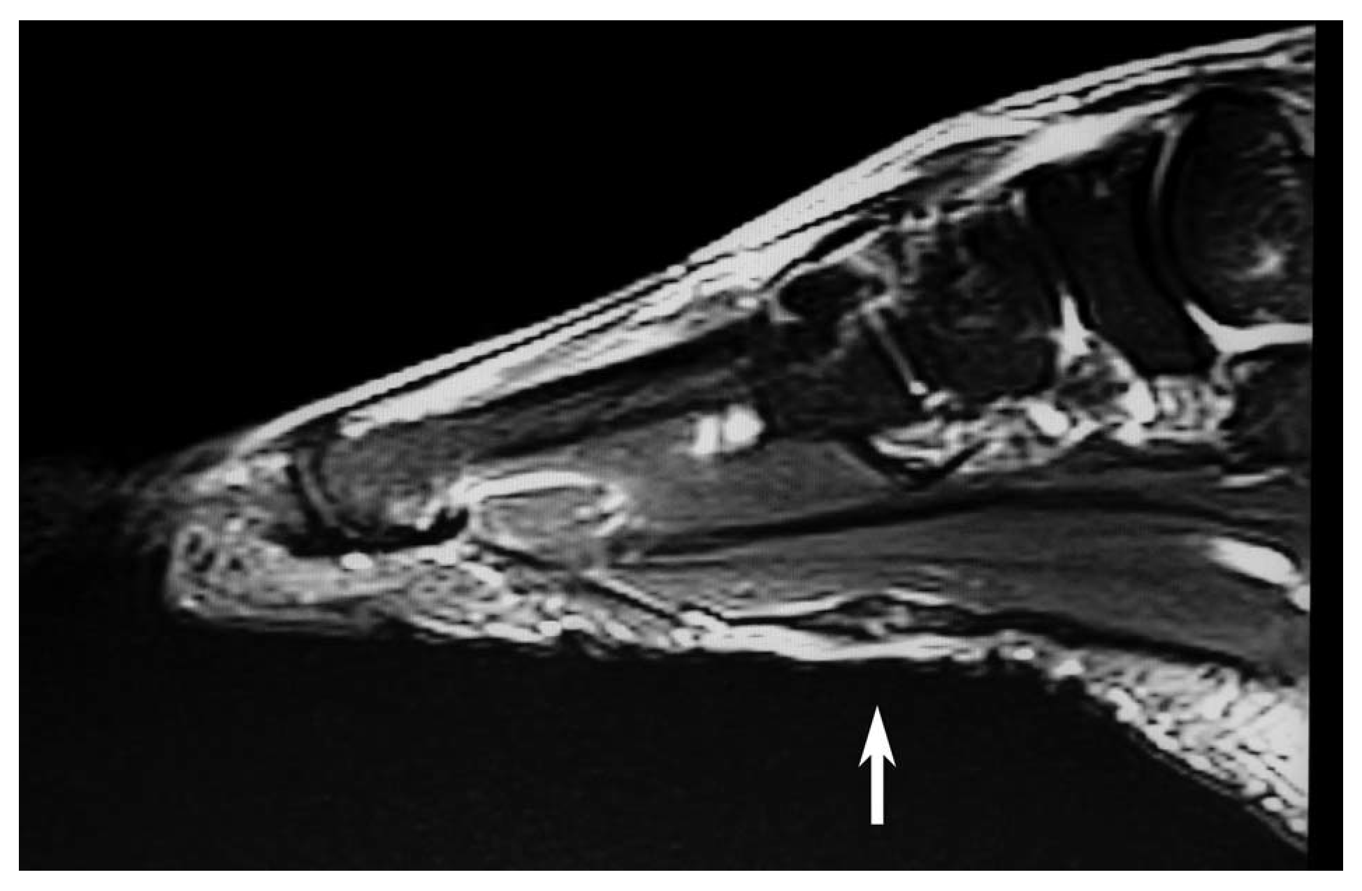
Physical exam revealed the mass was significantly larger (Fig. 1) and much more painful with palpation. There was some discoloration of the skin. An MRI was ordered to evaluate the mass. The MRI revealed a plantar fascia tear (Figs. 2 and 3). The tear measured 7.9 3 5.7 3 4.3 mm in sagittal, transverse, and coronal dimensions.
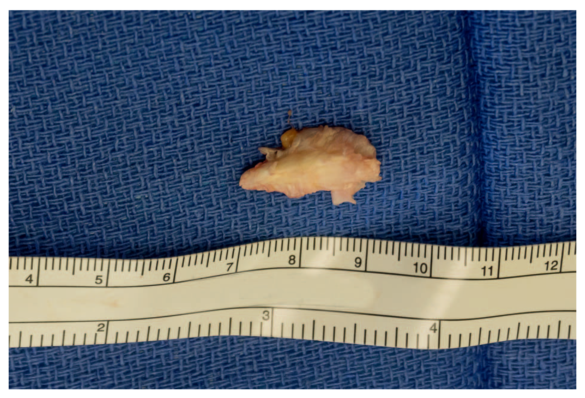
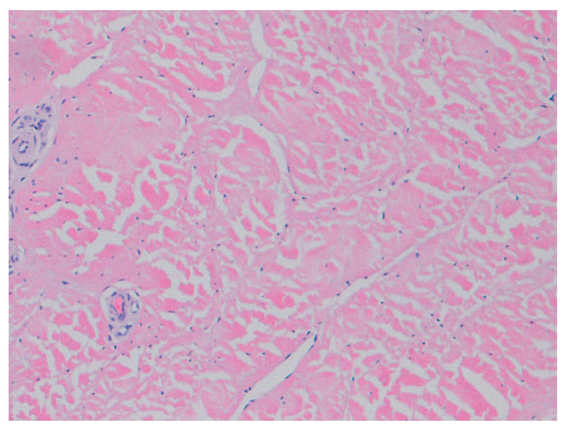
The patient underwent surgery to address the problem. The surgery was performed under general anesthesia, and a pneumatic tourniquet was used around the ankle. The area of the tear was incised with a linear incision, and the torn area of the plantar fascia was found to be thickened with scar tissue. The abnormal tissue was excised. It measured approximately 2 cm in length (Fig. 4). The pathologist’s analysis described the specimen as degenerate and disrupted collagen consistent with a tear (Fig. 5).
The plantar fascia was repaired with absorbable sutures. Following closure of the skin with simple interrupted sutures, a compression bandage was applied to the foot, and the patient was allowed to partially bear weight on her heel. The patient’s postoperative course was uneventful, other than requiring the usual postoperative care and physical therapy. Her pain in the area of the plantar fascia tear resolved completely, and at 5 months postoperatively, the problem has not reoccurred.
Figure 1.
Clinical presentation of the plantar foot showing a mass resembling a plantar fibroma.
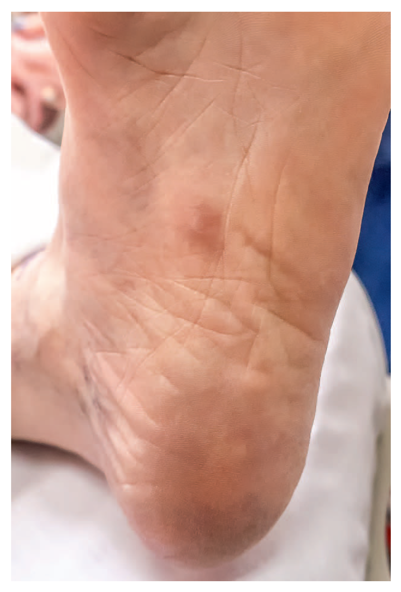
Figure 2.
Sagittal view of magnetic resonance image of plantar fascia tear.
Discussion
This article presents the first case of a midfoot plantar fascia tear in the absence of trauma. The tear in this case clinically resembled a fibroma. A fibroma is a benign encapsulated mass that grows slowly and is typically not painful. Histologically, a fibroma presents with an infiltrative pattern of growth with a proliferation of uniform, welldifferentiated spindle cells. In this case, the histological findings did not show such a pattern and they were consistent with a tear of the plantar fascia.
Figure 3.
Coronal view of magnetic resonance image of the plantar fascia tear.
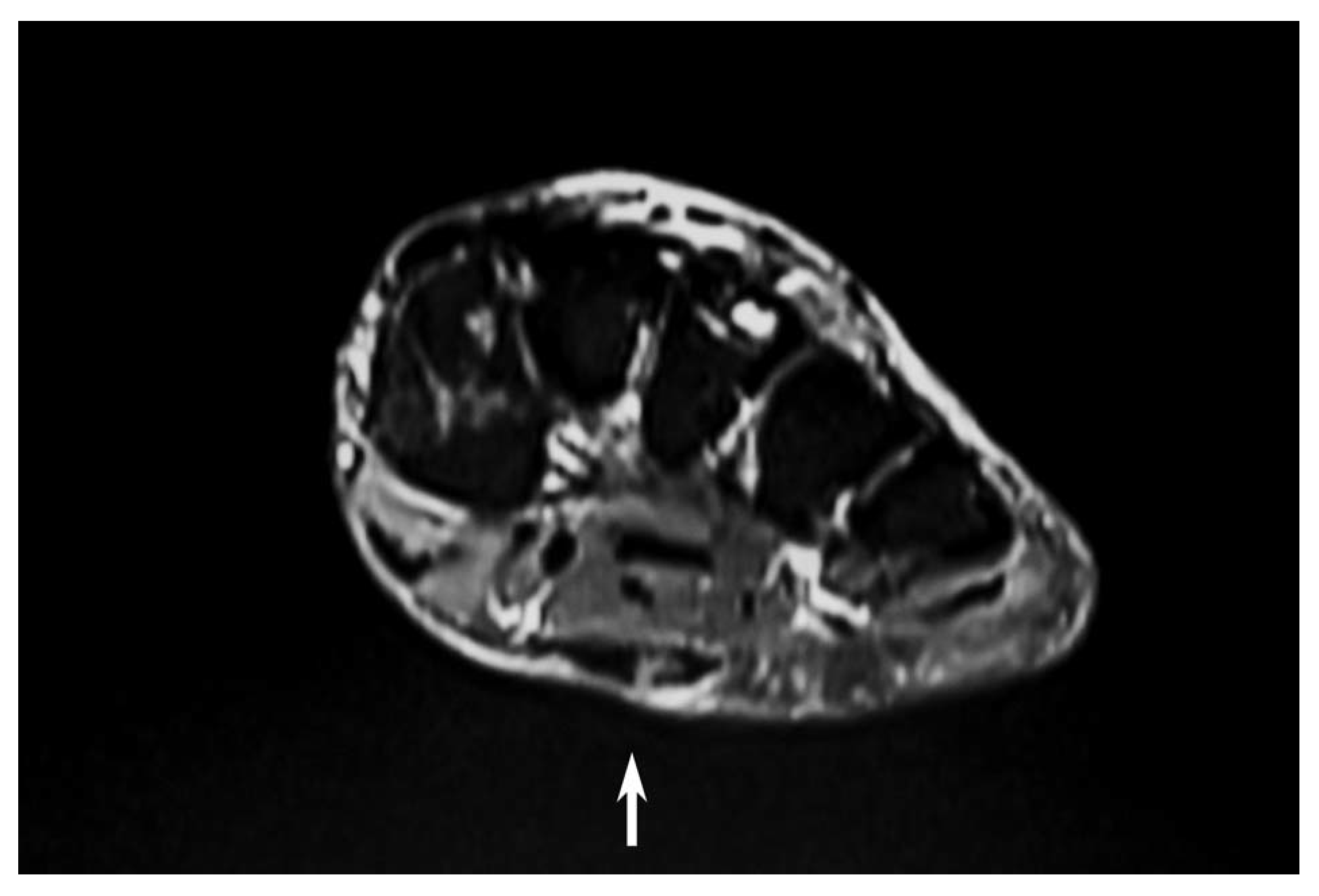
Figure 4.
Excised plantar fascia tear, measuring approximately 2 cm in length.
Tears of the plantar fascia often occur at or near the insertion of the plantar fascia into the calcaneus and have occurred spontaneously or related to trauma or corticosteroid injection. In the midfoot, all documented instances of tears have been trauma-related.[1,2,3,4,5,6,7,8]
Physicians should not assume that a bulge of the fascia is a plantar fibroma. Depending on the clinical findings and history, physicians may employ diagnostic imaging to assist in the diagnosis. Initially, a radiograph could evaluate the mass for any bone pathology, foreign body present, or calcification. Because of the soft-tissue nature of this problem, the physician should consider evaluating the mass with diagnostic ultrasound and MRI. On MRI, tears of the plantar fascia are easily distinguishable from fibromas, which present as round to oval and infiltrating, with the margins often well-defined.[12]
Figure 5.
Histological photomicrograph of surgical specimen.
In this case, the patient suggested that the physical therapist had torn the plantar fascia while performing deep tissue massage on her foot. No evidence substantiates this claim, and a literature search failed to find any such case.
Conservative options to consider when treating a plantar fascia tear include nonsteroidal anti-inflammatory medication, soaks, rest, custom-made foot orthotics, and physical therapy. Depending on the patient’s overall health and impairment from the plantar fascia tear, surgery may be an option.
Conclusions
Tears of the plantar fascia often occur in the heel area after trauma, may occur spontaneously, or may occur as a result of corticosteroid injection. However, physicians should be aware that tears may occur in other areas and may not always follow a traumatic episode. This case presented a plantar fascia tear that on clinical examination mimicked the appearance of a plantar fibroma. A thorough history and physical examination and MRI may be useful to evaluate the plantar fascia when a tear is suspected. Surgery is a viable option to address plantar fascia tears.
Financial Disclosures
None reported.
Conflicts of Interest
None reported.
References
- AHSTROM JP: Spontaneous rupture of the plantar fascia. Am J Sports Med 1988, 16, 306. [CrossRef] [Scilit] [PubMed]
- ALAGHA B, PADHIAR N: Partial tear of plantar fascia in a professional dancer. J Musculoskelet Res 2001, 18, 157.
- PAI VS: Rupture of the plantar fascia. J Foot Ankle Surg 1996, 35, 39. [CrossRef] [Scilit] [PubMed]
- NIELSON, J.H.; MICHELI, L.J. Acute plantar fascia rupture in a dancer. J Dance Med Sci 2004, 8, 116. [Google Scholar] [CrossRef] [Scilit]
- HERRICK, R.T.; HERRICK, S. Rupture of the plantar fascia in a middle-aged tennis player: A case report. Am J Sports Med 1983, 11, 95. [Google Scholar] [CrossRef] [Scilit] [PubMed]
- ACEVEDO, J.I.; BESKIN, J.L. Complications of plantar fascia rupture associated with corticosteroid injection. Foot Ankle Int 1988, 19, 91. [Google Scholar] [CrossRef] [Scilit] [PubMed]
- SELLMAN, J.R. Plantar fascia rupture associated with corticosteroid injection. Foot Ankle Int 1994, 15, 376. [Google Scholar] [CrossRef] [Scilit] [PubMed]
- LEE, H.S.; CHOI, Y.R.; KIM, S.W.; et al. Risk factors affecting chronic rupture of the plantar fascia. Foot Ankle Int 2014, 35, 258. [Google Scholar] [CrossRef] [Scilit] [PubMed]
- LEDDERHOSE G: Zur Pathologie der Aponeurose des Fusses und der Hand Langenbecks. Arch Klin Chir 55, 694, 1897.
- LEE, T.H.; WAPNER, K.L.; HECHT, P.J. Current concepts review: plantar fibromatosis. J Bone Joint Surg 1993, 75-A, 1080. [Google Scholar] [CrossRef] [Scilit] [PubMed]
- VEITH, N.T.; TSCHERNIG, T.; HISTING, T.; et al. Plantar fibromatosis—topical review. Foot Ankle Int 2013, 34, 1742. [Google Scholar] [CrossRef] [Scilit] [PubMed]
- KRANSDORF, M.J.; JELINEK, J.S.; MOSER, R.P.; et al. Magnetic resonance appearance of fibromatosis. Skeletal Radiol 1990, 19, 495. [Google Scholar] [CrossRef] [Scilit] [PubMed]
© 2019 American Podiatric Medical Association