


- To the Editor:
A forefoot varus defect is a common cause of over-pronation compensation. Traditional treatment for this condition employs the use orthotic posting, supporting the defect. In contrast to supporting a forefoot varus, an intervention to correct the condition by reducing the defect is absent in the current literature. As a result, I undertook an independent self-study to assess the hypothesis that a forefoot varus defect can be reduced, with improved efficiency of gait. The study took place over a 2-month period in 2013 using a rehabilitation regime emphasizing muscle balance and musculoskeletal alignment devices that I developed. The study involved my right foot, inflicted with a moderate to severe over-pronation with an associated forefoot varus, and a moderate to severe great toe bunion formation. The measured variables were ankle and first metatarsophalangeal (MTP) pain, one-legged standing balance time, forefoot eversion passive range of motion, and gait observation. Results following the intervention showed reduced ankle and first MTP pain, increased one-legged standing balance, an increase in forefoot eversion, enhanced supinated lateral heel strike, and medial forefoot push-off during gait, which was confirmed with shoe-wear pattern. The conclusion of the study supported the technique and training used to correct a forefoot varus. I propose a predictable realignment to the foot with use of the devices and specific treatment exercises, enhancing both pulley and windlass mechanisms that provide rigidity during the push-off phase of gait. Later observation of shoe-wear pattern show supinated function continued after the removal of semi-rigid rehabilitative shoe inserts. Additional study is recommended to further determine efficacy for a patient population.
Need for Effective Treatment
Over-pronation is a compensatory dysfunction attributable to alterations in normal foot alignment. The literature supports a forefoot varus as the most frequent cause of over-pronation compensation [1,2]. A forefoot varus occurs when the medial metatarsal heads of the foot are raised in relation to the lateral heads, in reference to a transverse plane of an inferior aspect of the calcaneus. With a forefoot varus condition, the lateral heads initially contact the surface before the rest of the forefoot. Over-pronation compensation must then occur, during weightbearing stance and gait, when the medial heads contact the surface [1,3].
Over-pronation leads to pes planus, with associated compensation that may progress up or down the kinetic chain, resulting in further dysfunction to include bunions [1,4], knock-knees [1,5], anteverted hips [1,6], and low back pain [1,7]. For many of those afflicted with over-pronation, the medical outcome is an expensive surgical repair, such as a joint replacement. It is therefore in the public’s best interests to develop a conservative, preventative treatment for over-pronation that reliably limits further musculoskeletal destruction that may lead to surgery in later life [1].
Literature Review
A question is asked by Mr. Speck [8] as to whether the varus angle can be changed. He proposes exercise, rather than fitting orthotics to fill in the gap under the defect, as a more reasonable way to manage the problem. He suggests that an effective exercise has been stretching by slightly inverting the heel while bringing the big toe to the floor (emphasis to avoid curling or gripping of the toes) [1,8].
Development of Treatment and Devices
Observation
My account in developing treatment and devices for my own pes planus condition was impacted by a motorcycle accident in the 1980s, in which my right ankle drastically everted, dislocating the talus from the mortis, fully rupturing the medial collateral ligament. Over the years, muscular support by the ankle supinators became weak and unstable, with a subluxing of the talocrual joint, splaying of the foot, and development of a bunion. A forefoot varus with hypermobile first-ray compensation was determined (coincidently, my left ankle developed pes planus with a forefoot varus, which may have been partially due to a distal fracture of the tibia/fibula following a separate motorcycle accident many years earlier) [1]. In this case, a moderate to severe over-pronation dysfunction was due to an unstable talus and a forefoot varus. In addition to pes planus with splaying of the foot and bunion formation, an internal rotated leg compensation developed. I experienced moderate to severe pain at the subtalar and first (MTP) joints, as well as occasional sharp knee pain. The dysfunction was compounded by a lack of ability to supinate the foot throughout the stance phase of gait, resulting in a vicious dysfunctional cycle.
I first observed a potential rehabilitation treatment with the use of devices when the pain of my subtalar and first MTP joints became profound. These pains subsided when my forefoot was stabilized on a nonskid floor, with the right leg externally rotated in relation to the ankle/foot, and the ankle maximally dorsiflexed. This movement provided significant relief of the bunion and ankle pain. On observing the great toe, the bunion had disappeared, the arch of the foot had returned, and the talus was fully locked into supination; “It felt good.” The observation lead me to theorize one could return the foot toward an improved mechanical advantage by placing the foot and lower leg into this rigid position. The locked rigid structure would give the advantage for return to supinated alignment through muscle balance techniques; thus, selective strengthening of appropriate muscles of supination would occur through exercises and functional activities, such as walking, stimulating adaptive shortening, while selective stretching would key in on lengthening the shortened muscles of pronation. The treatment/exercise regime needed to be designed for the outcome of recapturing supinated gait and return to a more functional biomechanics [1].
Devices
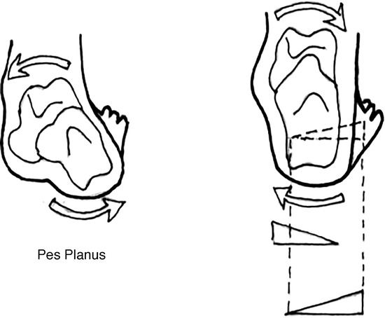
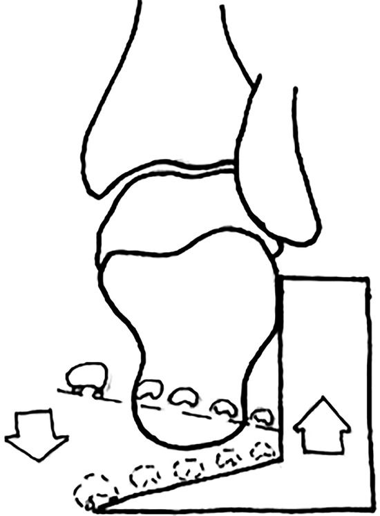
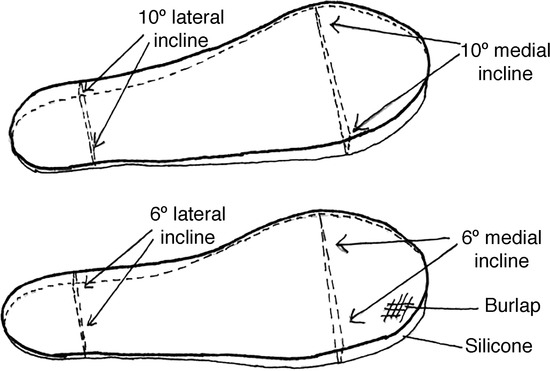
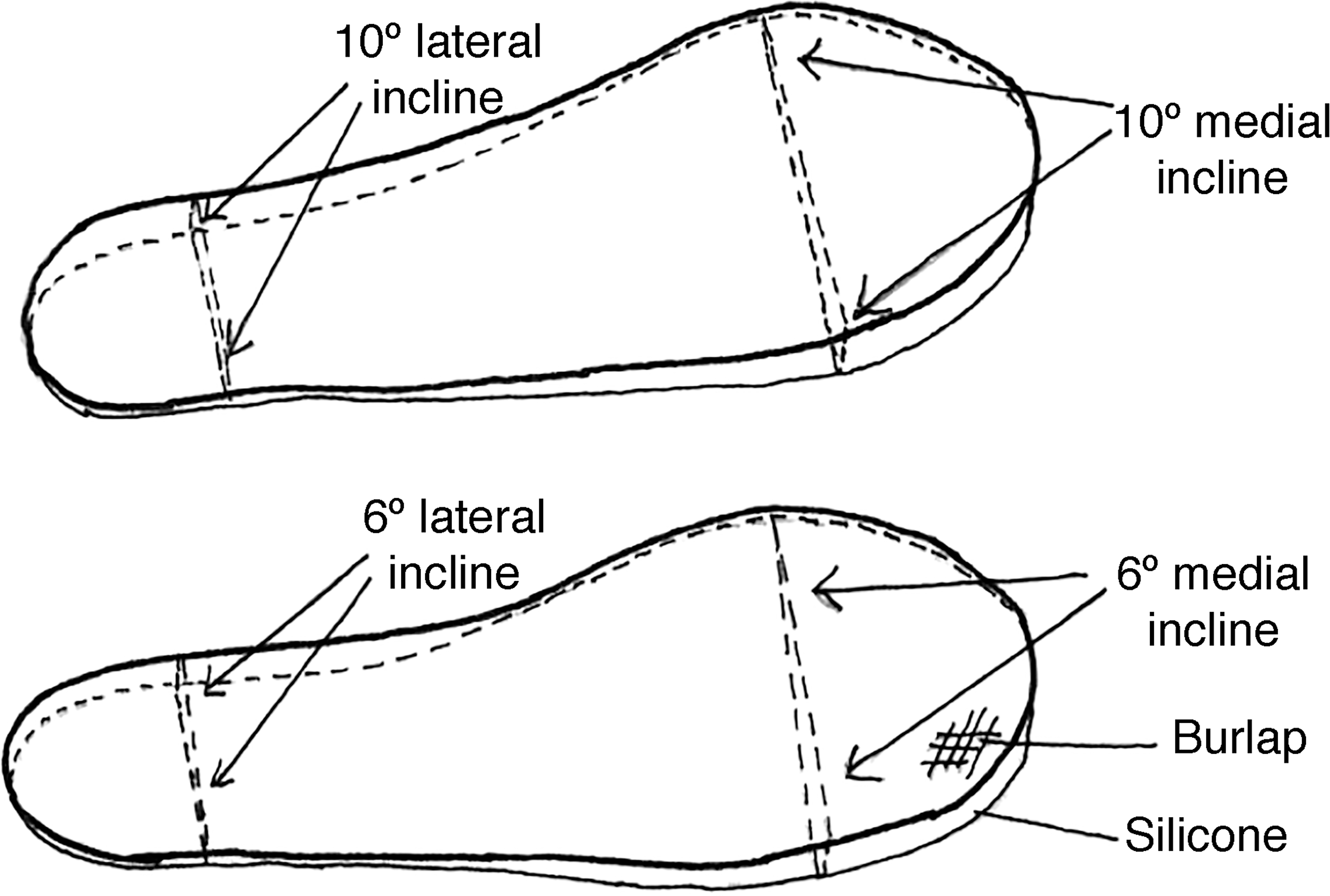
From the above, I developed devices to assist in rehabilitating my pes planus condition, with Figure 1 showing a basic idea concept involved in the rehabilitation. The devices included a stretch/mobilization device, as in Figure 2, to assist in reducing the forefoot varus and return of supinated function. The premise is to return an over-pronator to supinated function by reduction of forefoot varus, a common cause of over-pronation. This is accomplished by locking the ankle, foot, and lower leg into a rigid supinated alignment for stretch and mobilization of the forefoot varus. Thus, the locking would allow for specificity of exercise in reducing the varus condition. The stretching and mobilization of forefoot varus cannot be accomplished with the foot in a neutral or pronated condition, as “the loose bag-o-bones” identified with a non-supinated alignment eliminates the specificity of stretch/mobilization. I also developed rehabilitative foot inserts to enhance this supinated function, and return of weightbearing through the first ray on the push-off phase of gait. Figure 3 shows views of two right foot inserts of different inclined angles, wedged lateral to medial at the heel and medial to lateral at the forefoot. A custom calculation for the appropriate inclined angulation of the user’s heel corresponds to a stable foot platform for rehabilitation of the forefoot. I have also developed a combination insert using a custom molding of a users' supinated foot on the superior surface, with wedging on the inferior of the insert. Both inserts have been used with positive results [1,9].
Figure 1.
Illustration showing the concept for use of wedging underneath the user’s heel and forefoot to enhance supinated function.
Figure 2.
Illustration showing the concept for reducing a forefoot varus involving stretching, mobilization, and strengthening.
Figure 3.
Illustration showing the development of footwear inserts for rehabilitating an over-pronation condition.
Self-Study
In 2013, I conducted an 8-week self-study on my moderate to severe right foot over-pronation condition to further assess my theory for the treatment and device efficacy. Intervention involved treatment with a rehabilitative regime and devices I developed. Outcome measures included whether a forefoot varus angle can be changed, along with additional parameters. Posttreatment data indicates a reduction in forefoot varus, significant pain reduction of my ankle and bunion, increased lower-extremity proprioception, and an enhanced supinated gait with lateral heel strike and medial push-off, confirmed by gait observation and shoe wear pattern [1]
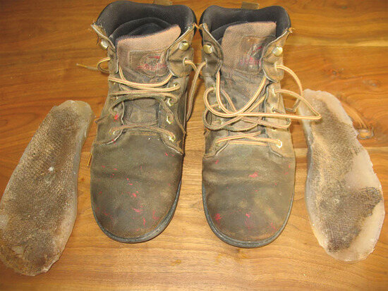
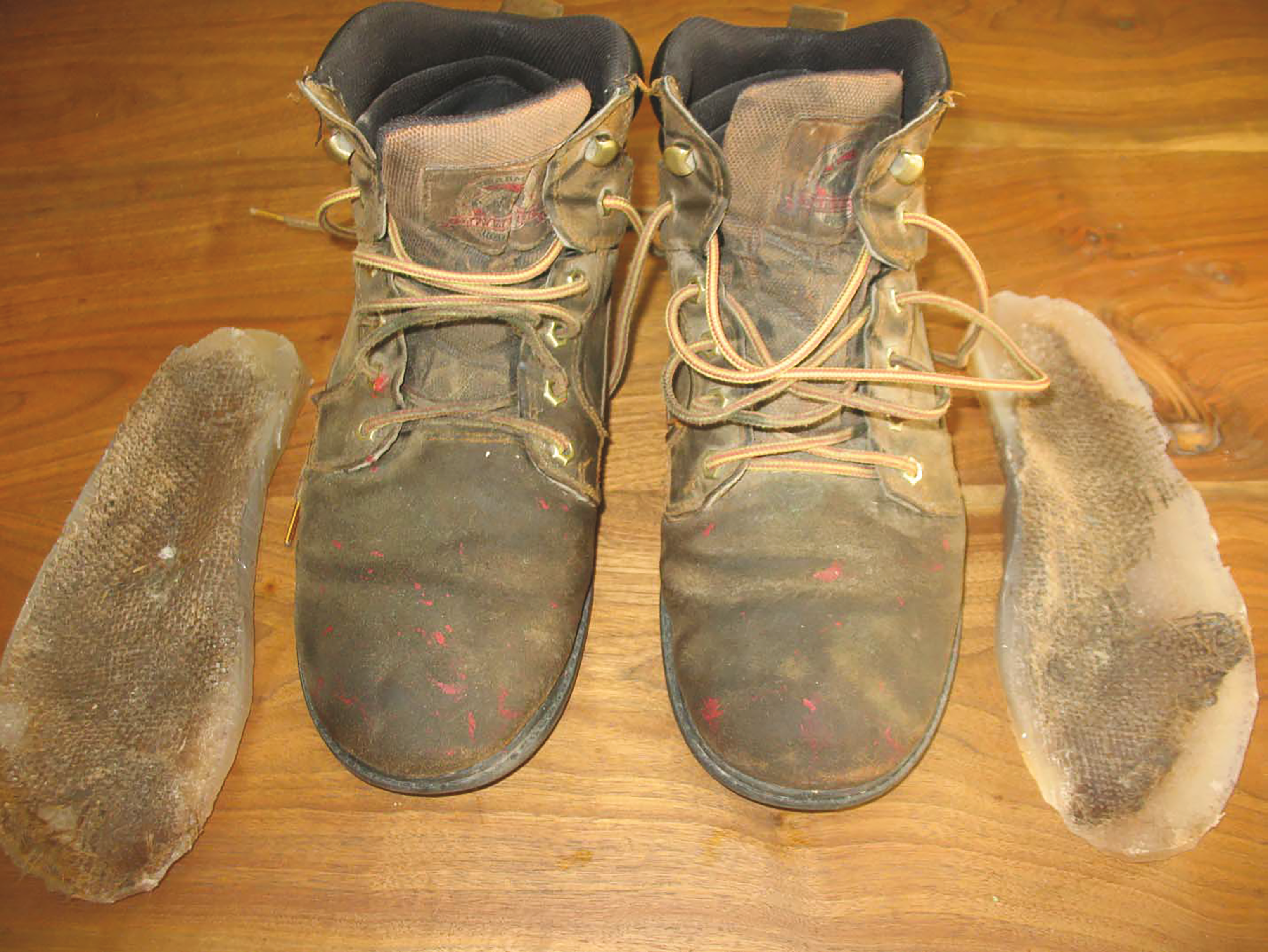
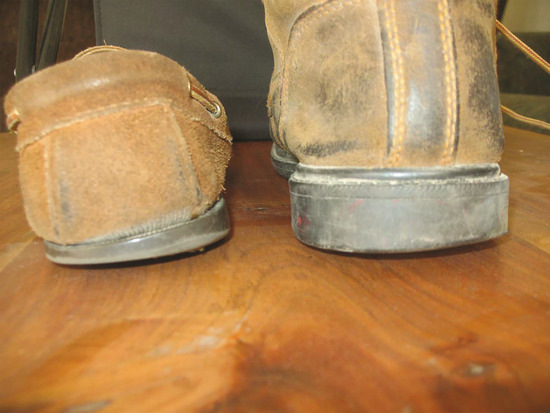
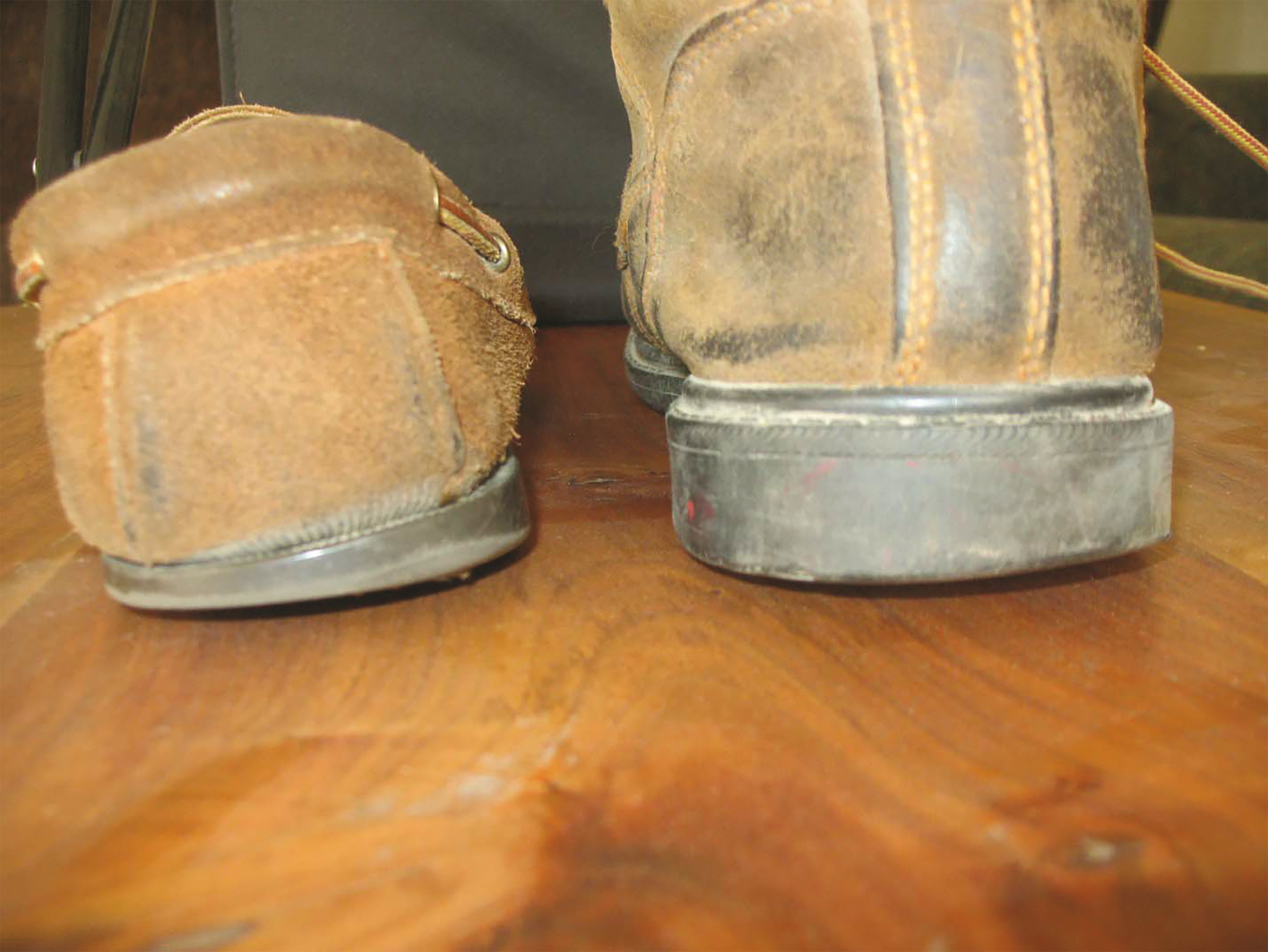
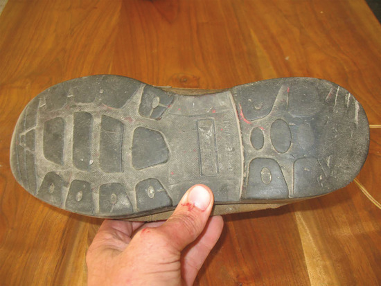
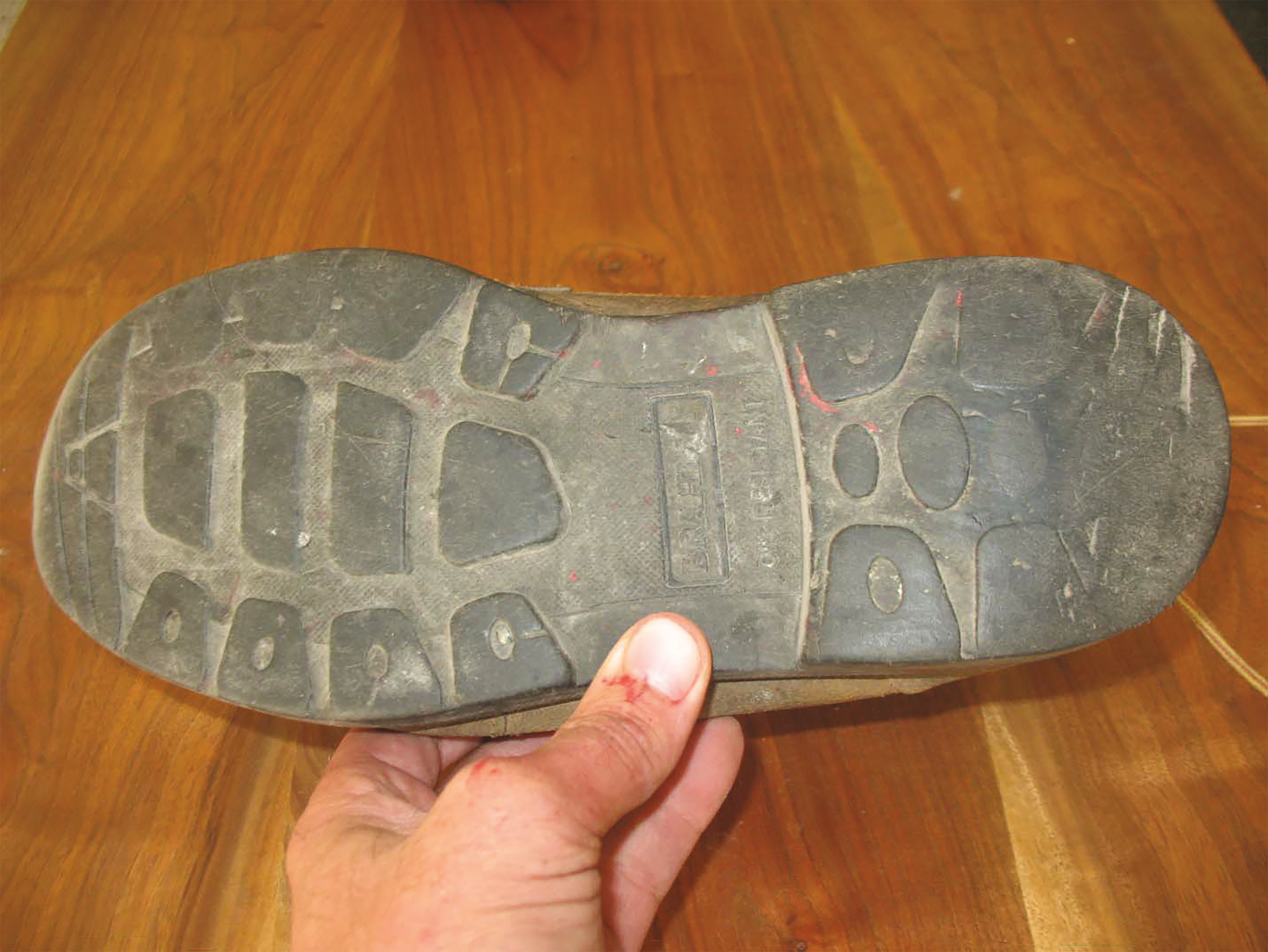
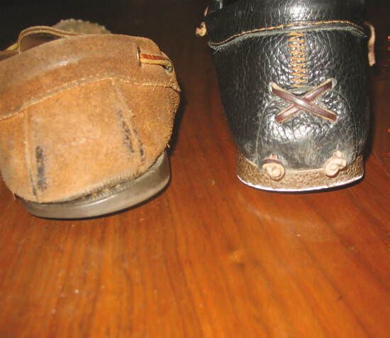
Figure 4 shows my boots with bilateral combination insert use; note the transverse wear region on the anterior boot surface. Figure 5 shows over-pronation wear of my right medial shoe heel without insert use, while my right boot shows wear of the lateral heel with insert use. Figure 6 shows medial wear of the first ray and large toe [1].
Figure 4.
Photo showing the use of combination rehabilitative inserts within boots. The superior surface displays a custom supinated molding of my foot. The inferior surface is inclined laterally at the heel and medially at the forefoot. Note the transverse wear of the anterior boot.
Figure 5.
Photos showing a comparison of my right shoe wear, without insert use, and my right boot using the inserts. Note the medial heel wear of the shoe and lateral boot wear.
Figure 6.
Photo showing the medial wear of the boot at the first ray region of the forefoot.
Discussion
Biomechanics of the Foot
The gait cycle is comprised of a stance phase and a swing phase. The stance phase is further subdivided into heel strike, mid-stance, and propulsion phases. On normal heel strike to early mid-stance, the foot pronates, which is a medial and downward movement of the ankle, or talus on the calcaneus, via the subtalar joint. This action provides for a torque converter, or change in force transmission, which enables a longitudinal force to be transferred through an inwardly rotating lower leg. These forces are dispersed through triplanar motion of the subtalar joint, providing for shock absorption [1,3].
Pronation following heel strike also causes adjustments in a person’s midfoot to slacken a specific muscle (peroneus longus) from a mechanism described as a midfoot pulley. This mechanism involves the peroneus longus as the cable, which originates at the lateral shin and inserts at the base of the first metatarsal. The cuboid is the “keystone” or pulley which the cable wraps underneath and attaches at the first ray insertion sight. Pronation of the subtalar joint releases the pulley from the cable on initial heel strike, causing the foot to become a “loose bag of bones” in order to adapt to terrain [1,3].
From mid-stance to push-off the subtalar joint supinates, tightening the cable onto the pulley. In effect, this action moves the head of the first metatarsal downward to create rigidity to the foot on push-off, producing resistance to weightbearing forces by stabilizing the first ray and big toe [1,3].
Another normal mechanism called the “windlass effect” occurs from heel-off to toe-off of the stance phase of gait. This supinated action of the subtalar joint results in a mechanical tightening of a broad band of ligaments and plantar aponeurosis of the foot to provide rigidity to the first ray and large toe on push-off. The head of the first metatarsal is the drum of the windlass, the aponeurosis is the cable, and the proximal phalanx of the big toe is the handle, winding the cable around the drum. The effect of the proximal phalanx extending during supination and push-off is to tighten the cable and move the head of the first metatarsal downward, remaining as a prominent resistance to all weightbearing pressure until the proximal phalanx was flexed. This “pronator twist “action of the forefoot provides a mechanical force strong enough to perform weightbearing resistance of the first ray and large toe, as confirmed by testing. Hicks [10] reports that the windlass mechanism contributes to about half of the full range of motion of the first ray, or 10 degrees of (plantarflexion) in nonweightbearing, while the lateral rays contribute to a lesser degree of about 5 degrees [1,10].
Neutral versus Supinated Alignment
The concept behind traditional orthotics eliminates normal biomechanical supination. This occurs due to subtalar joint neutral posting, as this neutral posting represents the midstance phase of gait, and a neutral alignment. In contrast, the literature supports supination of the foot as necessary for incorporation of the windlass and pulley mechanisms, which produce rigidity for the foot on push-off. Furthermore, pronation, and more profoundly over-pronation, provide for a “loose bag-of-bones” to adapt to terrain. Without the stabilization and rigidity of these normal mechanisms resulting from supination, the foot will continue to function as a loose bag-of-bones during gait phases when foot rigidity is necessary [1,3,9,10].
Advantages are apparent with a supinated foot as compared to a neutral alignment. The windlass and pulley mechanisms provide weightbearing resistance through supination. Thus an over-pronation condition, which develops a hypermobile first ray, has the enhanced weightbearing function of the aponeurosis and peroneus longus during supinated alignment. Hicks [10] reports that the effect of normal first-ray depression is to increase the range of motion over and above what the ankle movement can provide and to provide a foot that does not yield to increasing weightbearing forces, but which tends to flex and thrust downwards with an additional “flick” at take-off [10]. This is necessary for normal stabilization of the first ray and large toe on push-off. Conversely, literature supports that a subtalar joint neutral alignment compromises this same stability offered by the aponeurosis and peroneus longus [1,3,9,10].
Conclusions
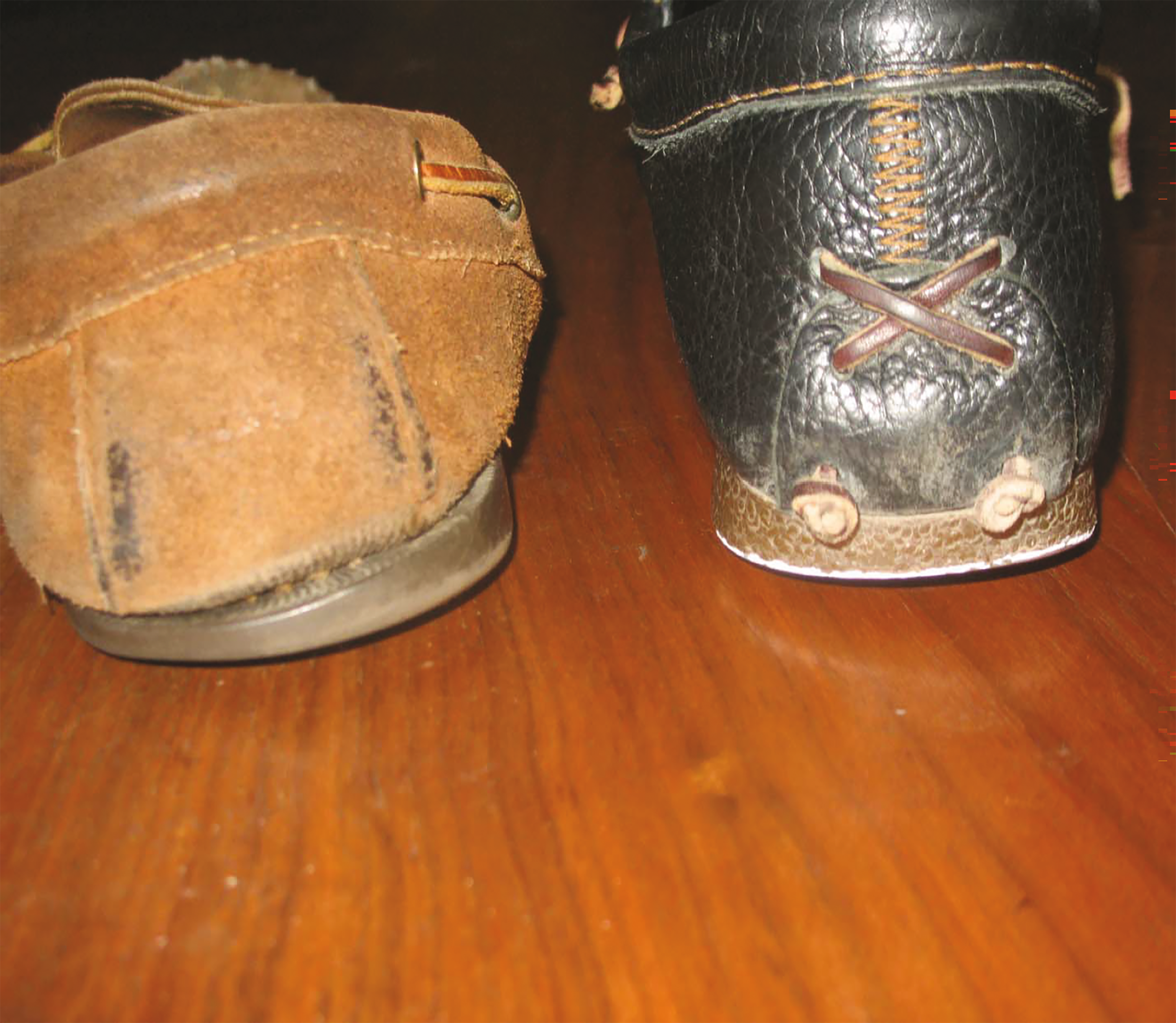
I am excited to report that I am now pain free, running up bluffs with a rigid weightbearing through the first ray on push-off bilaterally. My shoe-wear pattern now shows lateral heel strike, with medial first ray push-off without rehabilitative inserts (Fig. 7); my lower-extremity supinators have returned to a functional state. Thus in my serendipitous work and study, I have discovered that the use of the body’s normal mechanisms may assist in reestablishing a return to a more functional gait for those with this condition. You will see that the line of treatment pursued is different from the conventional, and I believe there is a more promising outcome with my approach for the multitude who suffer the short- and long-term effects of this condition.
Figure 7.
Photo showing a comparison between the right shoe heel wear before and after rehabilitation. The shoe on the left has marked medial over-pronation wear while the post-rehabilitation right shoe has lateral heel wear (enhanced with a white border).
The potential clinical applications my rehabilitative regime and devices include: an improvement of functional supinated dorsiflexion AROM (ankle equinus) [11]; a reduction or elimination of pain associated with over-pronation; a reduction, or reversal of, over-pronation dysfunction, hallux valgus formation, chondromalacia, malalignment syndrome, anteverted hips, and back dysfunction; and a return of a stable, weightbearing first ray on push-off from a flexible flat foot [1,9]
The scope of my concept includes elite athletic performance as another example of possible uses. Training in a supinated alignment with certain athletic events may be beneficial, as a rigid musculoskeletal structure provides the best scaffolding for muscular propulsion with certain athletic events. Thus, the range of uses may extend from treatment of injury to enhanced performance [1,9].
Funding
None reported.
Conflicts of Interest
The author is a patent-pending inventor.
Dual Publication
This article is a follow-up to “A Novel Approach to Treatment of Over-Pronation Dysfunction,” which was published in the October 2015 issue, Volume 27, Issue 4, pages 234-242, of the Journal of Orthopaedic Physical Therapy Practice. Sections of the text and figures are being used with permission from the Orthopaedic Section of the American Physical Therapy Association, Inc.
References
- Wiley, DE. A novel approach to treatment of over-pronation dysfunction. J Orthop Phys TherPrac 2015, 27, 234. [Google Scholar]
- Subotnick, S. Podiatric Sports Medicine, Futura; Mt. Kisco, NY; 1975.
- Root, ML; Weed, JH; Orien, WP. Normal and Abnormal Function of the Foot, Volume II, Clinical Biomechanics Corporation; Los Angeles; 1977.
- Hunter, S. Rehabilitation of Foot Injuries. Rehabilitation Techniques in Sports Medicine 1990, 342. [Google Scholar]
- D'Amico, J; Rubin, M. The influence of foot orthoses on quadriceps angle. JAPMA 1986, 76, 337. [Google Scholar]
- Ramig, D; Shadle, J; Watkins, A; et al. The foot and sports medicine: biomechanical foot faults as related to chondromalacia patellae. J Orthop Sports Phys Ther 1980, 2, 48. [Google Scholar]
- Botte, R. An interpretation of the pronation syndrome and foot types of patients with low back pain. JAPA 1981, 71, 243. [Google Scholar]
- Speck, J. Forefoot varus and over-pronation, can the varus angle be changed? Available at: http://www.somastruck.com (accessed on 12 April 2013).
- Wiley, DE. Method and apparatuses for positioning a user’s foot. US patent application 14/454,990. August 8, 2014.
- Hicks, JH. The plantar aponeurosis and the arch. The mechanics of the foot. J Anat 1954, 88, 25. [Google Scholar]
- Clifford, C. Understanding the biomechanics of equinus, Available at: http://www.podiatrytoday.com (accessed on 6 February 2016).
© 2017 American Podiatric Medical Association