


Abstract
Angioleiomyomas are benign tumefactions that originate from smooth muscle in vascular structures and are difficult to definitively diagnose preoperatively. Although these lesions are rarely encountered in the foot, the lower extremity is the most common site of occurrence. An angioleiomyoma typically manifests as a small, painful, solitary, mobile lesion. This case report describes a lateral retromalleolar para–Achilles tendon insertional location for a moderately sized immobile solid tumefaction in the subcutaneous tissues. The lesion was nonpainful and progressively enlarged over 5 years. An excisional biopsy was performed, and the nodular lesion was subsequently diagnosed histopathologically as an angioleiomyoma. Owing to the ambiguous nature of the clinical findings, angioleiomyoma should be included in the differential diagnosis of lower-extremity soft-tissue manifestations.
Angioleiomyomas are rare, small, benign smooth muscle tumors that arise from the muscular structures of small vessels. Nomenclature variations include angiomyoma, vascular leiomyoma, angioma, and dermal angioma. Rudolf Virchow [1] first identified this tumor as a leiomyoma in 1854. Leiomyomas were not again described in the medical literature until 1937 by Stout. [2] Morimoto [3] developed a two-armed classification system in 1973 that separated the lesions histologically and by site of origin. The histologic system classifies an angioleiomyoma as solid, venous, or cavernous, and the site of origin for these lesions is delineated to the head or an extremity. [3] Enzinger and Weiss [4] in 1995 published a classification system that organized the lesions into vascular, cutaneous, and deep soft-tissue presentations.
These lesions account for 5% of all benign soft-tissue neoplasms and may occur anywhere in the body. [5] Angioleiomyomas show a predilection for the extremities, with a preponderance of presentations in the lower extremity. Incidence rates of 50% to 70% are reported for lower-extremity presentations. [6] Despite the high incidence rate in the lower extremity, this lesion is rarely encountered in the foot. [4] Females show a greater occurrence rate than males. [4] Average age at presentation has been reported to be 30 to 70 years, with most cases diagnosed in the third and fourth decades of life. [7] The prevalence of this lesion in East Africa has been reported to be ten times that of the white population living in the same temperate climates, with trauma suspected as a primary causative factor. [5]
Angioleiomyoma typically manifests as a slow-growing, painful, solitary, mobile, nodular lesion that measures less than 2 cm at greatest diameter. Although the pathogenesis of angioleiomyoma is unknown, reported etiologic factors include minor trauma, venous stasis, and infection; hormonal fluctuation with estrogen is the most commonly cited. Patient self-identification of a soft-tissue mass is traditionally the presenting complaint. Pain and tenderness are the most impressive findings in the clinical setting, identified in 60% to 70% of presentations. [8,9] Pain associated with this lesion is thought to be related to active contraction of smooth muscle mediated by nerve fibers situated in the tumor parenchyma, which results in local areas of ischemia. [10]
Although a definitive diagnosis cannot be obtained from imaging, standard plain film radiographs, ultrasound studies, and magnetic resonance images (MRIs) are routinely used in the assessment of these lesions. Plain film radiography generally provides visualization of a noncalcified soft-tissue lesion. Ultrasonic examination routinely identifies a homogenous structure with well-defined margins. Typically, MRIs demonstrate a well-defined ovoid mass that can be found in the dermis, subcutaneous fat, or fascia. [9] Specifically, MRIs show signal intensity similar to muscle on T1-weighted images and higher signal intensity relative to muscle with a hypointense peripheral rim on T2-weighted images. [11]
A differential diagnosis for a well-demarcated, subcutaneous, nodular, soft-tissue mass of the lower extremity should include lipoma, ganglion cyst, schwannoma, synovial sarcoma, nodular synovitis, foreign body granuloma, pseudoaneurysm, neurofibroma, giant cell tumor, traumatic neuroma, hemangioma, hamartoma, angiolipoma, hiradenoma, glomus tumor, inclusion cyst, myxoid cyst, and, certainly, angioleiomyoma.
Marginal surgical excision of an angioleiomyoma is the treatment of choice, which also provides the opportunity for a definitive diagnosis. Recurrence of this lesion after surgical excision has been reported but is not common. [6] In the event of recurrence, repeated excision is mandated to exclude the possibility of malignant transformation into leiomyosarcoma and angioleiomyosarcoma, which are rare but have been reported to occur. [6,12]
Histologically, angioleiomyomas are benign lesions composed of smooth muscle with vascular channels, lacking cytologic atypia and mitotic activity. Immunohistochemical staining of these lesions expresses positively for desmin, muscle-specific actin, and α-smooth muscle actin, which provides the definitive diagnosis. [13] The histologic classification system described by Morimoto [3] includes the solid subtype, which has compacted smooth muscle with small vascular channels; the venous subtype, which has readily visualized thick muscular walls; and the cavernous lesion, which shows dilatation of the vascular structures with less smooth muscle. The Enzinger and Weiss [4] classification system notes that vascular lesions arise from the tunica media layer of small veins and arteries; cutaneous lesions are generally intradermal, arising from nonstriated muscle of erector pili; and deep soft-tissue lesions originate from vessels and unstriated muscles of the lower extremity. Angioleiomyoma can be found in the dermis, subcutaneous fat, or fascia.
Case Report
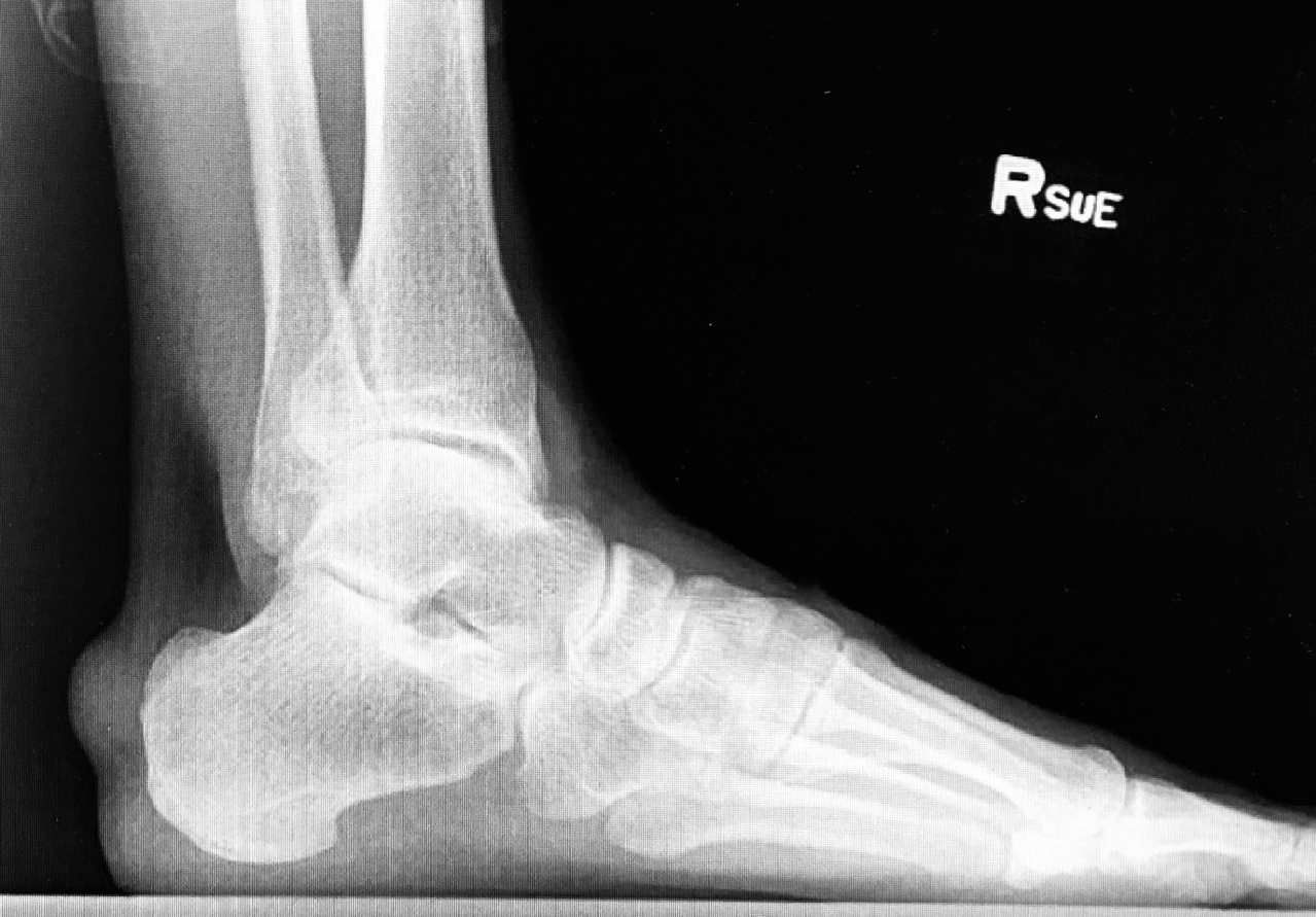
A well-developed and well-nourished 64-year-old white man was seen initially in the clinic with a nonpainful nodular lesion on the lateral posterosuperior heel. His medical history was remarkable for hyperlipidemia, gastroesophageal reflux disease, and glaucoma. The lesion had been gradually enlarging over the previous 5 years, with only minor discomfort experienced recently due to shoe wear. There was no relation of trauma or injury to the affected area. Clinical examination revealed a moderately sized, immobile solid tumefaction in the subcutaneous tissues in the retromalleolar space at the Achilles tendon lateral insertion. The lesion was nonpainful to direct palpation and range-of-motion examination. The patient related that the lesion had been progressively enlarging during the past 5 years. The gross dimension of the lesion on clinical examination was approximately 3.25 cm at greatest diameter. A radiographic study composed of two weightbearing views of the right foot revealed a focal soft-tissue nodule posterior to the calcaneus in the mediolateral exposure (Fig. 1). There was no evidence of intralesional mineralization, osseous erosion, or remodeling of the calcaneus associated with the identified soft-tissue lesion in the radiographic study. After discussion, MRI was deferred by the patient due to cost and his desire to simply have the lesion surgically excised. Surgical intervention was deemed the treatment of choice to alleviate the patient's inability to wear shoes and to definitively identify the pathologic lesion. An excisional biopsy identified an encapsulated benign-appearing nodular lesion in the subcuticular tissue adjacent to the Achilles tendon. The tumor was marginally excised, with the feeding vessel ligated proximally and distally. Proximally and distally oriented tags were maintained. The lesion had a gross appearance of a tan-gray rubbery mass that was well circumscribed. The tumor was submitted in toto for pathologic evaluation. Closure was accomplished using a plastic technique to address the redundant skin, with implantation of a cryopreserved amniotic membrane allograft to reduce postoperative incisional hypertrophy and Achilles tendon adhesions. The patient had an unremarkable postoperative course with minimal scarring. There has been no recurrence of the lesion in this patient at 1 year.
Figure 1.
Mediolateral weightbearing plain film radiographic image of the right foot depicting the posterior para–Achilles tendon soft-tissue lesion.
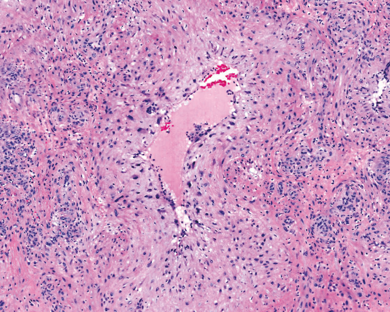
Histologically, the lesion was well circumscribed and symmetrical. At the periphery of the lesion, it was possible to identify the wall of a large vessel (Fig. 2). In the vessel there was a proliferation of several smaller vessels with thick walls embedded in a sclerotic stroma (Fig. 3). Higher magnification demonstrated the presence of prominent endothelial cells, some of them pleomorphic, which were interpreted as degenerative changes occurring in this neoplasm (Fig. 4).
Figure 2.
Scanning magnification of the lesion showing a fairly well-circumscribed neoplasm. At the periphery is the wall of a preexisting vessel (H&E, x25).
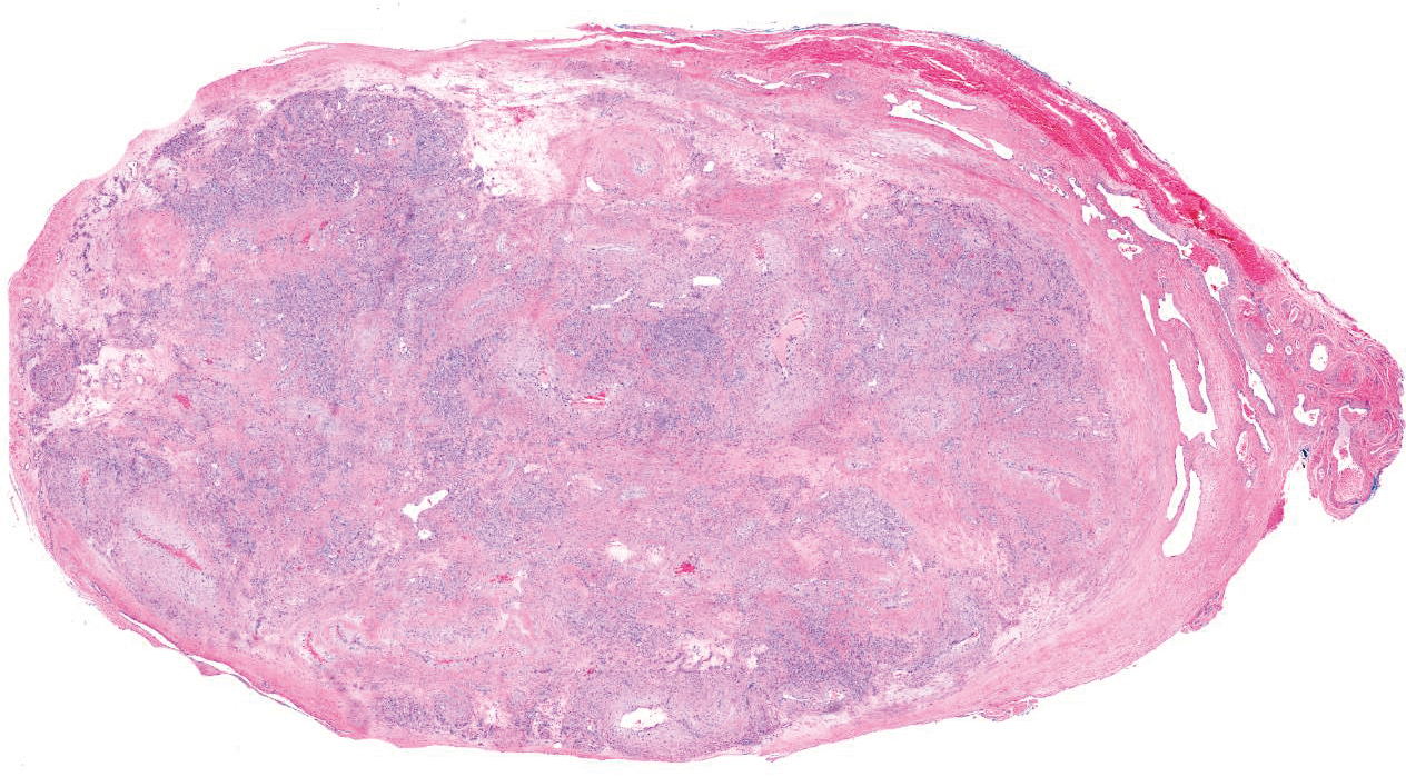
Figure 3.
The neoplasm was composed of uniform spindle cells embedded in a sclerotic stroma (H&E, x40).
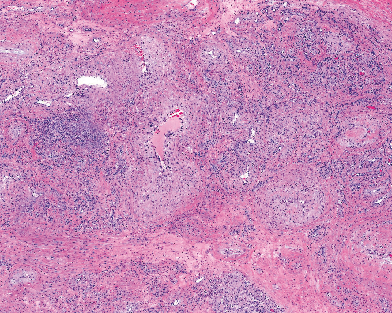
Figure 4.
At higher magnification, the endothelial cells are enlarged, with hyperchromatic nuclei and variable amounts of cytoplasm. Some of these changes are probably degenerative (H&E, x100).
Discussion
Angioleiomyomas are rare benign tumors that may occur anywhere in the body, with suspected histopathologic origins in the smooth muscle of vascular structures, specifically the tunica media of veins. The most common presentation of this tumor is generally accepted to be a solid, nodular, painful lower-extremity lesion measuring less than 2 cm at greatest diameter in a 30- to 40-year-old woman.
This case presentation demonstrates the ambiguity and variability associated with not only an angioleiomyoma but also other soft-tissue lesions in the lower extremity. The present patient presented with a solid, nodular, nonpainful lower-extremity lesion measuring more than 2 cm at greatest diameter in a 60- to 70-year-old man.
Imaging in this patient was limited to plain film radiography, which provided gross visualization of the lesion. An MRI was suggested to assess the extent of the lesion, the associated vascular structures involved, and possible osseous involvement. However, at the patient's request, the MRI was deferred due to the added expense of the study and his wish to proceed with the surgical excision. In this case, an MRI would certainly have been helpful but, in actuality, was not critical to the eventual outcome.
The diagnosis of angioleiomyoma was not considered in the differential diagnosis of this patient. Only after excision of the lesion and microscopic examination was the eventual definitive diagnosis of angioleiomyoma realized. The present case illustrates that angioleiomyomas can occasionally grow to a moderate size in older males without causing pain or tenderness. Due to the ambiguous nature of both the clinical presentation and imaging findings, it is difficult to preoperatively definitively diagnose an angioleiomyoma.
Conclusions
Angioleiomyomas should be included in the differential diagnosis of any lower-extremity soft-tissue mass regardless of age, sex, clinical findings, or imaging features.
Financial Disclosure
None reported.
Conflicts of Interest
None reported.
References
- Virchow R: Ueber Makroglossie und pathologische Neubildung quergestreifter Muskelfasern. Virchows Arch (Pathol Anat)7: 126, 1854.
- Stout AP: Solitary cutaneous and subcutaneous leiomyoma. Am J Cancer29: 435, 1937.
- Morimoto N: Angiomyoma (vascular leiomyoma): a clinicopathologic study. Med J Kagoshima Univ24: 663, 1973.
- Enzinger FM, Weiss SW: “Benign Tumors of Smooth Muscle,” inSoft Tissue Tumors, 4th Ed, edited bySWWeiss, JRGoldblum, p443, CV Mosby, St. Louis, 1995.
- Ramesh P, Annapureddy SR, Khan F, et al: Angioleiomyoma: a clinical, pathological and radiological review. Int J Clin Pract58: 587, 2004.
- Hachisuga T, Hashimoto H, Enjoji M: Angioleiomyoma: a clinicopathologic reappraisal of 562 cases. Cancer54: 126, 1984.
- Butler E, Hamil J, Seipel R, et al: Tumors of the hand: a ten year survey and report of 437 cases. Am J Surg100: 293, 1960.
- Freedman AM, Meland NB: Angioleiomyomas of the extremities: report of a case and review of the Mayo Clinic experience. Plast Reconstr Surg83: 328, 1989.
- Gupte C, Butt SH, Tirabosco R, et al: Angioleiomyoma: magnetic resonance imaging features in ten cases. Skeletal Radiol37: 1003, 2008.
- Hasegawa T, Seiki K, Yang P, et al: Mechanism of pain and cytoskeletal properties in angioleiomyomas: an immunohistochemical study. Pathol Int44: 66, 1994.
- Yoo HJ, Choi JA, Chung J, et al: Angioleiomyoma in soft tissue of extremities: MRI findings. AJR Am J Roentgenol192: 291, 2009.
- Herre D, Zimmermann A, Buchler U: Vascular leiomyoma in an index finger undergoing malignant transformation. J Hand Surg (Br)20: 484, 1995.
- Requena L, Sangueza OP: Cutaneous vascular proliferations, part III: malignant neoplasm: other cutaneous neoplasms with significant vascular component, and disorders erroneously considered as vascular neoplasms. J Am Acad Dermatol38: 143, 1998.
© 2017 American Podiatric Medical Association