


Abstract
We report on the rare case of multiple open interphalangeal dislocations. This uncommon pathologic condition results from dorsiflexion and axial loading of the digit. In the present patient, closed reduction was not possible. We review the literature on this pathologic condition and present a case of open irreducible dislocation of the lesser interphalangeal joints complicated by peripheral neuropathy and treated with resectional arthroplasty.
Dislocations of the lesser interphalangeal joints are extremely uncommon, resulting from dorsiflexion and axial loading of the digit.[1] To our knowledge, only eight case reports and two small retrospective studies exist in the English literature.[2-11] In most cases, closed reduction could not be achieved due to interposition of the plantar plate or surrounding soft tissues. Almost all of the individuals in the studies experienced dislocation of only one joint. In addition, the individuals represented in these studies were mostly young, active individuals without significant comorbidities. Each case was treated operatively by dislodging the interposed plantar plate and fixating as needed.
Presented herein is a case of an open, irreducible dislocation of the lesser interphalangeal joints complicated by peripheral neuropathy and treated with resectional arthroplasty. Unlike the studies discussed previously herein, the present patient's comorbidities limited our surgical treatment options.
Case Report
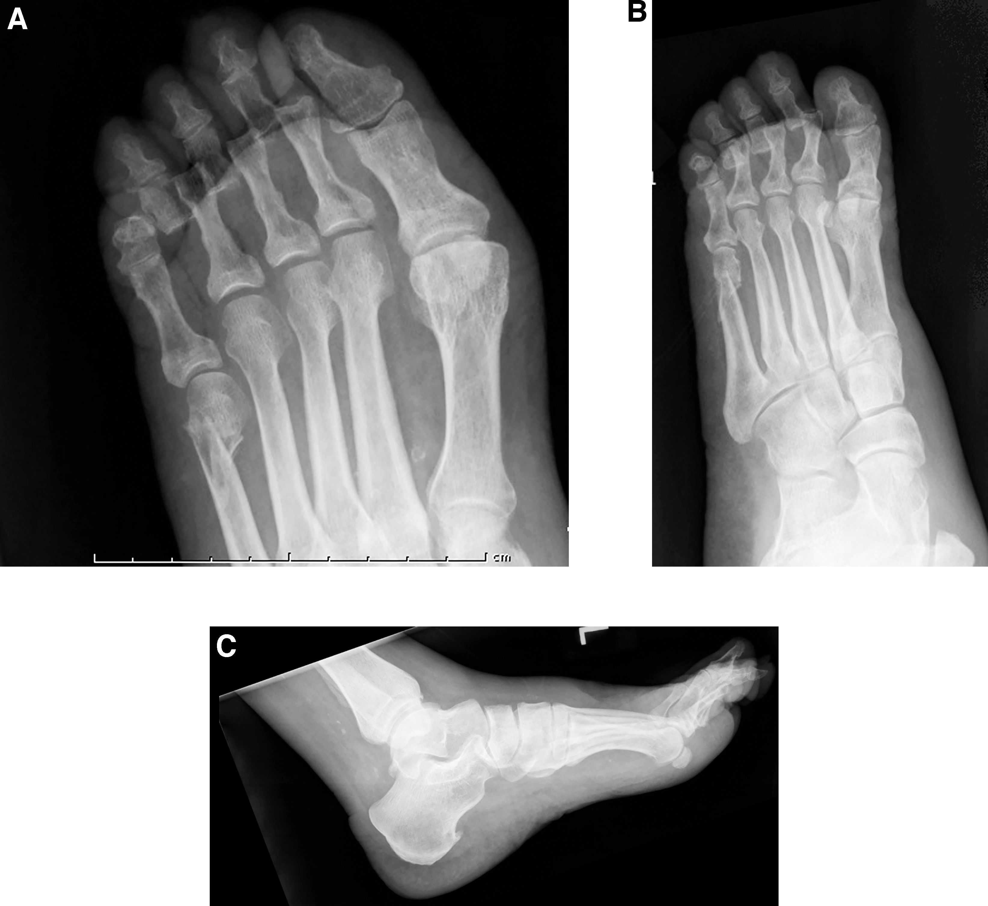
A 91-year-old man presented to the emergency department (ED) on January 23, 2015, with lower-extremity cellulitis. The patient's medical history was significant for peripheral neuropathy secondary to spinal stenosis and hypertension. Two days earlier, the patient experienced a fall in his home in which he landed prone, kicking the floor, with axial loading of his digits. Worsening erythema and edema of the left lower extremity subsequently developed. The patient presented to his primary-care physician on January 22, 2015, where amoxicillin and clavulanate potassium (Augmentin; GlaxoSmithKline, Research Triangle Park, North Carolina) and doxycycline were prescribed. Although referred to the ED by his primary-care physician, the patient did not present until the following day. At the time of presentation, the patient had a claw toe deformity of each of the lesser digits. On examination of the interdigital spaces, open dislocations of the interphalangeal joints were noted at the medial aspects of the third and fourth digits, with copious amounts of purulent discharge (Fig. 1). Bilateral pedal pulses were present, with a capillary fill time of less than 2 sec. The temperature gradient of the left lower extremity was reversed. Pedal radiographs revealed dislocation of the proximal interphalangeal joints of the second, third, and fourth toes with foreshortening and a metaphyseal fracture of the fifth metatarsal (Fig. 2). The patient met sepsis criteria at the time of ED presentation.
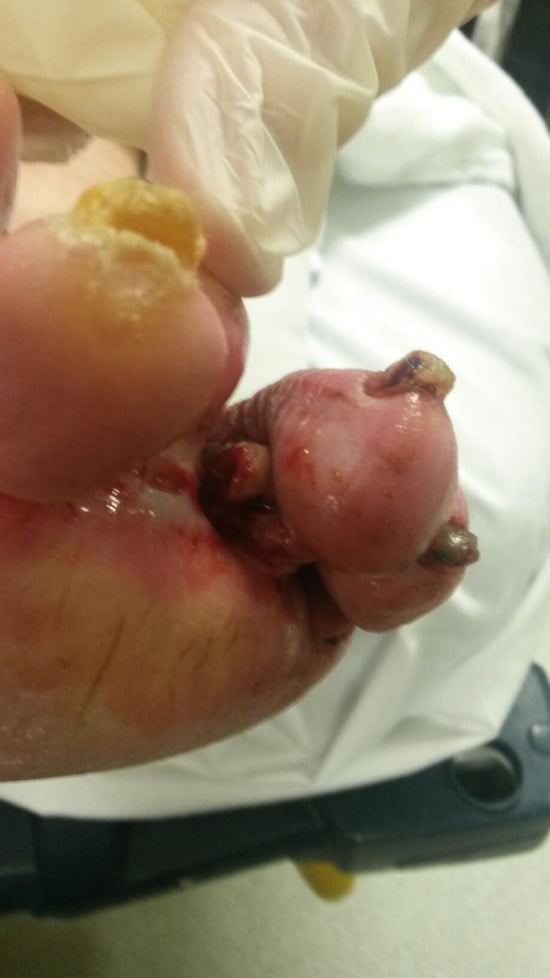
Figure 1.
Photograph of the affected limb at the time of presentation to the emergency department showing exposure of the distal aspect of the intermediate phalanx of the left fourth digit.
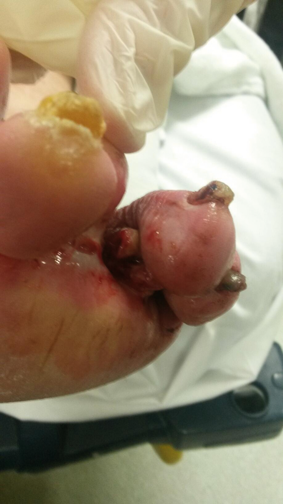
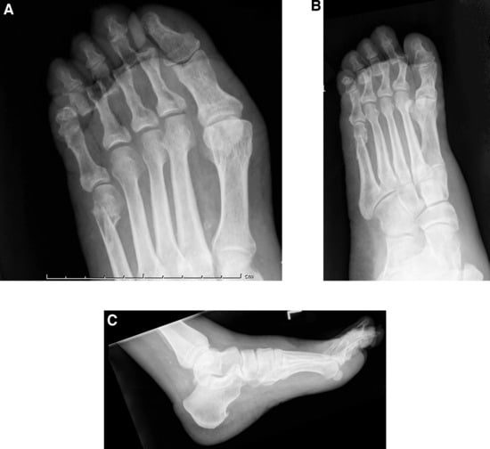
Figure 2.
Radiographs of the affected limb before medical intervention. A, Dorsoplantar view reveals marked dislocation of the second, third, and fourth proximal interphalangeal joints of the left foot. Cortical irregularity and medial migration of the left fifth metatarsal suggests metaphyseal fracture with mild displacement. B, Medial oblique view reveals plantar displacement of the dislocated second, third, and fourth proximal interphalangeal joints. C, Lateral view reveals no plantar displacement of the right fifth metatarsal fracture fragment.
The dislocated second digit was successfully reduced with abduction, distraction, and medial displacement. Despite several attempts, however, reduction of the open dislocations of the third and fourth digits was not attained. This was complicated by the patient's motor neuropathy, which had given rise to contracted claw toe deformities.
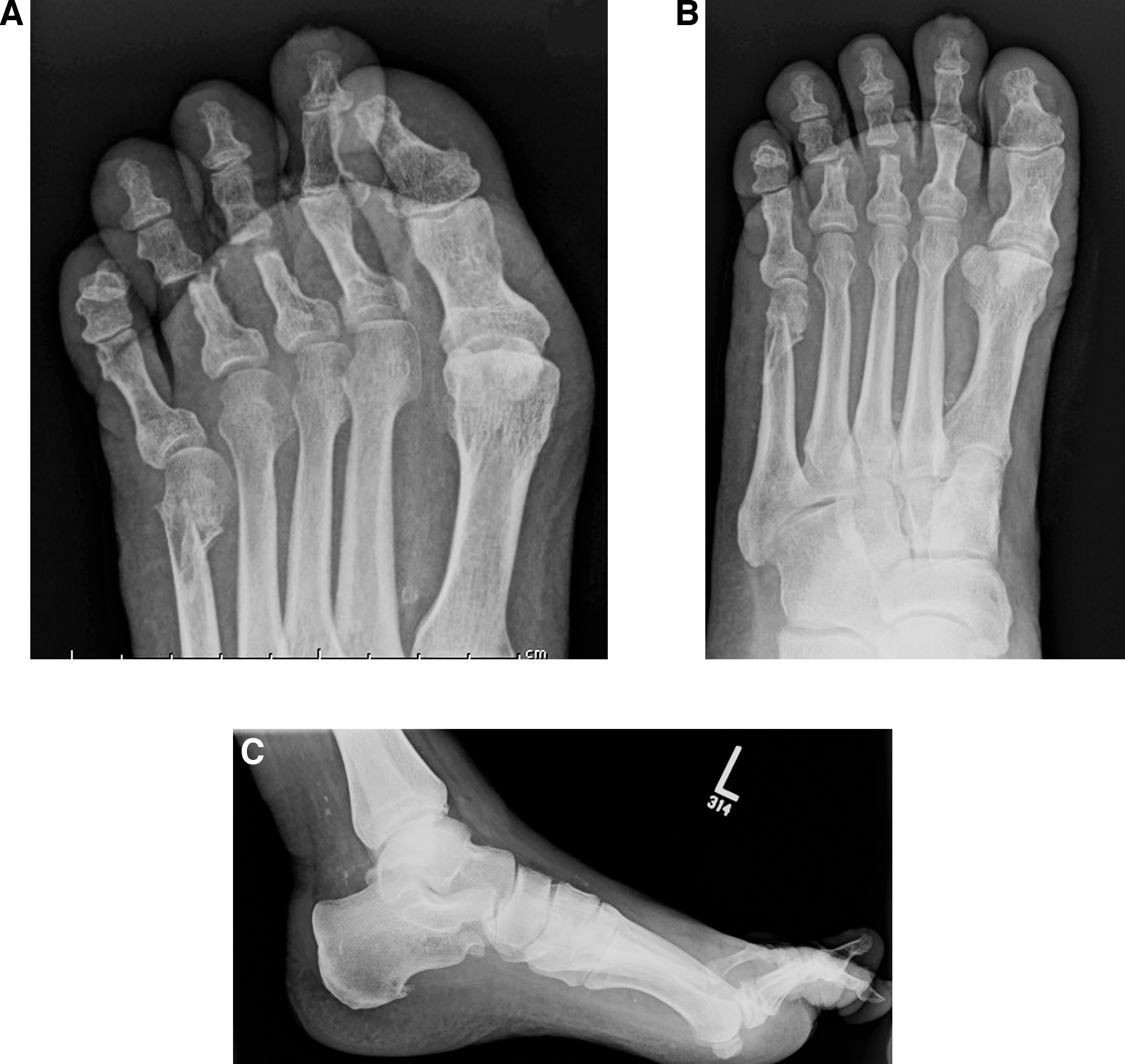
Because of the long-standing contaminated nature of the injury, complicated by the joint contractures, irrigation and debridement were performed at the bedside in the ED. To avoid the unnecessary risks of general anesthesia in an elderly individual, this conservative approach was undertaken. Under local anesthesia, the exposed heads of the proximal phalanges were resected using sterile bone-cutting forceps. Specimens of bone were submitted for culture and sensitivities. The wounds were irrigated with normal saline and were debrided of nonviable tissue. No soft-tissue dissection or release was necessary due to adequate exposure of phalangeal heads protruding from the wound. The digits were then easily reduced, and the wounds were allowed to heal by secondary intention (Fig. 3).
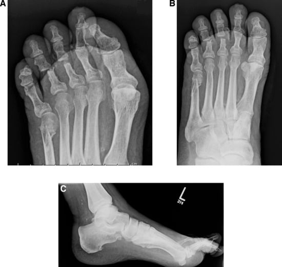
Figure 3.
Radiographs of the affected limb 1 day after medical intervention. A, Dorsoplantar view reveals adequate alignment in the transverse plane of the proximal and intermediate phalanges of the second, third, and fourth digits. B, Oblique view also reveals adequate alignment. C, Lateral view reveals adequate alignment in the sagittal plane of the second, third, and fourth proximal and intermediate phalanges.
The patient was admitted to the hospital and was given vancomycin and piperacillin-tazobactam for 5 days. Bone histopathologic analysis demonstrated necrotic changes. Bone cultures were positive for methicillin-sensitive Staphylococcus aureus, group B Streptococcus, and Proteus mirabilis. On the recommendation of the infectious disease specialist, the patient was to complete a 6-week course of vancomycin and oral ciprofloxacin therapy on hospital discharge.
At the time of the patient's 6-week follow-up visit, the wounds had completely reepithelialized. By the 8-month follow-up visit, the patient had made a full return to his baseline level of function. Throughout his recovery, he had not experienced any redislocations or vascular compromise.
Discussion
The current literature supports the use of surgical reduction of open dislocations due to interposition of the plantar plate limiting the efficacy of closed reduction.[7,9] These studies used general anesthesia or conscious sedation for their open reduction procedures.
In addition, current studies report treatment for younger individuals without other pathologic conditions of the lower extremity.[2-6,8,10] Owing to severe contractures of the third and fourth digits in the present patient, resectional arthroplasties were necessary to achieve adequate reduction. We believe that the patient's peripheral neuropathy was a complicating factor because contracted claw toe deformity is a known sequela to motor neuropathy, although the definitive etiology was never established.[12]
Of the reports of interphalangeal dislocations, two mention open dislocations without going into detail about how they were managed.[7,9] It may be assumed that they were treated in the usual manner with emergency irrigation and debridement. However, given the delay in treatment resulting in contamination of the present wound, secondary closure was required, with a course of intravenous antibiotic drug therapy for bone infection.
Conclusions
A case was presented of open interphalangeal dislocation treated by resectional arthroplasty to achieve reduction of the deformity. This treatment has not been previously described and serves as a viable option for the reduction of these uncommon injuries.
Funding
None reported.
Conflicts of Interest
None reported.
References
- Berkowitz MJ, Sanders RW: “Dislocations of the Foot,” inMann's Surgery of the Foot and Ankle, Vol 2, edited byMJCoughlin, CSaltzman, RBAnderson, p 1905, WB Saunders, Philadelphia,2014.
- Cordoba-Fernandez A: Management of nonreducible lesser toe interphalangeal dislocation: an unusual injury. JAPMA102: 252, 2012.
- Weinstein RN, Insler HP: Irreducible proximal interphalangeal dislocation of the fourth toe: a case report. Foot Ankle Int15: 627, 1994.
- Neubauer T, Wagner M, Quell M: Interphalangeal dislocation of the fourth toe with avulsion-fracture in a child: report of a case. Foot Ankle Int18: 175, 1997.
- Katayama M, Murakami Y, Takahashi H: Irreducible dorsal dislocation of the toe. J Bone Joint Surg Am70: 769, 1988.
- Stienstra JJ, Derner R: Closed reduction of a proximal interphalangeal joint dislocation. J Foot Surg29: 385, 1990.
- Yang IB, Sun KK, Sha WL, et al: Interphalangeal dislocation of toes: a retrospective case series and review of the literature. J Foot Ankle Surg50: 580, 2011.
- Johnson JM, Miller M: complex interphalangeal dislocation of the fourth toe after a sports injury: case report. Foot Ankle Int18: 668, 1997.
- Brunet JA, Tubin S: Traumatic dislocations of the lesser toes. Foot Ankle Int18: 406, 1997.
- Fugate DS, Thomson JD, Christensen KP: An irreducible fracture-dislocation of a lesser toe: a case report. Foot Ankle11: 317, 1991.
- Akahane M, Sada M, Tanaka Y: Irreducible DIP joint dislocation of the second toe. Foot11: 103, 2001.
- Bernstein RK: Physical signs of the intrinsic minus foot. Diabetes Care26: 1945, 2003.
© 2017 American Podiatric Medical Association