


Abstract
Superficial acral fibromyxoma is a benign and slow-growing solitary soft-tissue neoplasm. Since being described in 2001, more than 100 cases of superficial acral fibromyxoma on the foot have been reported worldwide, none of which have been reported in the podiatric medical literature. Only nine cases of superficial acral fibromyxoma have been reported with presentation on the plantar heel. We report an unusual case of a 47-year-old Jamaican woman with a painful, erythematous nodule on her right heel that was diagnosed as superficial acral fibromyxoma.
Superficial acral fibromyxoma (SAF) is a recently identified soft-tissue neoplasm that typically affects the fingers and toes. Fetsch et al [1] first recognized it as a distinct histopathologic entity in 2001, when they described a growing number of similar unclassified soft-tissue masses with a male predominance. Their aim was to define the clinicopathologic features and immunohistochemical findings in 37 patients with SAF. Clinically, they described a solitary mass appearing on a finger, palm of the hand, or, most commonly, a toe, with the hallux being the most affected site. [2-7] No cases were reported on the heel. Of the 37 cases, 20 involved the nail region and four caused a scalloping defect of the underlying bone. Patient age varied widely (range, 14–72 years; mean, 43 years), as did lesion size (range, 0.6–5.0 cm; mean, 1.75 cm) and period (range, 3 months to 30 years; mean, 3 years). Histologically, the tumors exhibited a proliferation of stellate-shaped and spindle fibroblast-like cells surrounded by one of three matrices (myxoid, myxocollagenous, or primarily collagenous). Lesions were described most commonly in the dermis, with some extending into the subcutis and, rarely, fascia or bone. Immunohistochemical analysis revealed reactivity to CD34 (21 of 23 patients), epithelial membrane antigen (18 of 25 patients), and CD99 (11 of 13 patients). Recurrence rates reported for 18 patients revealed three recurrences.
The first report of SAF on the heel occurred in 2008, when a description of 32 new cases described four on the heel. [2] Again showing a male predominance, most SAFs occurred on the hands and feet, with the hallux most commonly affected. All of the lesions were removed with local excision, and three cases of recurrence were reported out of 14 at a mean of 20 months (22%), none of which were on the heel. Different from the initial report, this paper described an increased number of capillary vessels and scattered mast cells in most patients. Also described were two lesions with nuclear atypia, neither resulting in metastasis.
Two additional features of SAF were described in 2008: a lipomatous component and CD10 expression. [8] One patient presented histologically with mature fat cells present throughout the lesion, possibly either from entrapment of subcutaneous adipocytes by spindle cells or simply due to fat cells in close proximity to the tumor site. In addition to the already described CD34, CD99, and epithelial membrane antigen, CD10 was described as strong in two of four patients, weak in one of four patients, and absent in one of four patients. In a single case study in 2007, Misago et al [9] also described a CD10 component along with CD34, CD99, and vimentin.
In 2012, Hollmann and Fletcher [10] published a series of 124 cases of SAF; 56 cases (45%) occurred on the foot. Most foot lesions (82%) occurred on the digits, most of which (96%) were in close proximity to the nail. This study is the second to describe heel occurrence. Although five lesions were noted to occur on the heel, these lesions were not described in depth. Nine of 25 patients (36%) with available radiographic studies had bony involvement. Ten of 42 patients (24%) had local recurrence after a mean of 27 months, all of which had positive margins at initial biopsy or excision.
The recommended diagnostic assessment for an unidentified soft-tissue lesion is radiography, magnetic resonance imaging (MRI), and biopsy. When encountered, SAF should be treated with local excision, with monitoring for recurrence. [1,2,4,8] Complete removal is crucial to prevent recurrence.
Herein, we introduce SAF to the podiatric medical literature with a rarely described occurrence on the plantar heel. The purpose is to review the identification, diagnosis, and treatment of SAF so that practitioners will be well equipped to differentiate it from similar pathologic conditions.
Case Report
A 47-year-old Jamaican woman presented to the Foot Center of New York (an affiliate of the New York College of Podiatric Medicine; New York, New York) with a painful, warm, erythematous papule on the plantar aspect of her right heel. Three years before the visit, the patient described a callous on her right heel. In the past year, the callous developed a central raised mass that increased in size over time. Past treatment included over-the-counter callous remover pads containing salicylic acid, 40%.
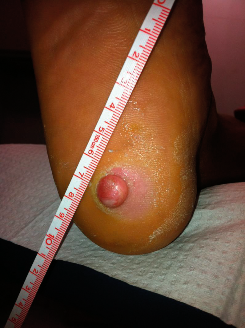
Her medical history was significant for anemia. She denied any history of surgery and cancer. She also denied tobacco or alcohol use. On physical examination, her light touch sensation, vibratory sensation, deep tendon reflexes, and protective sensation were all intact. Her dorsalis pedis and posterior tibial pulses were palpable bilaterally. The patient had a reversed temperature gradient, with the right foot warmer than the calf. The dermatologic examination showed a circular nodule with a 6.5 mm radius, raised 13 mm from the skin, with symmetrical, homogenous borders and consistent erythematous color. The mass was firmly adhered to the skin and did not move on palpation. The surrounding tissue was erythematous, macerated, and edematous (Fig. 1), possibly due to previous treatment with salicylic acid. Pain was elicited on palpation radially and centrally. On light debridement, no purulent or sanguineous discharge was observed.
Figure 1.
Superficial acral fibromyxoma on the right heel with a 6.5 mm radius, raised 13 mm from the skin.
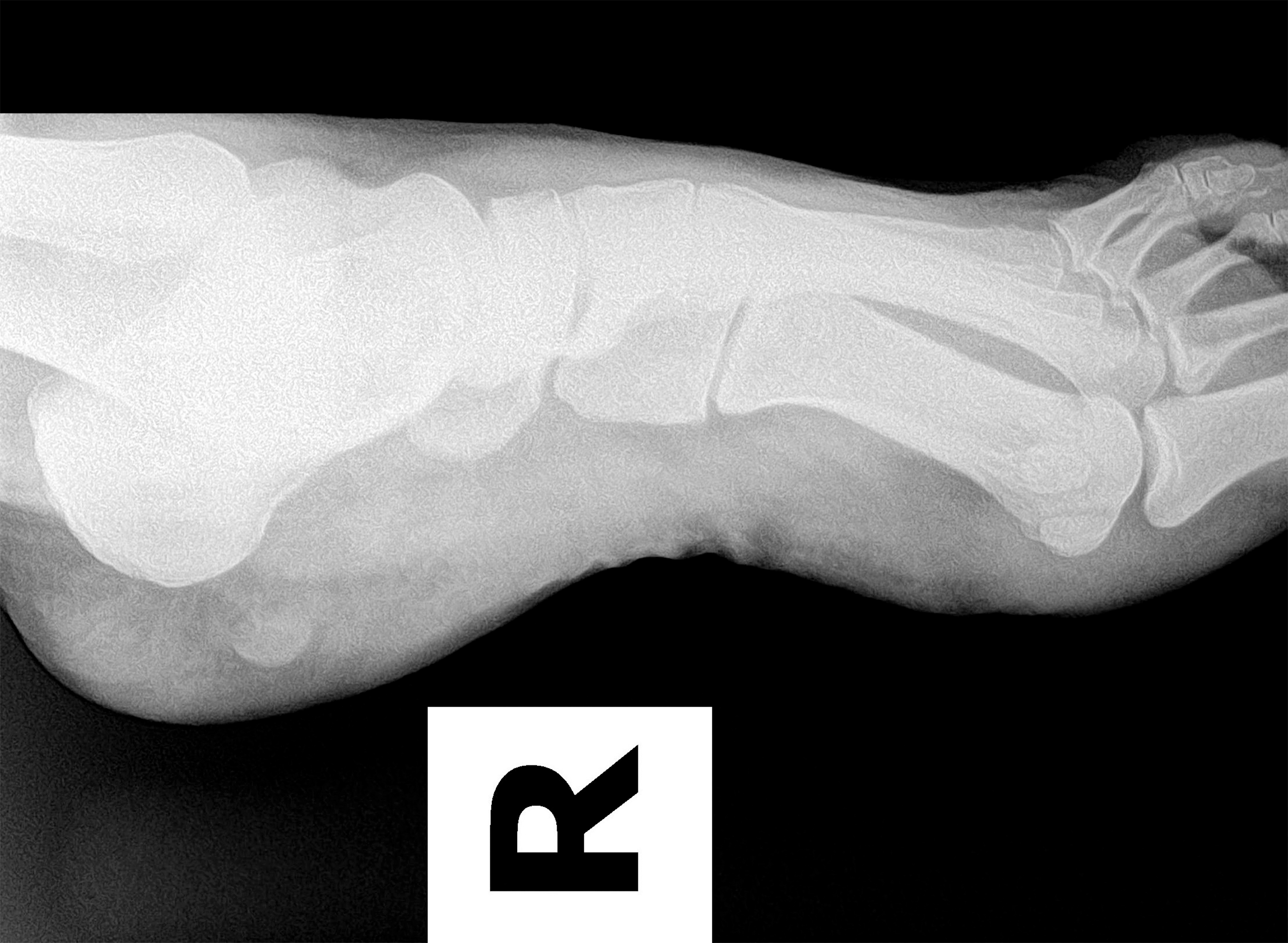
Radiographic evaluation revealed a normal underlying calcaneus. The lateral oblique view revealed soft-tissue edema to the calcaneal fat pad with a semicircular radiopaque lesion visible plantar to the posterocentral calcaneus (Fig. 2). Sonographic studies revealed a superficial vascularized mass that was well demarcated and hypoechoic. Neither MRI nor preoperative biopsy of the lesion was performed.
Figure 2.
Right foot medial oblique radiograph with a semicircular radiopaque lesion visible plantar to the posterocentral calcaneus.
Based on clinical appearance, the working differential diagnosis included eccrine poroma, pyogenic granuloma, and cavernous hemangioma. Far less likely possibilities included malignancies such as clear cell sarcoma and amelanotic melanoma. Owing to heel pain and an unknown diagnosis, excisional biopsy was scheduled. The lesion was off-loaded using a 0.25-inch felt horseshoe pad until the surgery date.
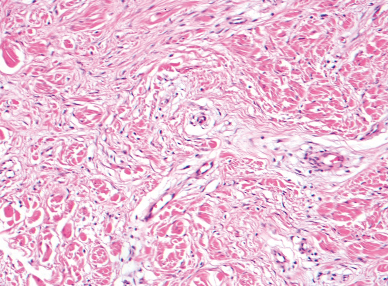
The patient was taken to the operating room, where an excisional biopsy was performed. An elliptical incision was made in a 3:1 pattern down to subcutaneous tissue. Owing to occurrence on the central plantar heel, the 3:1 incision was used to allow for primary closure. The lesion was removed as a single unit and was sent to the laboratory for identification. Frozen section identification was not available. Pathologic analysis revealed SAF (Fig. 3).
Figure 3.
Histopathologic analysis revealed spindle cells in a fibromyxoid stroma.
The postoperative course included nonweightbearing for 3 weeks with crutches and a surgical shoe. The patient developed an equinus gait on the right foot secondary to incisional pain that self-corrected by the 6-months postoperative follow-up visit. At 1-year follow-up, there was no evidence of recurrence.
Discussion
The clinical evaluation in this case revealed a soft-tissue tumor that was relatively large and well adhered to the skin. Ultrasonography showed that this mass was not fluid filled. This limited the differential diagnosis to eccrine poroma, pyogenic granuloma, cavernous hemangioma, clear cell carcinoma, and amelanotic melanoma.
Eccrine poroma is a benign tumor commonly seen on the plantar foot. [11] The lesion arises from the intraepidermal eccrine duct. Clinically, the lesion is similar to SAF in that both are solitary, slow growing, and well circumscribed. Eccrine poroma may occur as a plaque, appear as an ulceration, or present as a papule/nodule. The papule appearance is most similar to SAF. Both diagnoses should be considered in cases of chronic, growing lesions. Both are treated with complete excision to avoid recurrence, as any remaining cells may continue to proliferate. Pathologic analysis is the best way to differentiate the lesions. Ultrasound examination of eccrine poroma shows a well-defined lobulating mass in the subcutaneous fat layer and dermis. [12] Similar to SAF, color Doppler shows vascularity in and around the mass. Longstanding eccrine poromas may become malignant, transforming into eccrine porocarcinoma. Transformation of SAF into a malignant lesion is not well documented, but the possibility of these lesions becoming malignant and at different rates highlights the importance of confirming the correct diagnosis.
Pyogenic granuloma is a vascular hyperplasia sometimes seen on the lower extremities, and it is often secondary to trauma or infection. [13] Clinically, it appears as a red papule that increases in size over weeks or months. Although similar to SAF, this granuloma ulcerates through the skin surface, whereas SAF maintains an intact epidermis. Light debridement of the granuloma results in bleeding, whereas SAF shows no discharge. Pyogenic granulomas can be treated with curettage, surgical excision, or chemicals such as silver nitrate. Because pyogenic granulomas do not have an intact epidermis, the vascular mass is most often scraped off and cauterized. If SAF is misdiagnosed as a pyogenic granuloma, chemical treatments and curettage expose the patient to possible ulceration and infection due to skin damage.
Hemangiomas are benign cutaneous lesions with similar clinical signs to SAF. The most common types are capillary, which are small and occur in children, and cavernous, which are larger and can occur in adults. [14] These lesions are differentiated from SAF using ultrasound, MRI, and biopsy. Treatment includes observation, corticosteroid injection, laser therapy, and surgical excision. Hemangiomas are often observed because, unlike SAF, hemangiomas can undergo involution. Thus, misdiagnosing SAF as a hemangioma may lead to delayed surgical excision. It is unknown whether corticosteroid injection and laser treatment are effective for SAF.
Skin tumors can be diagnosed via punch, shave, incisional, or excisional biopsy. Shave biopsies are used mainly for epidermal lesions, and punch biopsies are used for lesions extending deeper than the epidermis. In this case, the lesion was painful and had been present for more than 1 year. Because the patient had no insurance and the lesion would need to be removed regardless of diagnosis, an excisional biopsy was performed.
Currently, no literature, to our knowledge, details the radiologic identification of SAF. In the course of treatment, practitioners evaluate soft-tissue tumors with radiographs, ultrasound, and MRI. This study shows that SAF can be seen on radiographs as a soft-tissue density, and as a hypoechoic vascularized mass on ultrasound. Further studies detailing the imaging characteristics are necessary.
Conclusions
This unusual dermatologic tumor occurs mostly on the hands and feet, but this is the first reported case in the podiatric medical literature. Reported occurrences on the foot are mostly on the digits and often include the nail. This case shows that the heel may be another common location for SAF. Correctly separating this diagnosis from similar lesions will expedite complete excision and prompt the practitioner to closely evaluate for recurrence.
Financial Disclosure
None reported.
Conflicts of Interest
None reported.
References
- Fetsch JF, Laskin WB, Miettinen M: Superficial acral fibromyxoma: a clinicopathologic and immunohistochemical analysis of 37 cases of a distinctive soft tissue tumor with a predilection for the fingers and toes. Hum Pathol32: 704, 2001.
- Al-Daraji WI, Miettinen M: Superficial acral fibromyxoma: a clinicopathological analysis of 32 tumors including 4 in the heel. J Cutan Pathol35: 1020, 2008.
- Kazakov D, Mentzel T, Burg G, et al: Superficial acral fibromyxoma: report of two cases. Dermatology205: 285, 2002.
- Meyerle C: Superficial acral fibromyxoma of the index finger. J Am Acad Dermatol50: 134, 2004.
- Andre J, Theunis A, Richert B, et al: Superficial acral fibromyxoma: clinical and pathological features. Am J Dermatopathol26: 472, 2004.
- Quaba O, Evans A, Al-Nafussi AA, et al: Superficial acral fibromyxoma. Br J Plast Surg58: 561, 2005.
- Guitart RJ, Laskin WB: Cellular digital fibromas: what about superficial acral fibromyxoma?J Cutan Pathol33: 762, 2006.
- Tardio JC, Butron M, Martin-Fragueiro L: Superficial acral fibromyxoma: report of 4 cases with CD10 expression and lipomatous component, two previously underrecognized features. Am J Dermatopathol30: 431, 2008.
- Misago N, Ohkawa T, Yanai T, et al: Superficial acral fibromyxoma on the tip of the big toe: expression of CD10 and nestin. J Eur Acad Dermatol Venereol22: 235, 2007.
- Hollmann TJ, Fletcher CD: Digital fibromyxoma (superficial acral fibromyxoma): a detailed characterization of 124 cases. Am J Surg Pathol36: 789, 2012.
- Wong MW, Tse GM: Eccrine poroma: a differential diagnosis in chronic foot lesions. Foot Ankle Int24: 789, 2003.
- Jin W, Kim GY, Lew BL, et al: Sonographic findings of an eccrine spiradenoma: case report and literature review. J Ultrasound Med27: 813, 2008.
- Lin RL, Janniger CK: Pyogenic granuloma. Cutis74: 229, 2004.
- Perumal V, Francisco CJ: Arteriovenous hemangioma of the foot: a case report. Foot Ankle Surg16: e61, 2010.
© 2017 American Podiatric Medical Association