


Abstract
The treatment of pilon tibia fractures is challenging. Anatomical reduction of the joint surface is essential. Excessive soft-tissue dissection may interfere with the blood supply and can result in nonunion. We sought to compare the outcomes of distal tibia fractures treated with medial locking plates versus circular external fixators. We retrospectively evaluated 41 consecutive patients with closed pilon tibia fractures treated with either minimally invasive locking plate osteosynthesis (n = 21) or external fixation (EF) (n = 20). According to the Ruedi and Allgower classification, 23 fractures were type B and 18 were type C. Soft-tissue injury was evaluated according to the Oestern and Tscherne classification. Time to fracture union, complications, and functional outcomes were assessed annually for 3 years with the American Orthopaedic Foot and Ankle Society (AOFAS) ankle score. Mean ± SD values in the plate group were as follows: age, 42.4 ± 14 years; union time, 19.4 ± 2.89 weeks (range, 12–26 weeks); and AOFAS ankle scores, 86.4 ± 2.06, 79.5 ± 1.03, and 77.9 ± 0.80 at 1, 2, and 3 years, respectively. Four patients in the plate group needed secondary bone grafting during follow-up. In the EF group (mean ± SD age, 40.7 ± 12.3 years), all of the patients achieved union without secondary bone grafting at a mean ± SD of 22.1 ± 1.7 weeks (range, 18–24 weeks). In the EF group, mean ± SD AOFAS ankle scores were 86.6 ± 1.69, 82.1 ± 0.77, and 79.7 ± 1.06 at 1, 2, and 3 years, respectively. There were no major complications. However, there were soft-tissue infections over the medial malleolus in five patients in the plate group and grade 1-2 pin-tract infections in 13 patients and grade 3 pin-tract infections in one patient in the EF group. Post-traumatic arthritis was detected in eight plate group patients and seven EF group patients. Minimally invasive plating and circular EF methods have favorable union rates with fewer complications.
Pilon tibia fractures are complex injuries constituting 1% of all lower-extremity fractures and 5% to 10% of tibia fractures.[1] These fractures often result from high-energy trauma leading to extensive soft-tissue damage and articular cartilage breakdown by supra-articular metaphyseal impaction.[2,3] Attempts at precise anatomical reduction and restoration of absolute stability with implants commonly lead to an unfavorable environment in which fracture healing is adversely affected.[4] Devitalization of bony fragments and surrounding soft tissue with the possible risks of infection and nonunion ensured the evolution of “biologically friendly” less invasive surgical techniques, and conventional open surgical procedures have been replaced by minimally invasive techniques during the past two decades. Percutaneous plating of the distal tibia with indirect reduction has become popular in the treatment of complex periarticular fractures because preservation of the local vascular supply with limited skin incision decreases the risk of nonunion and wound complications.[5,6]
External fixation (EF) has several mechanical and biological advantages in dealing with complex fractures through indirect reduction with ligamentotaxis.[7] Furthermore, extensive surgical dissection is avoided, fragment control is safely achieved, and early motion is secured with weightbearing, minimizing the risk of complications due to immobilization.[8]
The aim of the present study was to compare the functional outcomes of two less invasive surgical techniques for the treatment of closed distal tibia fractures with intra-articular extension. We report our mid-term clinical results of minimally invasive plate osteosynthesis (MIPO) and circular EF in these fractures.
Patients and Methods
This study was approved by the local scientific department of Bezmialem Vakif University Hospital, Instanbul, Turkey, and, therefore, has been performed in accordance with the ethical standards in the 1964 Declaration of Helsinki and its later amendments. Informed consent was provided by all of the study participants. Between January 1, 2007, and September 30, 2011, 41 patients with closed pilon tibia fractures treated with either MIPO or EF were evaluated retrospectively. There were 21 patients in the MIPO group and 20 in the EF group. The male to female ratio was 34 to 7. Twenty-three fractures were type B and 18 were type C according to the Ruedi and Allgower classification. The Oestern and Tscherne classification for soft-tissue injury in closed fractures was used (Table 1).[9] According to the classification, grade 0 describes a simple fracture with little or no soft-tissue injury. Grade 1 involves superficial abrasion or contusion with mild-to-moderate fracture comminution. Grade 2 injuries have deep contaminated abrasions, local contusional damage to skin or muscle, and moderately severe comminution. Grade 3 injuries have extensive contusion or crushing of skin or distraction of muscle and severe fracture patterns. Simple randomization was performed to maintain complete randomness of each individual to a particular group. Although there was no significant difference in the severity of soft-tissue injury, circular EF was the treatment of choice in patients with severe soft-tissue compromise, particularly involving the medial side of the distal tibia. Fracture comminution did not affect the treatment method itself. Twenty-four patients had experienced a high fall, 14 a traffic accident, and three a sports injury. Sixteen of the patients had accompanying musculoskeletal injuries, including vertebra, elbow, forearm, and contralateral calcaneus fractures, that were managed operatively. Patient characteristics are summarized in Table 1.
Table 1.
Distribution of Patients According to Treatment Group, Mechanism of Injury, and Additional Fractures
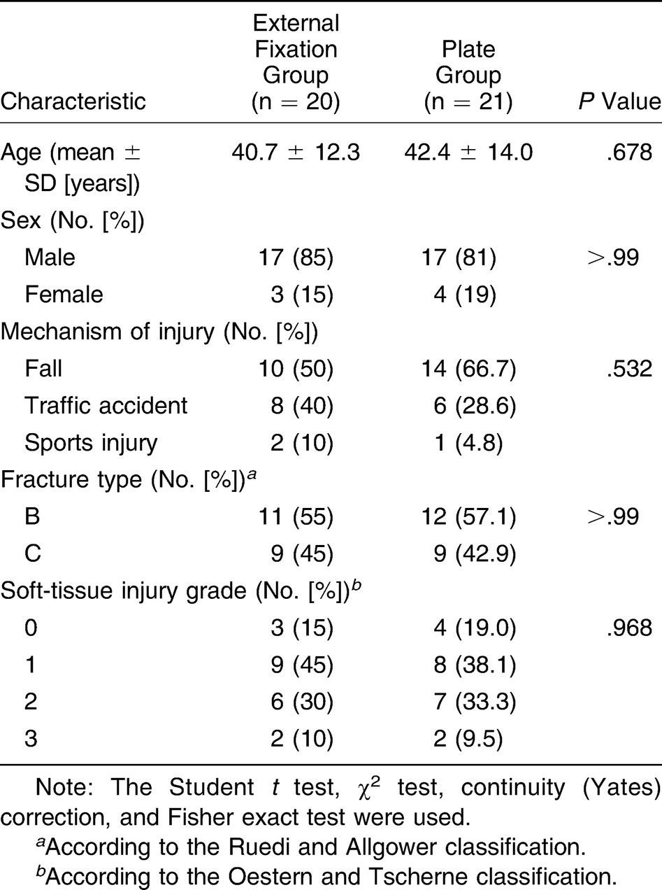
Skeletal traction via a calcaneal pin, elevation with a Thomas splint, and local anti-edema measures were applied to all of the patients on the day of hospital admission. Anteroposterior, lateral, and ankle mortise radiographic views were obtained for all of the patients. Computed tomography was necessary when radiographic interpretation of the joint was difficult.
Time to surgery differed owing to soft-tissue swelling, varying from 3 to 11 days. All of the operations were performed by the same surgery staff. On a radiolucent table, closed reduction with traction and manipulations were performed with the help of percutaneous clamps under fluoroscopy.
In the MIPO group, a 3.5-mm medial locking plate (TST, Istanbul, Turkey) was inserted subcutaneously through a 3-cm incision over the medial malleolus and was locked with at least four screws across fracture sites to have a bridging effect on the supra-articular metaphyseal junction.
In the EF group, a proximal ring with two tensioned pins was placed at the tip of the fibular head. Then, 2 cm proximal to the fracture, a second ring was applied and connected with four rods.[1,10,11] Just proximal to the ankle joint, a third ring was inserted with olive wires and pins for reduction and alignment under traction and was connected with four rods to the EF system (Tasarimmed, Istanbul, Turkey). Joint distraction and maintenance were achieved with a calcaneal foot-ring insertion with pins and wires.
Fibular plating was necessary in 18 patients to restore fibular length, especially in distal third fractures. The talocrural angle was used for confirmation.
All of the patients were followed up until clinical and radiographic union was achieved. The first visit was at 3 weeks, and subsequent visits were conducted every 3 months for at least 1 year if not complicated. Union delay was defined as no clinical and radiographic healing after 6 months. After 24 weeks, patients who needed secondary bone grafting were considered to have union delay and were excluded from the calculation of mean union time. The anterior iliac crest was the site of autogenous cancellous bone graft harvest that was performed in residual metaphyseal defects and union delay.
Postoperatively, a soft ankle brace allowing joint motion was applied for 3 weeks in the MIPO group. Partial weightbearing with crutches was allowed after 6 weeks. Full weightbearing was encouraged after bone healing was achieved.
In the EF group, weightbearing with support was encouraged as early as possible, and the foot ring was removed at the third week postoperatively to allow joint motion. Daily dressing of pins was recommended to avoid pin-tract infection. Once radiographic consolidation occurred, the EF device was removed, and an ankle-foot orthosis was used for an additional 3 weeks.
Functional outcomes were assessed with the American Orthopaedic Foot and Ankle Society (AOFAS) ankle scoring system, with scores recorded annually for 3 years. This scoring system assesses the ankle, subtalar, and calcaneocuboid joints and focuses on three distinct categories: pain, function, and alignment. Post-traumatic arthritis was described as painful range of motion with radiographic evidence of a narrowed joint space.
Statistical Analysis
When assessing the results obtained in this study, a statistical analysis software program (IBM SPSS Statistics for Windows, Version 22; IBM Corp, Armonk, New York) was used. Kolmogorov-Smirnov test parameters were used to assess the suitability of the normal distribution of study data. Data were analyzed using descriptive statistical methods (mean, standard deviation, and frequency) and two groups of comparisons between Student t test parameters showing normal distribution for comparing quantitative data, and the comparison between the two groups of parameters in a normal-distribution Mann-Whitney U test was used. In the comparison of qualitative data, the χ2 test, Fisher exact χ2 test, and continuity (Yates) adjustments were used. The P < .05 level was evaluated.
Results
There were no statistically significant differences between groups regarding fracture types and soft-tissue injury patterns (Table 1). In the MIPO group, the mean ± SD patient age was 42.4 ± 14 years (range, 17–73 years). Of 21 patients, 17 were male and four were female. Twelve of the fractures were type B, and the remaining nine were type C according to the Ruedi and Allgower classification (Fig. 1). Mean ± SD follow-up was 39.3 ± 3.8 months (range, 36–51 months). Mean ± SD time to surgery was 6.5 ± 1.5 days (range, 5–11 days), depending on the soft-tissue conditions (Table 2). Mean ± SD union time was 19.4 ± 2.9 weeks (range, 12–26 weeks). All of the fractures except four united without the need for secondary bone grafting. These four patients were treated with cancellous bone autografting after 24 weeks due to residual metaphyseal bone defect, and union was achieved in all. Twelve patients had fibular plating to restore the talocrural angle. Full weightbearing without crutches was seen at a mean ± SD of 25.6 ± 1.85 weeks. Five patients had superficial wound infection over the medial malleolus that was responsive to oral antibiotic drug therapy. Four patients had hardware removal due to ankle pain after a minimum of 14 months. Eight patients had post-traumatic arthritis at the end of the second year (Fig. 2). Annual analysis of AOFAS ankle scores revealed mean ± SD scores of 86.4 ± 2.06, 79.5 ± 1.03, and 77.9 ± 0.80 at years 1, 2, and 3, respectively (Table 3).
Figure 1.
A 34-year-old patient with an intra-articular distal tibia fracture.
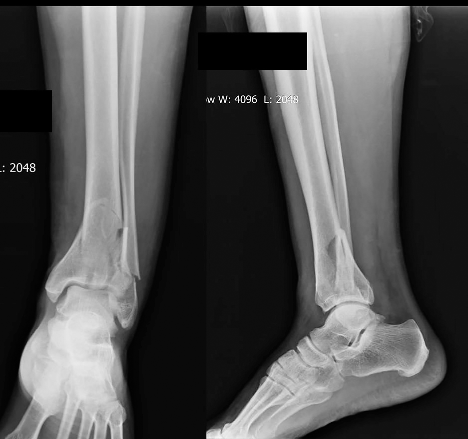
Table 2.
Time to Surgery, Union Time, and Need for Fibular Plating and Grafting
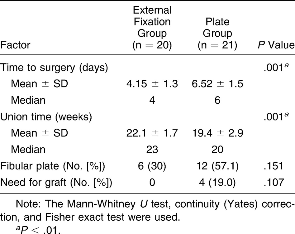
Figure 2.
A, Early postoperative anteroposterior view showing acceptable joint surface reconstruction. B, Anteroposterior view of the patient at 3-week follow-up. C and D, Periarticular sclerosis was seen on anteroposterior and lateral radiographs of the patient 6 months postoperatively.
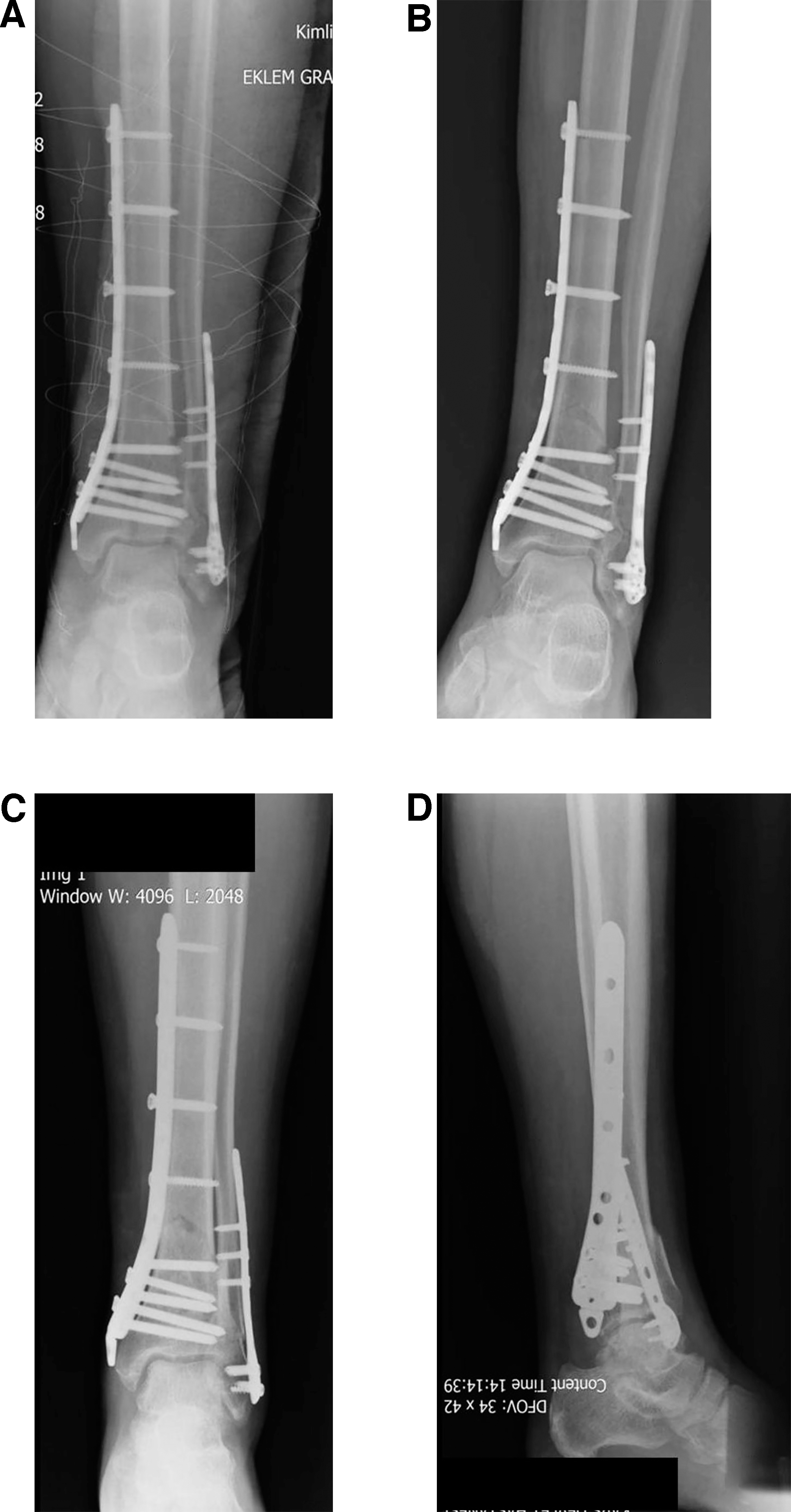
Table 3.
Follow-up Time, AOFAS Ankle Scores, and Postoperative Conditions
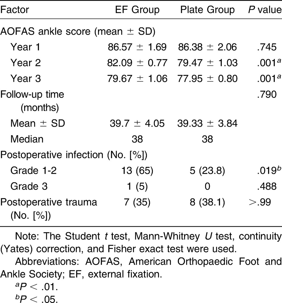
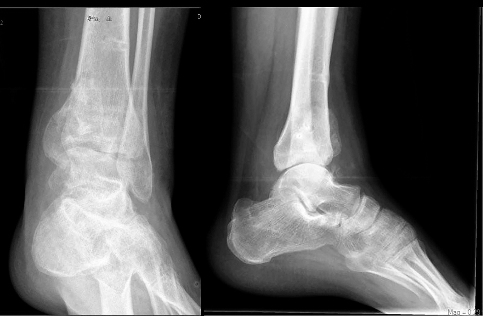
In the EF group, the mean ± SD patient age was 40.7 ± 12.3 years (range, 18–64 years). The male to female ratio was 17 to 3. Eleven patients had type B fractures and nine had type C according to the Ruedi and Allgower classification (Figs. 3 and 4). Mean ± SD follow-up time was 39.7 ± 4.05 months (range, 36–49 months). Mean ± SD time to surgery was 4.15 ± 1.3 days (range, 3–7 days) (Fig. 5 and Table 2). Fracture consolidation was achieved in all of the patients without secondary bone grafting at a mean ± SD of 22.1 ± 1.7 weeks (range, 18–24 weeks). Fibular plating was necessary in six patients. Full weightbearing without crutches was achieved at a mean of 19.3 weeks. Mean ± SD fixator removal time was 26.2 ± 3.6 weeks (range, 20–33 weeks). Thirteen patients had superficial pin-tract infections treated with oral antibiotics. At 3-month follow-up, one patient had a deep pin-tract infection on the distal ring of the EF system that was treated with local debridement, parenteral antibiotic drugs, and revision pinning. Post-traumatic arthritis was obvious in seven patients by the end of the second year (Fig. 6). Mean ± SD AOFAS ankle scores were 86.6 ± 1.69, 82.1 ± 0.77, and 79.7 ± 1.06 at years 1, 2, and 3, respectively (Table 3).
Figure 3.
A 30-year-old patient with an intra-articular distal tibia fracture.
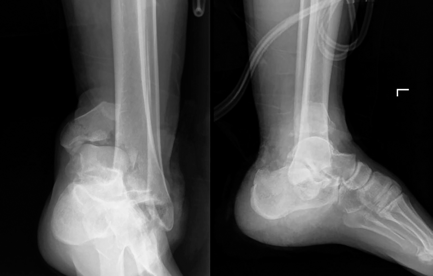
Figure 4.
Computed tomographic scan showing the comminuted fracture of the tibial plafond.
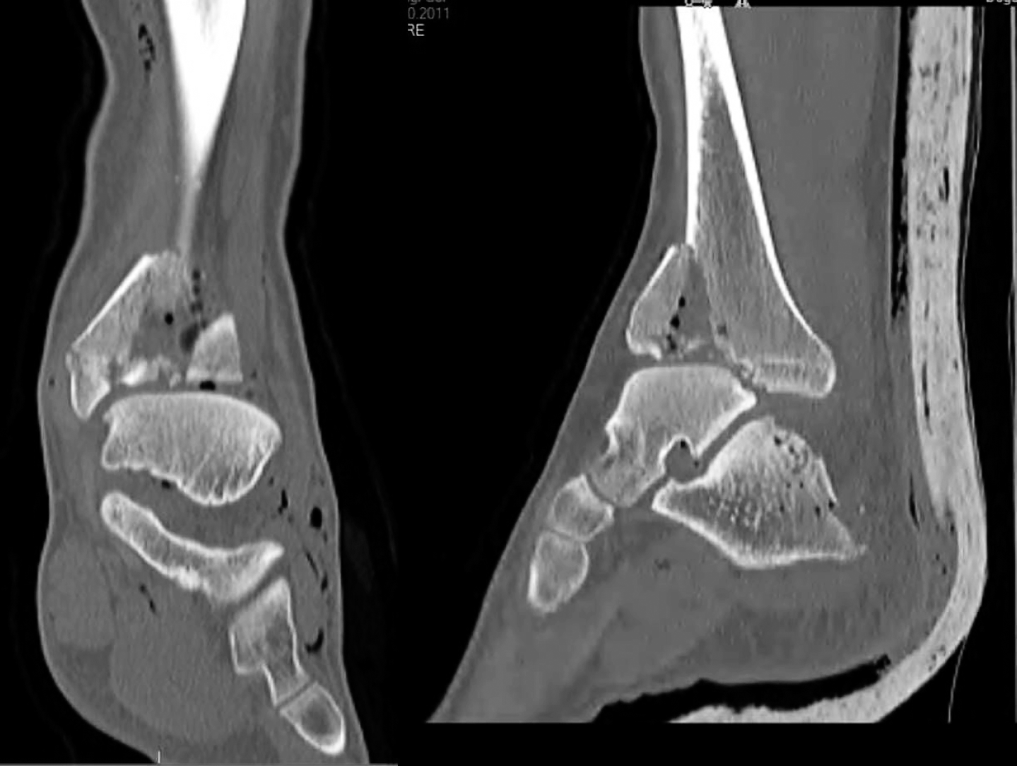
Figure 5.
With the application of circular external fixation and ligamentotaxis, acceptable joint surface reconstruction was obtained.
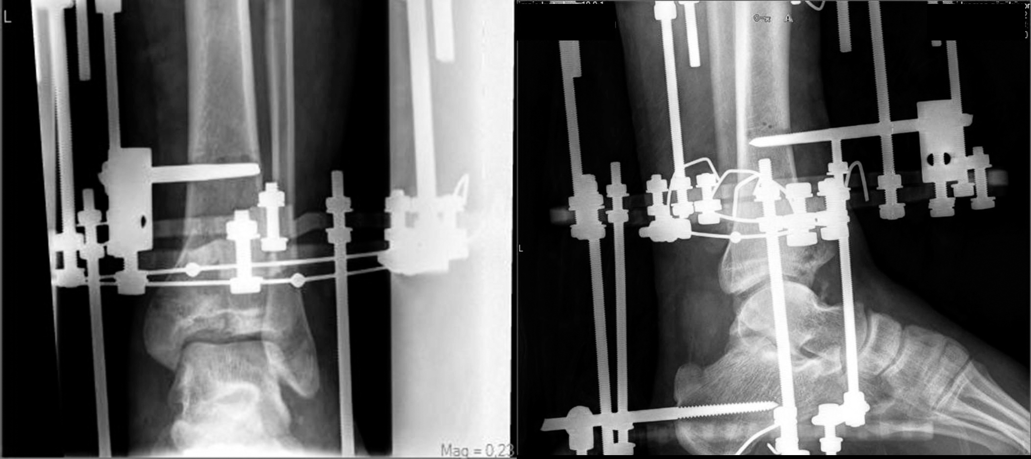
Figure 6.
Anteroposterior and lateral radiographs showing arthrosis and joint space narrowing 6 months postoperatively.
Discussion
Management of distal tibial periarticular fractures is still challenging.[12] Comminution of the fracture with metaphyseal fragments, damage to articular cartilage, and poor soft-tissue coverage deteriorate the injury.[13,14] Conventional surgical techniques may fail, thereby complicating the fracture by devitalization of fragments, increasing the rate of nonunion and infection with further harm to the soft-tissue envelope.[3,15]
For the past two decades, minimally invasive surgery for complex distal tibial injuries has resulted in favorable outcomes with low complication rates.[13,16] Improvements in implant technology and a better understanding of fracture biomechanics have provided more biological solutions to these complex injuries. Percutaneous plating and EF are the preferred methods for the management of pilon fractures with high-energy trauma and poor soft-tissue coverage.
Using the principles of biological fixation, MIPO demonstrated higher union rates than conventional techniques.[4,6,17] Although it is technically demanding, with its indirect reduction methods and risk of neurovascular injury, MIPO allows stable fixation and early mobilization. Reduced tissue devitalization and preservation of fracture hematoma can promote early healing with decreased wound complications.
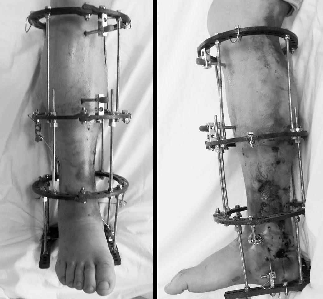
The circular EF system preserves the endosteal and periosteal vascular supply, controls even small metaphyseal or articular fragments by olive wires, and allows compression of the fracture site and deformity correction during the process (Fig. 7).[7,15] However, it is an intraoperative fluoroscopy-dependent technique with demanding experience.
Figure 7.
External fixation treatment is a better surgical option in patients with soft-tissue problems.
Because distal tibia fractures are mostly related to high-energy trauma, soft-tissue conditions play a major role in the choice of surgical treatment. Borelli et al[18] demonstrated that conventional open reduction and internal fixation disrupted the blood supply in the metaphyseal region of the tibia. To reduce soft-tissue complications, Krettek et al[19] introduced percutaneous plating, which is biologically less hazardous for distal tibia fractures. Recent studies found that MIPO had both clinical and theoretical advantages, including less surgical trauma, lower rates of infection and wound problems, and better fracture healing.[4-6]
Circular EF helps to control comminuted fragments with small wires and to compress the fracture site, providing a mechanically stable environment that allows early mobilization.[8,20] Furthermore, ankle joint distraction could be achieved via a foot ring, maintaining the capsuloligamentous traction that is required to promote proteoglycan synthesis and normal chondrocyte metabolism.[7]
In the present study, we reported on a retrospective cohort of patients with complex type B and C (according to the Ruedi and Allgower classification) pilon fractures treated by MIPO versus circular EF. We found low complication rates in both groups, but no statistically significant differences were noted between the two treatment modalities with respect to union, infection rates, and functional outcomes.
Leonard et al[17] reported on 26 high-velocity intra-articular distal tibia fractures treated with percutaneous plating before 36 hours; outcomes were excellent in 83% at mean follow-up of 28 months. They emphasized that early intervention limited further soft-tissue damage, decreased bacterial growth, and improved blood supply and venous return in the limb.[17]
In a prospective randomized study, Wyrsch et al[21] reported that EF produced similar outcomes with lower complication rates compared with traditional techniques. Watson et al[22] retrospectively evaluated 107 pilon tibia fractures and indicated that the worse the initial soft-tissue injury was, the poorer the overall function tended to be, regardless of the initial fracture pattern. It is important not to cause further soft-tissue damage in the management of these complex injuries.
Vidyadhara and Rao[1] treated 21 complex pilon tibia fractures with Ilizarov EF and reported that all of the fractures united by approximately 21 to 25 weeks without the need for secondary grafting, and AOFAS ankle scores were good to excellent in 17 of the patients. Only four patients had post-traumatic arthritis, and seven patients had superficial pin-tract infection that was responsive to oral antibiotic drug therapy.
Infection is the most important early postoperative complication, and it may require further intervention to the extent of arthrodesis or amputation.[22] Traditional surgical techniques for pilon fractures are prone to infection because extensive soft-tissue damage favors bacterial growth and potential wound problems. Theoretically, both percutaneous plating and EF have lower infection rates.[3,8,13] Although we had a substantial number of superficial infections in both groups, all of the cases responded to oral antibiotic drug therapy. One deep pin-tract infection required removal of the wire, local debridement, parenteral antibiotic drug therapy, and revision pinning. In open primary plating, infection was reported in up to 50% of the patients, whereas Ahmad et al[4] reported an infection rate of 27% in 18 patients with pilon fractures treated by MIPO. Blauth et al[13] found no significant differences in infection among primary plating, staged minimally invasive plating, and EF in 51 patients with pilon fractures. In a retrospective study of 14 patients with pilon fractures treated by percutaneous reduction and Ilizarov EF, Bacon et al[2] reported osteomyelitis in more than 20%, whereas McDonald et al[20] did not report any deep infection with Ilizarov EF. Although pin-tract infection remains the most common problem with any type of EF,[8] we did not encounter osteomyelitis in the present series. Post-traumatic arthritis is one of the major long-term disabilities after high-energy pilon fractures, and the degree of arthritis is strongly correlated with the clinical outcome.[7]
The initial cartilage damage and definitive fracture reduction are hypothesized to be of major influence in the evolution of fractures. Murray et al[23] studied cartilage damage in plafond fractures using molecular methods and concluded that increased chondrocyte death with disturbances in the cartilaginous extracellular matrix and metabolism lead to a degeneration cascade after severe joint injuries. Limited painful range of motion with a radiographically narrowed ankle joint yielded a diagnosis of post-traumatic arthritis in six patients in the MIPO group, whereas only two patients had arthritis in the EF group by the end of the second year, correlating with annual AOFAS ankle scores. Joint distraction with EF in the management of distal tibia fractures may play a protective role against post-traumatic arthritis.
The limitations of this study are the low number of patients, unequal distribution of the groups, lack of a control group, and retrospective design without protocol. Thus, type 2 errors may be expected in these results. Functional status was assessed by the AOFAS ankle scoring system, which included maximum walking distance, walking surfaces, pain, and activity limitations. Return to work status, which is an important outcome in work and health research, was not addressed in this study. It may have a predictive value in the assessment of full recovery. That might also be regarded as a limitation.
Conclusions
Management of complex intra-articular distal tibia fractures remains a challenge. Based on the literature and the present results, the circular EF and percutaneous plating with closed reduction techniques seem to be acceptable surgical options. Both methods have favorable union rates with fewer complications.
Financial Disclosure
None reported.
Conflicts of Interest
None reported.
References
- Vidyadhara S, Rao SK: Ilizarov treatment of complex tibial pilon fractures. Int Orthop30: 113, 2006.
- Bacon S, Smith WR, Morgan SJ, et al: A retrospective analysis of comminuted intra-articular fractures of the tibial plafond: open reduction and internal fixation versus external Ilizarov fixation. Injury39: 196, 2008.
- Stufkens SAS, van den Bekerom MPJ, Kerkhoffs GMMJ, et al: Long-term outcome after 1822 operatively treated ankle fractures: a systematic review of the literature. Injury42: 119, 2011.
- Ahmad MA, Sivaraman A, Zia A, et al: Percutaneous locking plates for fractures of the distal tibia: our experience and a review of the literature. J Trauma72: 81, 2012.
- Francois J, Vandeputte G, Verheyden F, et al: Percutaneous plate fixation of fractures of the distal tibia. Acta Orthop Belg70: 148, 2004.
- Shanmugam C, Rahmatalla A, Maffulli N: Percutaneous fixation of distal tibial fractures using locking plates. Tech Orthop22: 162, 2007.
- Bozkurt M, Ocguder DA, Ugurlu M, et al: Tibial pilon fracture repair using Ilizarov external fixation, capsuloligamentotaxis, and early rehabilitation of the ankle. J Foot Ankle Surg47: 302, 2008.
- Leung F, Kwok HY, Pun TS, et al: Limited open reduction and Ilizarov external fixation in the treatment of distal tibial fractures. Injury35: 278, 2004.
- Oestern HJ, Tscherne H: “Pathophysiology and Classification of Soft Tissue Injuries Associated with Fractures,” inFractures with Soft Tissue Injuries, p 1, edited byHTscherne, LGotzen, Springer-Verlag, Berlin,1984.
- Israelite CL, Blyakher AA: Tibial pilon fractures: the Ilizarov method. Oper Tech Orthop6: 208, 1996.
- Danoff JR, Saifi C, Goodspeed DC, et al: Outcome of 28 open pilon fractures with injury severity-based fixation. Eur J Orthop Surg Traumatol25: 569, 2015.
- Calori GM, Tagliabue L, Mazza E, et al: Tibial pilon fractures: which method of treatment?Injury41: 1183, 2010.
- Blauth M, Bastian L, Krettek C, et al: Surgical options for the treatment of severe tibial pilon fractures: a study of three techniques. J Orthop Trauma15: 153, 2001.
- Tarkin IS, Clare MP, Marcantonio A, et al: An update on the management of high-energy pilon fractures. Injury39: 142, 2008.
- Vasiliadis ES, Grivas TB, Psarakis SA, et al: Advantages of the Ilizarov external fixation in the management of intra-articular fractures of the distal tibia. J Orthop Surg Res4: 35, 2009.
- Probe RA: Minimaly invasive fixation of tibial pilon fractures. Oper Tech Orthop11: 205, 2001.
- Leonard M, Magill P, Khayyat G: Minimally-invasive treatment of high velocity intra-articular fractures of the distal tibia. Int Orthop33: 1149, 2009.
- Borrelli J, Prickett W, Song E, et al: Extra osseous blood supply of the distal tibia and the effects of different plating techniques: human cadaveric study. J Orthop Trauma16: 691, 2002.
- Krettek C, Schandelmaier P, Tscherne H: Distale femurfraktturen: transartikulare rekonstruktion, perkutane plattenosteosynthese und retrograde nagelung. Unfallchirurg99: 2, 1996.
- McDonald MG, Burgess RC, Bolano LE, et al: Ilizarov treatment of pilon fractures. Clin Orthop Relat Res325: 232, 1996.
- Wyrsch B, McFerran MA, McAndrew M: Operative treatment of fractures of the tibial plafond: a randomized, prospective study. J Bone Joint Surg Am78: 1646, 1996.
- Watson JT, Moed BR, Karges DE, et al: Pilon fractures: treatment protocol based on severity of soft tissue injury. Clin Orthop375: 78, 2000.
- Murray MM, Zurakowski D, Vrahas MS: The death of articular chondrocytes after intra articular fracture in humans. J Trauma56: 128, 2004.
© 2017 American Podiatric Medical Association