


Abstract
Background: Wedged insoles have been used to treat knee pathologies and to prevent injuries. Although they have received much attention for the study of knee injury, the effects of wedges on ankle joint biomechanics are not well understood. This study sought to evaluate the immediate effects of lateral and medial wedges on knee and ankle internal joint loading and center of pressure (CoP) in men during walking. Methods: Twenty-one healthy men walked at 1.4 m/sec in five footwear conditions: neutral, 6° (LW6) and 9° (LW9) lateral wedges, and 6° (MW6) and 9° (MW9) medial wedges. Peak internal knee abduction moments and angular impulses, internal ankle inversion moments and angular impulses, and mediolateral CoP were analyzed. Analysis of variance with post hoc analysis and Pearson correlations were performed to detect differences between conditions. Results: No differences in internal knee joint loading were found between neutral and any of the wedge conditions. However, as the wedge angle increased from medial to lateral, the internal ankle inversion moment (LW6: P = .020; LW9: P < .001; MW6: P = .046; MW9: P < .001) and angular impulse (LW9: P = .012) increased, and the CoP shifted laterally (LW9: P < .001) and medially (MW9: P < .001) compared with the neutral condition. Conclusions: Neither lateral nor medial wedges were effective in altering internal knee joint loading during walking. However, the greater internal ankle inversion moment and angular impulse observed with lateral wedges could lead to a higher risk of ankle injury. Thus, caution should be taken when lateral wedges need to be prescribed.
Wedged insoles can be easily integrated into an individual's daily activities and have been applied to promote health benefits, provide comfort, and prevent injuries [1], as well as for the management of knee pathologies such as knee osteoarthritis [2-5] and patellofemoral pain syndrome[6-8]. Wedging is the most common form of conservative treatment for a lower-limb injury because it is simple, inexpensive, and noninvasive [5,9-11]. However, its effects from a biomechanical perspective are still not fully understood. Previous studies have applied an average wedge angle of 5° to 15° [2-5,12,13]; however, the wedging degree necessary to induce relevant biomechanical changes in gait is still unclear. For example, it is known that small wedge angles do not change the internal knee abduction (valgus) moments or the position of center of pressure (CoP) during walking [10,14]. On the other hand, higher wedge angles have been suggested to cause discomfort [11,13]. Thus, it is paramount to better understand the dose-effect relationship between the wedge angle and biomechanical variables such as joint moments to optimize care.
Moments represent the tendency of a force to rotate an object when that force is applied at a given distance away from an axis of rotation. In the context of human joints of the lower extremity, these forces can be of external origin (eg, ground reaction forces [12] or other applied forces) or internal [15] (muscles, ligaments, soft tissues, etc). Thus, joint moments are often classified as either external or internal based on the most appropriate interpretation. Mathematically, they are often defined as equal and opposite of each other [16], and, thus, many researchers use the terms interchangeably. In this article, based on typical biomechanical convention, all moments referred to are in reference to internal moments unless specifically stated otherwise.
One way to decrease frontal plane knee joint loading is to shift the CoP laterally, decreasing the lever arm between the ground reaction force vector and the knee joint center in the frontal plane (Fig. 1) [12,17]. The application of a lateral wedge has been shown to be effective in shifting the CoP laterally, thus reducing the internal knee abduction moment in healthy individuals and in those with knee osteoarthritis [2,5,17]. However, a negative biomechanical response in some people with knee osteoarthritis [4] and nonresponse in healthy people [18] to lateral wedges have also been detected.
Figure 1.
Proposed mechanism of action: medial, neutral, and lateral wedged conditions.
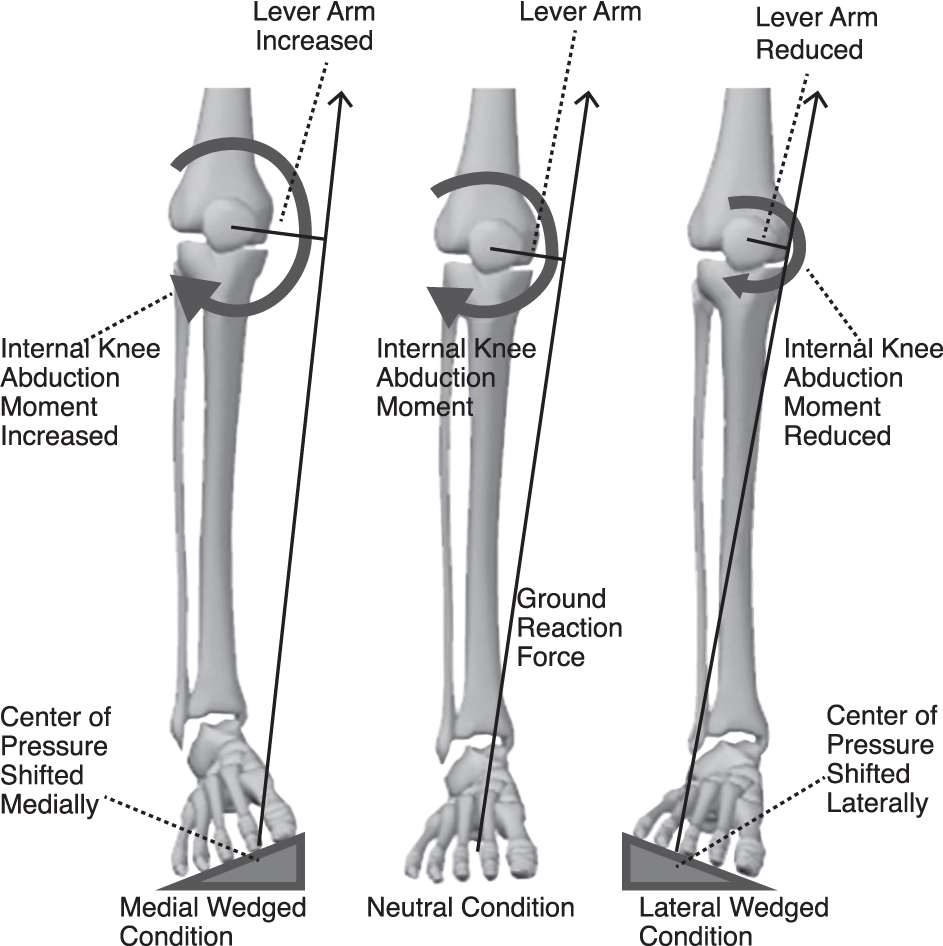
Contrary to lateral wedges, the use of medial wedges shifts the CoP medially, thus increasing the internal knee abduction moment (Fig. 1) [19]. In fact, higher external knee adduction (varus) moments were found when medial wedges were tested in healthy individuals [18,20] Boldt et al [21]. also reported that internal knee abduction moments increased by 3.3% when medial wedges were used in patients with patellofemoral pain syndrome and in healthy individuals. Because increases to the internal knee abduction moment have been related to various knee pathologies, the effects of medial wedges should be carefully investigated.
Although the use of wedges has been associated with some benefits in terms of knee joint loading, the effects on the ankle joint have received very little attention. A study by Bennell et al [22] reported that lateral wedged insoles may cause pain in the foot as well as discomfort. In addition, lateral wedges have been related to an increased external subtalar joint valgus moment while decreasing external knee adduction moments [2,5,10]. As lateral wedges decrease knee joint loading, they may increase ankle joint loading, which can predispose this joint to an injury. Because higher internal ankle inversion moments may be related to a higher risk of injury [23], the effects of wedges on the ankle joint should also be examined.
Considering the lack of data in the literature, the purpose of this study was to evaluate the immediate effects of lateral and medial wedges on knee and ankle internal loading in healthy men during walking. It was hypothesized that lateral wedged insoles decrease internal knee frontal plane moments and angular impulses but increase internal ankle inversion moments and impulses. It was also hypothesized that medial wedges reverse this effect. Finally, it was hypothesized that lateral wedges shift the CoP laterally and medial wedges shift the CoP medially.
Methods
Participants
Twenty-one healthy men were recruited for this study (mean ± SD: age, 25 ± 3.4 years; height, 178.1 ± 6.7 cm; and mass, 73.0 ± 9.1 kg). All of the participants were free of lower-extremity injury within the past 6 months and had normal knee joint alignment, as determined by an experienced physiotherapist. Testing the wedges in healthy people minimized the effects of pain as a confounder variable, which could alter the movement pattern and consequently minimize the effect of the wedge. Individuals who presented with previous surgery that could alter gait patterns or a muscular or neurologic condition that could influence lower-limb function were excluded from this study. Before the test, participants signed a written consent form approved by the Conjoint Health Research Ethics Board at the University of Calgary (Calgary, Alberta, Canada).
Wedged Insoles
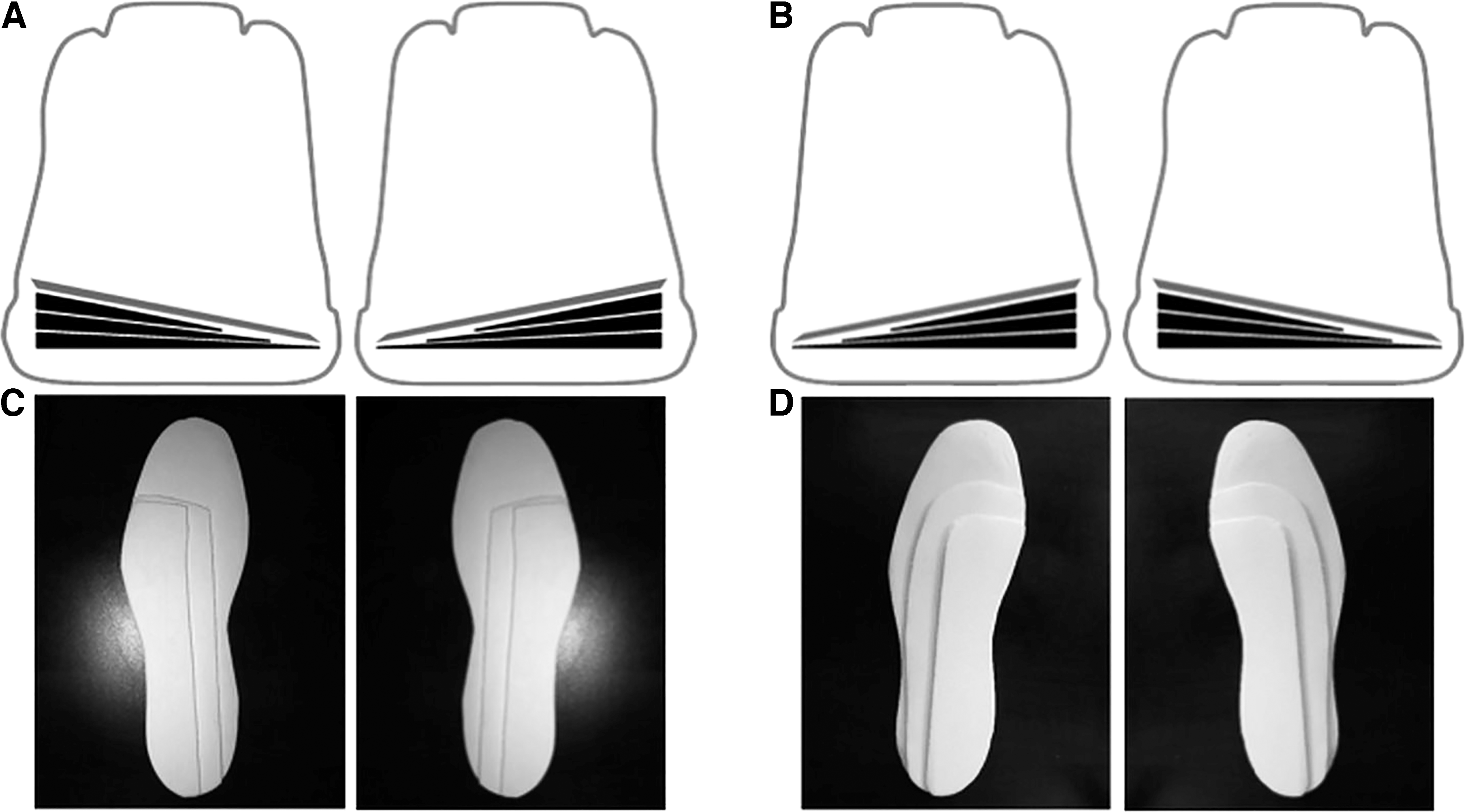
In this study, five different footwear conditions were evaluated. A standardized neutral shoe (Aegis 2.0; Adidas International, Herzogenaurach, Germany) was chosen as the neutral control shoe that was compared against four different wedged conditions: 1) a 6° lateral wedge (LW6), 2) a 9° lateral wedge (LW9), 3) a 6° medial wedge (MW6), and 4) a 9° medial wedge (MW9). This range of wedge angles was chosen because wedges of smaller degrees have been shown to have no influence on knee loading [10,14]. The wedges were constructed by cutting insert-shaped pieces of approximately 3° ethylene vinyl acetate (SOLFLEX Firm [60]; Vittoria Phoenix Inc, Vittoria, Ontario, Canada) and then sanding them either medially or laterally. The wedges were created by placing additional layers on top of one another (Fig. 2). Given that full-length wedged insoles have been demonstrated to be more efficient in changing external knee adduction moments than heel wedges [3], the wedges used in this study ran the entire length of the foot. The design of the insoles has been shown in a previous study (Fig. 2) [19]. Wedge conditions were randomly assigned during testing and were inserted bilaterally under the sock liner of a neutral control shoe.
Figure 2.
A, Posterior view of 9° lateral wedge (LW9) insoles inserted into neutral control shoes. B, Posterior view of 9° medial wedge (MW9) insoles inserted into neutral control shoes. C, Example of LW9 insoles. D, Example of MW9 insoles.
Gait Analysis
Three retroreflective markers were secured to the right shank and right foot (shoe) of each participant. In addition, anatomical markers were attached to the femoral epicondyles and the lateral and medial malleoli to determine the joint centers during a standing anatomical calibration trial. Eight high-speed cameras (Motion Analysis Corp, Santa Rosa, California) with a sampling frequency of 240 Hz were used to record the three-dimensional positions of each marker. Simultaneously, a force platform (Kistler Instrument AG, Winterthur, Switzerland) embedded in the floor collected ground reaction force data at a frequency of 2400 Hz. The participants had to perform at least five valid trials in each of the five footwear conditions. A valid trial was considered one in which the individual landed with the right foot near the middle of the force platform and the walking speed was within ±5% of the target speed of 1.4 m/sec, which was monitored using two photocells separated by 1.9 m. Participants were allowed as many practice trials as necessary to familiarize themselves with the procedures and the test conditions.
Kinematic and kinetic data were analyzed using Kintrak software (Motion Analysis Corp) and were filtered using fourth-order low-pass Butterworth filters with cutoff frequencies of 8 and 50 Hz, respectively. To calculate knee and ankle internal joint moments, a standard inverse dynamics approach was used [15]. Knee abduction angular impulses were calculated as the integral of internal knee abduction moments with respect to time [24,25]. Furthermore, ankle inversion angular impulses were also calculated based on the integral of the frontal plane ankle moment curve.
The first peak internal knee abduction moment was defined as the maximum value during the first half of the stance phase. The internal ankle inversion moment and mediolateral CoP position were obtained at the time the first peak internal knee abduction moment occurred. By transforming the CoP position in the force plate coordinate system to the laboratory coordinate system and then subtracting this value from the ankle joint center position in the laboratory coordinate system, the mediolateral CoP position relative to the ankle joint center was determined. All time-series curves were normalized to percentage of stance phase (0%–100%) with increments of 1%.
Statistical Analysis
The discrete variables were extracted from each trial, and the average value for each shoe condition and participant was obtained. Means and standard deviations were calculated for each condition. Repeated-measures analysis of variance (ANOVA) was performed in R 2.15.1 (R Foundation, Vienna, Austria) to determine statistically significant differences across conditions, and post hoc analysis was performed using pairwise comparisons with Holm adjustment. The significance level for all statistical analyses was set at α = 0.05 and the effect size estimates were computed using partial eta squared (ηp2). A ηp2 of 0.1, 0.25, and 0.4 were considered small, medium, and large effect sizes, respectively to determine the proportion of total variability attributable to the independent variable [26]. In addition, Pearson correlations were applied to assess the relation between the CoP and changes in knee and ankle internal joint moments.
Results
The ANOVA revealed a main effect for wedges on the first peak internal knee abduction moments (F4,100 = 5.26; P < .001; ηp2 = 0.174) and knee abduction angular impulses (F4,100 = 3.981, P = .005; ηp2 = 0.137). Post hoc analysis showed no differences between the neutral and lateral or between the neutral and medial wedge conditions for internal knee abduction moments and impulses. However, a decrease in internal knee abduction moments was found with LW9 compared with MW6 (P = .018) and MW9 (P = .001) and also with LW6 relative to MW9 (P = .022) conditions (Fig. 3). Knee abduction angular impulses also significantly decreased with the LW9 compared with the MW9 condition (P = .011) (Table 1).
Figure 3.
Mean time-normalized series average of the knee abduction moment (A) and ankle inversion moment (B) curves across participants for each condition tested. LW6, 6° lateral wedge; LW9, 9° lateral wedge; MW6, 6° medial wedge; MW9, 9° medial wedge.
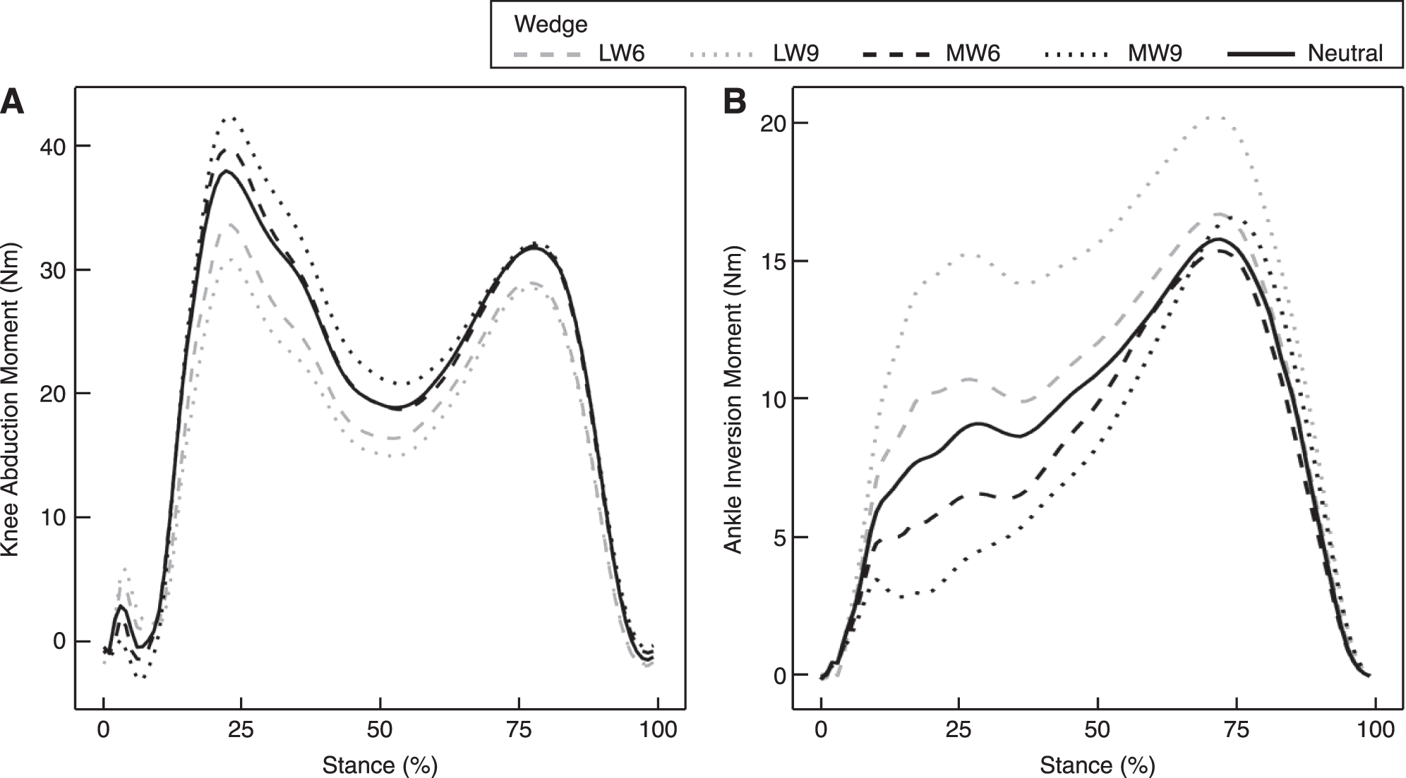
Table 1.
Knee Abduction Moment, Knee Abduction Angular Impulse, Ankle Inversion Moment, Ankle Inversion Angular Impulse, and Center of Pressure for the Conditions Tested
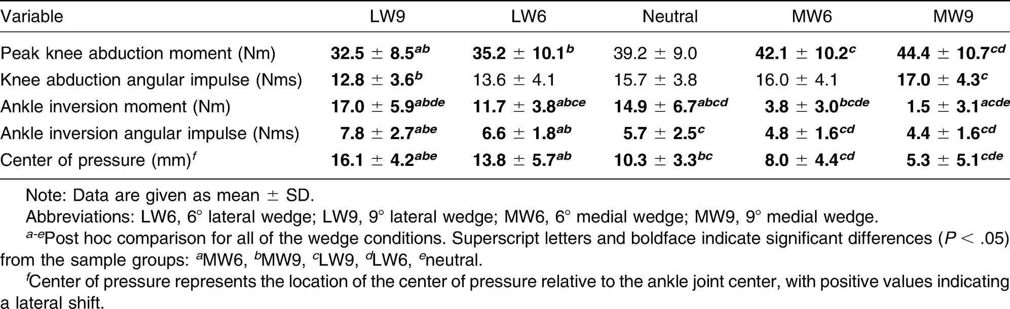
The internal ankle inversion moments showed a significant difference between wedge conditions (F4,100 = 31.2; P < .001; ηp2 = 0.555) (Fig. 3). In the post hoc test, the results demonstrated that the lateral wedges increased and the medial wedges decreased the internal ankle inversion moments relative to the neutral condition (LW6: P = .020; LW9: P < .001; MW6: P = .046; MW9: P < .001). In addition, significant differences between lateral and medial wedges were also found. The ANOVA for the ankle inversion impulses showed a significant main effect across wedges (F4,100 = 8.931; P < .001; ηp2 = 0.263). Post hoc analysis demonstrated that the ankle inversion impulse increased with the LW9 relative to the neutral condition (P = .012) (Table 1).
Center of pressure position revealed an ANOVA main effect across wedge conditions (F4,100 = 18.67; P < .001; ηp2 = 0.427), where larger wedge angles showed a greater shift in CoP compared with lower wedge angles. The post hoc test showed that lateral wedges shifted the CoP laterally and medial wedges shifted the CoP medially relative to the neutral condition (LW9: P < .001; MW9: P = .003) (Table 1).
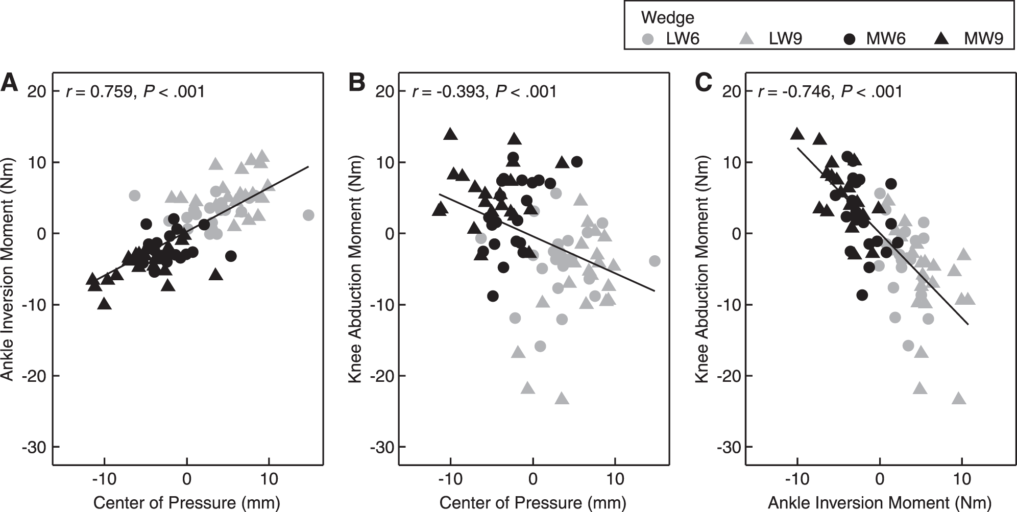
A significant higher and positive correlation was observed between the CoP and the internal ankle inversion moment (r = 0.759; P < .001) compared with the smaller and negative correlation for the CoP relative to the internal knee abduction moment (r = −0.393; P < .001). In addition, the internal ankle inversion moment was negatively correlated with the internal knee abduction moment (r = −0.746; P < .001) (Fig. 4).
Figure 4.
Scatterplots depicting the relationships between the following biomechanical variables: center of pressure and ankle inversion moment (A), center of pressure and knee abduction moment (B), and ankle inversion moment and knee abduction moment (C). All of the axes show changes relative to the neutral condition. LW6, 6° lateral wedge; LW9, 9° lateral wedge; MW6, 6° medial wedge; MW9, 9° medial wedge.
Discussion
This study evaluated the immediate effects of lateral and medial wedges on the knee and ankle joints in healthy men during walking. The results showed that although no changes were found in internal knee moments and impulses when lateral and medial wedged insoles were compared with the neutral condition, a significant difference was found for the internal ankle inversion moment and angular impulse and for the CoP compared with the neutral condition.
The use of lateral wedges decreased internal knee abduction moments, and medial wedges increased them. However, the differences were observed only when higher degrees of lateral wedges were compared with higher medial wedge conditions. In fact, a study by Schmalz et al [20] also found an effect of wedging on the external knee adduction moments only when a higher wedge angle (14° of medial wedge) was compared with no wedge in healthy people during walking. Although the higher wedges resulted in greater changes in internal knee joint loading, it has previously been reported that increased wedge angles may cause discomfort [13,27]. In addition, decreased external knee adduction moments with lateral wedges have been shown in studies involving healthy men and women [27-30] and in studies of only healthy women [5] during walking. However, as has been described by Park et al [31], internal knee joint loading patterns may be altered by hormonal effects. Therefore, a study conducted with only male individuals was chosen to reduce the possibility of observing biomechanical effects related to hormonal issues.
Referring to internal ankle inversion moments, differences were found across wedge conditions relative to the neutral condition. Lateral wedges increased internal ankle inversion moments and medial wedges decreased them during walking. A higher external subtalar joint valgus moment was also found when 5° to 15° lateral wedges were tested during walking [2,5,10]. Moreover, ankle inversion angular impulses showed significant changes between higher lateral wedges compared with the neutral condition. Lateral wedges increased the ankle angular impulse, which could lead to greater loading at the ankle joint, and medial wedges would, in turn, diminish it. The effects of wedges in relation to angular impulses have been previously reported, but only in the knee joint during walking [30] or running [19]. Because angular impulses represent the total load throughout the stance phase, different than the peak moments that represent the joint loading at only a particular time in the stance phase, angular impulses may provide additional information to better understand the effects of wedges in the ankle joint during walking.
Clinically, wedges have been indicated as a conservative approach to treat knee pathologies. Nevertheless, although lateral wedges decreased internal knee abduction moments, they also increased internal ankle joint loading, as observed in this study, which may enhance the risk of ankle injuries. Therefore, this should be considered when lateral wedges are prescribed. In this study, because only the immediate effects of wedges were tested, future research is necessary to better understand the long-term effects of wedged insoles and their associated effects on knee and ankle internal joint loadings and ankle injuries.
Systematic changes in the CoP were also observed in this study, where lateral wedges shifted the CoP laterally and medial wedges shifted the CoP more medially. Specifically, higher wedge angles were related to greater shift of the CoP compared with the neutral condition during walking. Kakihana et al [10] reported that when a 6° lateral wedge was applied in healthy adults during walking, the CoP shifted laterally, reducing the internal knee abduction moments. Despite the fact that the CoP significantly changed with lateral and medial wedges, it did not influence the internal knee joint loading. The higher positive correlation of the CoP with the ankle joint compared with the knee joint may suggest that the degree of wedging was not sufficient to affect the knee joint.
There are some limitations in this study. First, only healthy individuals with asymptomatic knee joints were tested. The effects of wedges in people who present with a knee pathology such as knee osteoarthritis or patellofemoral pain syndrome could be more exacerbated as a compensation to mitigate the pain level during walking. Second, a longer-term intervention could elucidate further the benefits of wedged footwear for musculoskeletal injury as opposed to the immediate effects observed in the present study. Nevertheless, a study of the immediate effects of wedges on gait parameters in healthy individuals is necessary to enhance our understanding of the underlying mechanism behind these interventions. Last, it is possible that an increased effect of either lateral or medial wedges on internal knee abduction moments compared with no wedge would be achieved if a larger sample size were tested because a trend was observed. Hence, this needs to be further addressed in future studies.
Conclusions
The results of this study show that neither lateral nor medial wedges were effective in altering internal knee frontal plane moments and impulses compared with a neutral condition in healthy men during walking. On the other hand, higher degrees of lateral wedges increased and of medial wedges decreased internal ankle joint loading during walking. Therefore, because the results suggest that lateral wedges increased the internal loading in the ankle joint, their prescription should be carefully analyzed. Future studies are needed to address the optimal prescription of wedges aimed at altering knee and ankle joint loads during walking.
Acknowledgment
Dr. R. Neil Humble for construction of the wedged insoles, and Adidas International for supplying the footwear used for the laboratory experiment.
Financial Disclosure
This study was financially supported by a scholarship from Fundação de Amparo à Pesquisa do Estado de São Paulo (FAPESP #14/13502-7) to Claudiane A. Fukuchi, PT.
Conflict of Interest
None reported.
References
- Skou ST, Hojgaard L, Simonsen OH: Customized foot insoles have a positive effect on pain, function, and quality of life in patients with medial knee osteoarthritis. JAPMA103: 50, 2013.
- Butler RJ, Barrios JA, Royer T, et al: Effect of laterally wedged foot orthoses on rearfoot and hip mechanics in patients with medial knee osteoarthritis. Prosthet Orthot Int33: 107, 2009.
- Hinman RS, Bowles KA, Payne C, et al: Effect of length on laterally-wedged insoles in knee osteoarthritis. Arthritis Rheum59: 144, 2008.
- Hinman RS, Payne C, Metcalf BR, et al: Lateral wedges in knee osteoarthritis: what are their immediate clinical and biomechanical effects and can these predict a three-month clinical outcome?Arthritis Rheum59: 408, 2008.
- Kakihana W, Akai M, Nakazawa K, et al: Effects of laterally wedged insoles on knee and subtalar joint moments. Arch Phys Med Rehabil86: 1465, 2005.
- Barton CJ, Menz HB, Crossley KM: Effects of prefabricated foot orthoses on pain and function in individuals with patellofemoral pain syndrome: a cohort study. Phys Ther Sport12: 70, 2011.
- Gross MT, Foxworth JL: The role of foot orthoses as an intervention for patellofemoral pain. J Orthop Sports Phys Ther33: 661, 2003.
- Collins N, Crossley K, Beller E, et al: Foot orthoses and physiotherapy in the treatment of patellofemoral pain syndrome: a randomised clinical trial. Br J Sports Med43: 169, 2009.
- Crenshaw SJ, Pollo FE, Calton EF: Effects of lateral-weged insoles on kinetics at the knee. Clin Orthop Relat Res375: 185, 2000.
- Kakihana W, Akai M, Yamasaki N, et al: Changes of joint moments in the gait of normal subjects wearing laterally wedged insoles. Am J Phys Med Rehabil83: 273, 2004.
- Erhart JC, Mündermann A, Mündermann L, et al: Predicting changes in knee adduction moment due to load-altering interventions from pressure distribution at the foot in healthy subjects. J Biomech41: 2989, 2008.
- Reeves ND, Bowling FL: Conservative biomechanical strategies for knee osteoarthritis. Nat Rev Rheumatol7: 113, 2011.
- Kerrigan DC, Lelas JL, Goggins J, et al: Effectiveness of a lateral-wedge insole on knee varus torque in patients with knee osteoarthritis. Arch Phys Med Rehabil83: 889, 2002.
- Fukuchi C, Worobets J, Wannop JW, et al: A small integrated lateral wedge does not alter knee joint moments during walking. Footwear Sci4: 207, 2012.
- Winter DA: “Kinetics: Forces and Moments of Force,”inBiomechanics and Motor Control of Human Movement, 3rd Ed, Wiley, Hoboken, NJ, 2005.
- Walter JP, D'Lima DD, Colwell CWJr, et al: Decreased knee adduction moment does not guarantee decreased medial contact force during gait. J Orthop Res28: 1348, 2010.
- Shelburne KB, Torry MR, Steadman JR, et al: Effects of foot orthoses and valgus bracing on the knee adduction moment and medial joint load during gait. Clin Biomech (Bristol, Avon)23: 814, 2008.
- Nester CJ, van der Linden ML, Bowker P: Effect of foot orthoses on the kinematics and kinetics of normal walking gait. Gait Posture17: 180, 2003.
- Lewinson RT, Fukuchi CA, Worobets JT, et al: The effects of wedged footwear on lower limb frontal plane biomechanics during running. Clin J Sport Med23: 208, 2013.
- Schmalz T, Blumentritt S, Drewitz H, et al: The influence of sole wedges on frontal plane knee kinetics, in isolation and in combination with representative rigid and semi-rigid ankle-foot-orthoses. Clin Biomech (Bristol, Avon)21: 631, 2006.
- Boldt AR, Willson JD, Barrios JA, et al: Effects of medially wedged foot orthoses on knee and hip joint running mechanics in females with and without patellofemoral pain syndrome. J Appl Biomech29: 68, 2012.
- Bennell KL, Bowles KA, Payne C, et al: Lateral wedge insoles for medial knee osteoarthritis: 12 month randomised controlled trial. BMJ342: d2912, 2011.
- Kristianslund E, Bahr R, Krosshaug T: Kinematics and kinetics of an accidental lateral ankle sprain. J Biomech44: 2576, 2011.
- Stefanyshyn DJ, Stergiou P, Lun VM, et al: Knee angular impulse as a predictor of patellofemoral pain in runners. Am J Sports Med34: 1844, 2006.
- Thorp LE, Sumner DR, Block JA, et al: Knee joint loading differs in individuals with mild compared with moderate medial knee osteoarthritis. Arthritis Rheum54: 3842, 2006.
- Portney LG, Watkins MP: “Power and Sample Size,”inFoundations of Clinical Research: Applications to Practice, 3rd Ed, Pearson, Prentice Hall, NJ, 2009.
- Tipnis RA, Anloague PA, Laubach LL, et al: The dose-response relationship between lateral foot wedging and the reduction of knee adduction moment. Clin Biomech29: 984, 2014.
- Kang JW, Park HS, Na CK, et al: Immediate coronal plane kinetic effects of novel lateral-offset sole shoes and lateral-wedge insole shoes in healthy individuals. Orthopedics36: 165, 2013.
- Fisher DS, Dyrby CO, Mündermann A, et al: In healthy subjects without knee osteoarthritis, the peak knee adduction moment influences the acute effect of shoe intervention designed to reduce medial compartment knee load. J Orthop Res25: 540, 2007.
- Jones RK, Zhang M, Laxton P, et al: The biomechanical effects of a new design of lateral wedge insole on the knee and ankle during walking. Hum Mov Sci32: 596, 2013.
- Park SK, Stefanyshyn DJ, Ramage B, et al: Relationship between knee joint laxity and knee joint mechanics during the menstrual cycle. Br J Sports Med43: 174, 2009.
© 2016 American Podiatric Medical Association