


Abstract
Onychomycosis is a fungal infection, and, as such, one of the goals of treatment should be eradication of the infective agent. Despite this, in contrast to dermatologists, many podiatric physicians do not include antifungals in their onychomycosis treatment plans. Before initiating treatment, confirmation of mycologic status via laboratory testing (eg, microscopy with potassium hydroxide preparation, histopathology with periodic acid–Schiff staining, fungal culture, and polymerase chain reaction) is important; however, more podiatric physicians rely solely on clinical signs than do dermatologists. These dissimilarities may be due, in part, to differences between specialties in training, reimbursement patterns, or practice orientation, and to explore these differences further, a joint podiatric medicine–dermatology roundtable was convened. In addition, treatment options have been limited owing to safety concerns with available oral antifungals and relatively low efficacy with previously available topical treatments. Recently approved topical treatments—efinaconzole and tavaborole—offer additional options for patients with mild-to-moderate disease. Debridement alone has no effect on mycologic status, and it is recommended that it be used in combination with an oral or topical antifungal. There is little to no clinical evidence to support the use of lasers or over-the-counter treatments for onychomycosis. After a patient has achieved cure (absence of clinical signs or absence of fungus with minimal clinical signs), lifestyle and hygiene measures, prophylactic/maintenance treatment, and proactive treatment for tinea pedis, including in family members, may help maintain this status.
More than merely a cosmetic concern, onychomycosis is an often painful [1,2] fungal infection most frequently of the toenails that has significant effects on quality of life [3,4] and has the potential to lead to serious complications. [5,6] Risk factors include male sex, [7] advanced age, [7,8] family or personal history of onychomycosis, [8,9] tinea pedis, [8] nail trauma, [9] and fomite exposure (eg, communal showers and pool decks). [8] Patients with diabetes, [9,10] peripheral vascular disease, [9,11] immunosuppression, [12,13] or psoriasis [8,14] are also at increased risk for developing the disease. Onychomycosis is currently estimated to affect 4.3% of the general population in North America/Europe, [15] but as the population ages and the prevalence of diabetes and peripheral vascular disease increases, the prevalence of onychomycosis is expected to rise.
In an average month, podiatric physicians see and treat more than five times as many patients with onychomycosis as dermatologists, [16] yet it seems that the bulk of the peer-reviewed medical literature is directed at dermatologists. Likewise, there are also striking differences between specialties in approach to diagnosis and treatment, with podiatric physicians relying on clinical diagnosis [16,17] and using debridement [17,18] much more often than dermatologists. With the goal of exploring these differences and how to leverage them to optimize patient care regardless of which specialty provides it, a roundtable meeting of experts from dermatology and podiatric medicine was convened. This article highlights the findings of that discussion as it pertains to practicing podiatric physicians' management of onychomycosis. A companion piece from the dermatology perspective has also been published and underscores the need for greater understanding and cooperation between the specialties. [19]
Diagnosis
Reports have shown that podiatric physicians perform confirmatory laboratory testing much less frequently than do dermatologists, [16,17] suggesting that podiatric physicians base their diagnosis solely on clinical signs more often than do dermatologists. The presence of clinical signs is highly predictive [20,21]; however, laboratory testing should be used to confirm clinical suspicion of onychomycosis and rule out other potential causes of nail dystrophy. [22-24] It is also important to inquire whether the patient has signs of fungal infection or lesions elsewhere on the body because these can be important clues when considering differential diagnoses. If lesions are not limited to the feet, a podiatric physician should refer the patient to a dermatologist for additional evaluation for fingernail onychomycosis or other skin and nail disorders (eg, chronic dermatitis, nail psoriasis, and lichen planus). Furthermore, if nail discoloration is limited to one nail, especially if black or brown, it is essential to rule out localized trauma or neoplasm in this situation. In addition, consultation or referral to dermatology or internal medicine may be helpful in diagnosing underlying or coexisting conditions that affect nail appearance (eg, psoriasis, peripheral vascular disease, and diabetes).
Several laboratory tests are available for confirming fungal infection based on nail samples, including direct microscopy using potassium hydroxide (KOH) preparation, histopathology using periodic acid–Schiff staining, fungal culture, dermatophyte test medium, and polymerase chain reaction. [25] The tests with the most rapid results (within a few days), KOH and periodic acid–Schiff, are specific but are not able to distinguish between dermatophyte, nondermatophyte mold, or yeast infections. With a little more time (1-2 weeks), dermatophyte test medium can detect dermatophytes, but not molds, yeasts, or specific fungal species.
Fungal culture and polymerase chain reaction can be used to confirm the presence of fungi and to determine fungus type, and they are the only tests that can identify fungal species. Fungal culture is sometimes thought of as the gold standard for onychomycosis diagnosis, but its utility is limited by a high rate of false-negative results, variable sensitivity, high cost, and extended time to availability of results (3-4 weeks). To increase sensitivity, fungal culture is often combined with KOH or periodic acid–Schiff. [26,27] Currently, molecular techniques such as polymerase chain reaction are fairly expensive, but as they become more cost-effective, these techniques may supplant fungal culture as the gold standard for onychomycosis confirmatory testing owing to their speed (results available in a few days), sensitivity, robustness in the presence of contaminants/less viable fungi, and ability to characterize less common causative agents and mixed infections.
Definition of Cure
In addition to diagnosis, laboratory tests are used in conjunction with clinical improvement to evaluate treatment efficacy. In clinical studies, various criteria are used to define antifungal efficacy, including mycologic, clinical, and complete cure. Mycologic cure most often refers to negative culture and negative KOH findings, and occasionally it refers to negative culture findings in addition to negative periodic acid–Schiff findings. Complete cure usually refers to completely clinically clear nail (0% nail involvement) plus mycologic cure; this end point, as it applies to a single target toenail (most often the great toenail), is preferred by the US Food and Drug Administration (FDA) to assess antifungal efficacy. However, a completely clear nail may be unattainable in some patients because onychomycosis frequently occurs in nails that are already abnormal because of chronic trauma or other diseases or owing to permanent nail changes caused by damage or scarring of the nail matrix or nail bed with severe, chronic, or long-term infection. [28] A more practical end point may be a definition where the nail is not 100% clear but there is significant improvement, so-called clinical cure or effective cure. This intermediate end point has been defined differently across clinical trials, but many have defined it as 10% or less nail plate involvement plus mycologic cure.
Scher et al [24] proposed that the criteria for cure should be 100% absence of clinical signs (mycologic confirmation not required) or negative mycologic results with minimal clinical signs (ie, <10% nail area affected by distal subungual hyperkeratosis or onycholysis, nail plate thickening that does not improve due to a comorbid condition, or both). Perhaps just as important is that they proposed a definition of noncure that includes positive mycologic findings or 1 or more of the following clinical signs (even in the presence of negative mycologic findings): 1) greater than 10% nail involvement, 2) white/yellow or orange/brown patches/streaks in the nail plate or nail bed, 3) lateral onycholysis with debris, or 4) hyperkeratosis on the lateral nail plate/nail fold.
Treatment
Onychomycosis rarely resolves without treatment, and, if left untreated, it can spread to other nails and other parts of the body, as well as to other people. [29-31] In addition, onychomycosis is associated with potentially serious complications, including secondary infections and ulcers, particularly in diabetic patients. [5,6]
Oral Antifungals
Currently available oral treatments widely used for onychomycosis include terbinafine, itraconazole, and fluconazole, although fluconazole is not FDA approved for onychomycosis treatment. [28] Despite the efficacy of oral antifungals, [32-34] their use is limited by the potential for serious safety issues, including hepatotoxicity (terbinafine and fluconazole, although concerns about acute liver toxicity with terbinafine may have been overemphasized35), cardiac issues (itraconazole), and drug-drug interactions (terbinafine, itraconazole, and fluconazole). [32,33,36] Furthermore, some patients have an exaggerated fear of systemic agents, which may preclude use. Whether real or exaggerated, these safety concerns are often greatest in individuals who are most susceptible to onychomycosis and its complications, including the elderly and patients with diabetes, immunosuppression, or peripheral vascular disease.
Topical Antifungals
Topical treatment minimizes the potential for systemic adverse effects and drug-drug interactions. [28] However, topical ciclopirox has relatively low efficacy and a lacquer formulation that is difficult for patients to use, [37] and topical amorolfine is not approved in the United States. [28] Efforts to develop formulations with improved efficacy have resulted in recent FDA approval of two new topical treatments: efinaconazole 10% solution and tavaborole 5% solution.
Tavaborole is a broad-spectrum oxaborole antifungal agent with low molecular weight, permitting good nail plate penetration. [38] In pivotal clinical trials of tavaborole in patients with mild-to-moderate distal subungual onychomycosis (20%–60% nail involvement, ≥3 mm clear nail, and positive mycologic findings), the mycologic cure rate was 31% to 36% versus 7% to 12% with vehicle; complete or almost complete cure (≤10% nail involvement along with mycologic cure), 15% to 18% versus 2% to 4% with vehicle; and complete cure, 7% to 9% versus 1% to 2%. [39] The only adverse events that occurred in 1% or more of tavaborole-treated patients and more frequently with tavaborole than with vehicle were application site exfoliation, erythema, and dermatitis and ingrown toenail. [39]
Efinaconazole, the first triazole antifungal agent to be specifically developed for topical treatment of onychomycosis of the toenail, has broad-spectrum activity against dermatophyte, nondermatophyte mold, and yeast species [40] and low systemic exposure. [41] In two pivotal phase 3 clinical trials of efinaconazole in patients with mild-to-moderate distal subungual onychomycosis (20%–50% nail involvement, no dermatophytoma or matrix/lunula involvement, ≥3 mm clear nail, <3 mm thick, and positive mycologic findings), the mycologic cure rate was 53% to 55% versus 17% with vehicle, the complete/almost complete cure rate (defined as ≤5% nail involvement along with mycologic cure) was 23% to 26% versus 7% to 8%, and the complete cure rate was 15% to 18% versus 3% to 6%. [42] Adverse events (>1%) observed for efinaconazole in these trials included ingrown toenail, application site dermatitis, application site vesicles, and application site pain. [42]
Over-the-Counter Treatments
There is little to no clinical evidence supporting the efficacy of over-the-counter agents and home remedies, such as foot soaks with bleach or hydrogen peroxide, salicylic acid, mentholated vapor rub, and tea tree oil. [43]
Laser Therapy
Several lasers are FDA cleared for the temporary cosmetic improvement of nail appearance in onychomycosis. [28] The mechanism of action for laser therapy is not clearly understood, but similar to topical treatment, laser treatment is locally administered and unlikely to produce systemic adverse effects. [28] Several small, mostly uncontrolled studies have demonstrated improved nail appearance by objective and subjective measures [44-52]; however, only one study has reported mycologic cure rates, and none have reported complete cure rates. Because data from clinical studies have not been sufficient to prove benefits beyond cosmetic effects, laser therapy is not recommended for eradication of fungal infections at this time. [53]
Debridement
Debridement is the traditional podiatric medical approach to treating onychomycosis. When used as monotherapy, it has been shown to markedly reduce toenail thickness and improve foot-related quality of life, but it does not impact fungal disease. [54] When used in combination with a systemic antifungal agent (terbinafine), debridement incrementally and significantly increased patient-reported treatment satisfaction and reduced symptom frequency [55] as well as numerically (but not statistically significantly) increased complete, mycologic, and clinical cure rates compared with terbinafine alone. [56]
Implementing a Treatment Plan
Treatment patterns differ dramatically between podiatric medicine and dermatology. Podiatric physicians use debridement much more often than do dermatologists, and dermatologists are much more likely to prescribe a systemic antifungal. [17,18] This marked preference for debridement among podiatric physicians supports the position that they are especially focused on relieving discomfort and improving cosmetic appearance. It is important, however, that mycologic cure be among the goals of any onychomycosis treatment plan because regardless of nail thickness and appearance, without a mycologic cure, patients will never be cleared of the infection.
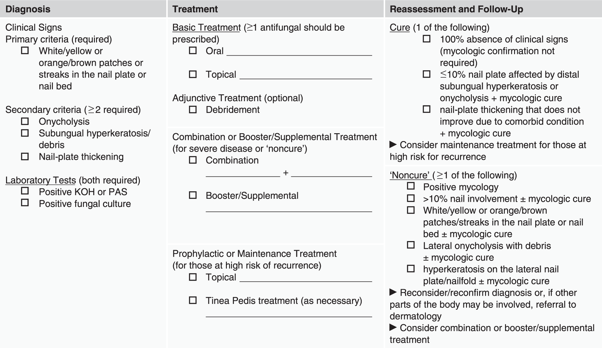
A comprehensive onychomycosis treatment plan should be designed with the intention to clear the infection as well as to improve nail appearance and reduce discomfort (Fig. 1). Several clinical signs and patient characteristics have been suggested as helping to guide these treatment decisions by helping to determine the choice of therapy, length of treatment, and duration of follow-up. [24,57] These poor prognosis factors have not been well studied; however, two small clinical studies have shown that advanced age, [58] male sex, [58] matrix/lunula involvement, [58] greater than 50% nail involvement, [54] significant lateral disease/onycholysis, [58] longitudinal streaking (dermatophytoma/spikes), [58] slow nail growth, [58] and infection caused by nondermatophyte molds or yeasts or mixed infection [54] reduced the likelihood of cure under the particular clinical settings of those studies.
Figure 1.
Onychomycosis treatment plan. KOH, potassium hydroxide; PAS, periodic acid–Schiff.
Basic Treatment
For patients with mild-to-moderate (<50%–60% nail involvement) onychomycosis, topical therapy should be sufficient to achieve clinical cure. In addition, pediatric patients, patients taking multiple medications, patients with contraindications for systemic therapy, and pregnant women are good candidates for topical treatment. [57]
For patients with greater than 60% nail involvement, poor prognosis factors, or peripheral vascular disease, treatment success may be more likely with systemic treatment given its greater efficacy. In addition, booster/supplemental treatment [59] or combination treatment [60-62] may be required to achieve cure in these patients. If the condition still does not improve, the diagnosis should be reconsidered or reconfirmed.
Adjunctive Treatment
Debridement should be considered as an adjunct treatment for any patient with concerns about nail appearance, pain, or other symptoms. Adjunctive debridement may be especially beneficial in elderly patients who have thick nails or who are unable to maintain proper nail care owing to low flexibility or poor eyesight.
Prophylactic/Maintenance Treatment
In patients who have a history of frequent relapse, the use of a topical antifungal as maintenance treatment may reduce the risk of recurrence. [63,64]
Follow-up: Minimizing the Risk of Relapse and Recurrence
As in any disease that requires long-term care, realistic expectations for treatment duration and outcome, as well as the benefits and risks of various treatment options, should be clearly discussed with patients. [28] Even after mycologic cure, mycologic relapse or recurrence occurs in approximately one-fifth of patients (22%) within 3 years, [65] increasing to 34% at 5 years [66] and 43% at 7 years. [59]
Lifestyle and hygiene measures can minimize the risk of recurrence due to new infection or re-infection, [57] and, as mentioned earlier, ensuring mycologic cure may reduce the risk of relapse of clinical signs. Similar precautionary hygiene measures should be taken to avoid the spread of infection among families and those living in close quarters owing to increased risk of transmission. [30,67] For patients who are at greater risk for recurrence due to comorbid conditions (eg, diabetes and psoriasis), referral to an internist or dermatologist (as appropriate) should be considered because the risk of recurrence may be reduced with proper management of the comorbid condition.
Conclusions
Regardless of specialty, the goals of any onychomycosis treatment plan should include eradication of the fungal infection because without mycologic cure, patients are at risk for relapse followed by continued discomfort and risk of complications. By itself, debridement has no effect on mycologic status, and the data for the effect of laser treatment are sparse and not definitive. Oral antifungals are effective for achieving mycologic cure and improving nail appearance (ie, percentage of involvement), but their use may be limited by safety concerns. Newer topical antifungals, including efinaconazole and tavaborole, offer safer alternatives without sacrificing efficacy. When making a diagnosis, it is important to confirm clinical suspicion of onychomycosis (ie, mycologic status) before initiating therapy to rule out other potential causes of nail dystrophy. It is also important for a podiatric physician to confirm that the disease is limited to the toenails before proceeding with treatment; if it is not, patients should be referred to dermatology for further evaluation. Beyond such referrals, greater information sharing and collaboration between podiatric medicine and dermatology will help optimize patient care for onychomycosis by highlighting best practices regardless of discipline. We hope that this paper, along with its companion piece, serve as the initial steps toward greater cooperation.
Acknowledgment
The content of this article is based on the proceedings of a roundtable meeting attended by each of the authors held May 19, 2014, in Chicago, IL, and sponsored by Valeant Pharmaceuticals North America LLC (Bridgewater, NJ). All roundtable and manuscript content was developed independent of the funding source. Jennifer Jaworski, MS, a full-time employee of Prescott Medical Communications Group (Chicago, IL), assisted with preparation of the manuscript at the direction of the authors with financial support from Valeant.
Financial Disclosure
None reported.
Conflicts of Interest
Dr. Vlahovic has received financial compensation for consultancy services from Valeant Pharmaceuticals North America LLC. Dr. Joseph has received financial compensation for consultancy services and as a speaker's bureau member from Valeant Pharmaceuticals North America LLC, Anacor Pharmaceuticals Inc, and PharmaDerm (a division of Fougera Pharmaceuticals Inc). He has also received financial compensation for consultancy services from Meiji Seika Pharma Co Ltd. Dr. Scher has received financial compensation for consultancy services from Valeant Pharmaceuticals North America LLC, Galderma Pharmaceuticals LP, Medimetriks Pharmaceuticals Inc, Meiji Seika Pharma Co Ltd, Moe Medical Devices LLC, and Topica Pharmaceuticals Inc. Dr. Tosti has received financial compensation for consultancy services from Valeant Pharmaceuticals North America LLC. Dr. Plasecia has received financial compensation for consultancy services from Valeant Pharmaceuticals North America LLC. Dr. Pariser has received financial compensation for consultancy services from Valeant Pharmaceuticals North America LLC and research grants from Anacor Pharmaceuticals. Dr. Markinson has received financial compensation for consultancy services from Valeant Pharmaceuticals North America LLC and Anacor Pharmaceuticals Inc. Dr. Markinson has also received consultant and speaking fees from PharmaDerm.
Dr. Joseph was not involved in the peer-review or acceptance of this paper.
References
- Drake LA, Scher RK, Smith EB, et al: Effect of onychomycosis on quality of life. J Am Acad Dermatol 38: 702, 1998.
- Elewski BE: The effect of toenail onychomycosis on patient quality of life. Int J Dermatol 36: 754, 1997.
- Lubeck DP: Measuring health-related quality of life in onychomycosis. J Am Acad Dermatol 38: S64, 1998.
- Lubeck DP, Patrick DL, McNulty P, et al: Quality of life of persons with onychomycosis. Qual Life Res 2: 341, 1993.
- Boyko EJ, Ahroni JH, Cohen V, et al: Prediction of diabetic foot ulcer occurrence using commonly available clinical information: the Seattle Diabetic Foot Study. Diabetes Care 29: 1202, 2006.
- Roujeau JC, Sigurgeirsson B, Korting HC, et al: Chronic dermatomycoses of the foot as risk factors for acute bacterial cellulitis of the leg: a case-control study. Dermatology 209: 301, 2004.
- Gupta AK, Jain HC, Lynde CW, et al: Prevalence and epidemiology of onychomycosis in patients visiting physicians' offices: a multicenter Canadian survey of 15,000 patients. J Am Acad Dermatol 43: 244, 2000.
- Sigurgeirsson B, Steingrimsson O: Risk factors associated with onychomycosis. J Eur Acad Dermatol Venereol 18: 48, 2004.
- Svejgaard EL, Nilsson J: Onychomycosis in Denmark: prevalence of fungal nail infection in general practice. Mycoses 47: 131, 2004.
- Gupta AK, Konnikov N, MacDonald P, et al: Prevalence and epidemiology of toenail onychomycosis in diabetic subjects: a multicentre survey. Br J Dermatol 139: 665, 1998.
- Gupta AK, Gupta MA, Summerbell RC, et al: The epidemiology of onychomycosis: possible role of smoking and peripheral arterial disease. J Eur Acad Dermatol Venereol 14: 466, 2000.
- Güleç AT, Demirbilek M, Seckin D, et al: Superficial fungal infections in 102 renal transplant recipients: a case-control study. J Am Acad Dermatol 49: 187, 2003.
- Gupta AK, Taborda P, Taborda V, et al: Epidemiology and prevalence of onychomycosis in HIV-positive individuals. Int J Dermatol 39: 746, 2000.
- Gupta AK, Lynde CW, Jain HC, et al: A higher prevalence of onychomycosis in psoriatics compared with non-psoriatics: a multicentre study. Br J Dermatol 136: 786, 1997.
- Sigurgeirsson B, Baran R: The prevalence of onychomycosis in the global population: a literature study. J Eur Acad Dermatol Venereol 28: 1480, 2014.
- Koshnick RL, Lilly KK, St Clair K, et al: Use of diagnostic tests by dermatologists, podiatrists and family practitioners in the United States: pilot data from a cross-sectional survey. Mycoses 50: 463, 2007.
- Schein JR, Gause D, Stier DM, et al: Onychomycosis: baseline results of an observational study. JAPMA 87: 512, 1997.
- Lubeck DP, Schein JR, Gause D, et al: Health-related quality of life in patients with toenail onychomycosis: data from a 9-month observational study. J Clin Outcomes Manag 6: 37, 1999.
- Scher RK, Tosti A, Joseph WS, et al: Onychomycosis diagnosis and management: perspectives from a joint dermatology-podiatry roundtable. J Drugs Dermatol 14,2015, in press.
- Garcia-Doval I, Cabo F, Monteagudo B, et al: Clinical diagnosis of toenail onychomycosis is possible in some patients: cross-sectional diagnostic study and development of a diagnostic rule. Br J Dermatol 163: 743, 2010.
- Fletcher CL, Hay RJ, Smeeton NC: Onychomycosis: the development of a clinical diagnostic aid for toenail disease: part I. Establishing discriminating historical and clinical features. Br J Dermatol 150: 701, 2004.
- Allevato MA: Diseases mimicking onychomycosis. Clin Dermatol 28: 164, 2010.
- Rich P, Elewski B, Scher RK, et al: Diagnosis, clinical implications, and complications of onychomycosis. Semin Cutan Med Surg 32(suppl 1): S5, 2013.
- Scher RK, Tavakkol A, Sigurgeirsson B, et al: Onychomycosis: diagnosis and definition of cure. J Am Acad Dermatol 56: 939, 2007.
- Tchernev G, Penev PK, Nenoff P, et al: Onychomycosis: modern diagnostic and treatment approaches. Wien Med Wochenschr 163: 1, 2013.
- Karimzadegan-Nia M, Mir-Amin-Mohammadi A, Bouzari N, et al: Comparison of direct smear, culture and histology for the diagnosis of onychomycosis. Australas J Dermatol 48: 18, 2007.
- Lawry MA, Haneke E, Strobeck K, et al: Methods for diagnosing onychomycosis: a comparative study and review of the literature. Arch Dermatol 136: 1112, 2000.
- Lipner S, Scher RK: Onychomycosis: current and future therapies. Cutis 93: 60, 2014.
- Daniel CRIII, Gupta AK, Daniel MP, et al: Two feet-one hand syndrome: a retrospective multicenter survey. Int J Dermatol 36: 658, 1997.
- Ghannoum MA, Mukherjee PK, Warshaw EM, et al: Molecular analysis of dermatophytes suggests spread of infection among household members. Cutis 91: 237, 2013.
- Szepietowski JC, Reich A, Garlowska E, et al: Factors influencing coexistence of toenail onychomycosis with tinea pedis and other dermatomycoses: a survey of 2761 patients. Arch Dermatol 142: 1279, 2006.
- Lamisil [package insert]. East Hanover, NJ: Novartis Pharmaceuticals Corporation; 2013.
- Sporonox [package insert]. Titusville, NJ: Janssen Pharmaceuticals Inc; 2012.
- Scher RK, Breneman D, Rich P, et al: Once-weekly fluconazole (150, 300, or 450 mg) in the treatment of distal subungual onychomycosis of the toenail. J Am Acad Dermatol 38: S77, 1998.
- Yan J, Wang X, Chen S: Systematic review of severe acute liver injury caused by terbinafine. Int J Clin Pharm 36: 679, 2014.
- Diflucan [package insert]. New York, NY: Pfizer; 2014.
- Gupta AK, Joseph WS: Ciclopirox 8% nail lacquer in the treatment of onychomycosis of the toenails in the United States. JAPMA 90: 495, 2000.
- Elewski BE, Tosti A: Tavaborole for the treatment of onychomycosis. Expert Opin Pharmacother 15: 1439, 2014.
- Kerydin [package insert]. Palo Alto, CA: Anacor Pharmaceuticals; 2014.
- JoSiu WJ, Tatsumi Y, Senda H, et al: Comparison of in vitro antifungal activities of efinaconazole and currently available antifungal agents against a variety of pathogenic fungi associated with onychomycosis. Antimicrob Agents Chemother 57: 1610, 2013.
- Jarratt M, Siu WJ, Yamakawa E, et al: Safety and pharmacokinetics of efinaconazole 10% solution in healthy volunteers and patients with severe onychomycosis. J Drugs Dermatol 12: 1010, 2013.
- Jublia [package insert]. Bridgewater, NJ: Valeant Pharmaceuticals North America LLC; 2014.
- Elewski B, Pariser D, Rich P, et al: Current and emerging options in the treatment of onychomycosis. Semin Cutan Med Surg 32(suppl 1): S9, 2013.
- Landsman AS, Robbins AH, Angelini PF, et al: Treatment of mild, moderate, and severe onychomycosis using 870- and 930-nm light exposure. JAPMA 100: 166, 2010.
- Landsman AS, Robbins AH: Treatment of mild, moderate, and severe onychomycosis using 870- and 930-nm light exposure: some follow-up observations at 270 days. JAPMA 102: 169, 2012.
- Zhang RN, Wang DK, Zhuo FL, et al: Long-pulse Nd:YAG 1064-nm laser treatment for onychomycosis. Chin Med J (Engl) 125: 3288, 2012.
- Hochman LG: Laser treatment of onychomycosis using a novel 0.65-millisecond pulsed Nd:YAG 1064-nm laser. J Cosmet Laser Ther 13: 2, 2011.
- Kozarev J, Vižintin Z: Novel laser therapy in treatment of onychomycosis. J Laser Health Acad 2010: 1, 2010.
- Kozarev J: ClearSteps—laser onychomycosis treatment: assessment of efficacy 12 months after treatment and beyond. J Laser Health Acad 2011: S07, 2011.
- Kimura U, Takeuchi K, Kinoshita A, et al: Treating onychomycoses of the toenail: clinical efficacy of the sub-millisecond 1,064 nm Nd:YAG laser using a 5 mm spot diameter. J Drugs Dermatol 11: 496, 2012.
- Waibel J, Wulkan AJ, Rudnick A: Prospective efficacy and safety evaluation of laser treatments with real-time temperature feedback for fungal onychomycosis. J Drugs Dermatol 12: 1237, 2013.
- Kalokasidis K, Onder M, Trakatelli MG, et al: The effect of Q-switched Nd:YAG 1064 nm/532 nm laser in the treatment of onychomycosis in vivo. Dermatol Res Pract 2013: 379725, 2013.
- Gupta AK, Simpson FC: Laser therapy for onychomycosis. J Cutan Med Surg 17: 301, 2013.
- Malay DS, Yi S, Borowsky P, et al: Efficacy of debridement alone versus debridement combined with topical antifungal nail lacquer for the treatment of pedal onychomycosis: a randomized, controlled trial. J Foot Ankle Surg 48: 294, 2009.
- Potter LP, Mathias SD, Raut M, et al: The impact of aggressive debridement used as an adjunct therapy with terbinafine on perceptions of patients undergoing treatment for toenail onychomycosis. J Dermatolog Treat 18: 46, 2007.
- Jennings MB, Pollak R, Harkless LB, et al: Treatment of toenail onychomycosis with oral terbinafine plus aggressive debridement: IRON-CLAD, a large, randomized, open-label, multicenter trial. JAPMA 96: 465, 2006.
- Elewski BE, Rich P, Tosti A, et al: Onchomycosis: an overview. J Drugs Dermatol 12: s96, 2013.
- Sigurgeirsson B: Prognostic factors for cure following treatment of onychomycosis. J Eur Acad Dermatol Venereol 24: 679, 2010.
- Gupta AK, Cooper EA, Paquet M: Recurrences of dermatophyte toenail onychomycosis during long-term follow-up after successful treatments with mono- and combined therapy of terbinafine and itraconazole. J Cutan Med Surg 17: 201, 2013.
- Avner S, Nir N, Henri T: Combination of oral terbinafine and topical ciclopirox compared to oral terbinafine for the treatment of onychomycosis. J Dermatolog Treat 16: 327, 2005.
- Baran R, Sigurgeirsson B, de Berker D, et al: A multicentre, randomized, controlled study of the efficacy, safety and cost-effectiveness of a combination therapy with amorolfine nail lacquer and oral terbinafine compared with oral terbinafine alone for the treatment of onychomycosis with matrix involvement. Br J Dermatol 157: 149, 2007.
- Lecha M: Amorolfine and itraconazole combination for severe toenail onychomycosis: results of an open randomized trial in Spain. Br J Dermatol 145(suppl 60): 21, 2001.
- Piraccini BM, Sisti A, Tosti A: Long-term follow-up of toenail onychomycosis caused by dermatophytes after successful treatment with systemic antifungal agents. J Am Acad Dermatol 62: 411, 2010.
- Sigurgeirsson B, Olafsson JH, Steinsson JT, et al: Efficacy of amorolfine nail lacquer for the prophylaxis of onychomycosis over 3 years. J Eur Acad Dermatol Venereol 24: 910, 2010.
- Tosti A, Piraccini BM, Stinchi C, et al: Relapses of onychomycosis after successful treatment with systemic antifungals: a three-year follow-up. Dermatology 197: 162, 1998.
- Sigurgeirsson B, Olafsson JH, Steinsson JB, et al: Long-term effectiveness of treatment with terbinafine vs itraconazole in onychomycosis: a 5-year blinded prospective follow-up study. Arch Dermatol 138: 353, 2002.
- Gazes MI, Zeichner J: Onychomycosis in close quarter living review of the literature. Mycoses 56: 610, 2013.
© 2016 American Podiatric Medical Association