


Abstract
The soleal sling may be a site of tibial nerve entrapment. Objective diagnosis of this syndrome is difficult with current nerve conduction study techniques, magnetic resonance imaging, and neurosensory testing. Diagnostic ultrasound is ideally suited to visualize the tibial nerve statically and dynamically as it enters the soleal sling, thus making an objective diagnosis of soleal sling impingement much easier.
Nerve entrapment may be a frequent and overlooked cause of foot and ankle pain. Plantar foot pain may be particularly distressing to the patient. Some plantar foot pain may be attributed to compression injuries to the nerves at fibro-osseous tunnel sites of the foot and ankle. Foot nerve entrapments most often described in the medical literature include Morton's entrapment and tarsal tunnel compression injuries to branches of the tibial nerve complex. However, tarsal tunnel release and Morton's excision can have a significant failure rate. [1,2,3] More recently, much has been published on the anatomy of the plantar tunnels of the foot as the more likely source of entrapment in tarsal tunnel syndrome and on decompression instead of excision of the proper digital nerve branch in Morton's entrapment. [4,5] Although very successful, even thorough release of these areas still has a failure rate.
It has been proposed that some symptoms associated with peripheral neuropathies are, in fact, the result of a double-crush phenomenon. Research has indicated that so-called length-dependent neuropathies, such as diabetic peripheral sensory neuropathy, on occasion, may be the result of this double crush. [6] Nerves that are susceptible to crush seem to have anatomical sensory loss as opposed to global sensory loss consistent with a purely length-dependent pattern. [7]
The soleal sling has emerged as a likely proximal source of primary or secondary crush or entrapment of the tibial nerve. [8,9] The distal symptoms are consistent with the double-crush theory of Upton and McComas. [10] The double-crush phenomenon is much like a garden hose with one person standing on the hose near the source, limiting the flow of water. A second person could then stand on the hose further downstream, causing enough compression along the course to completely occlude the flow of water. This combination of two people standing on the hose is analogous to the double-crush phenomenon in the peripheral nervous system.
Anatomy
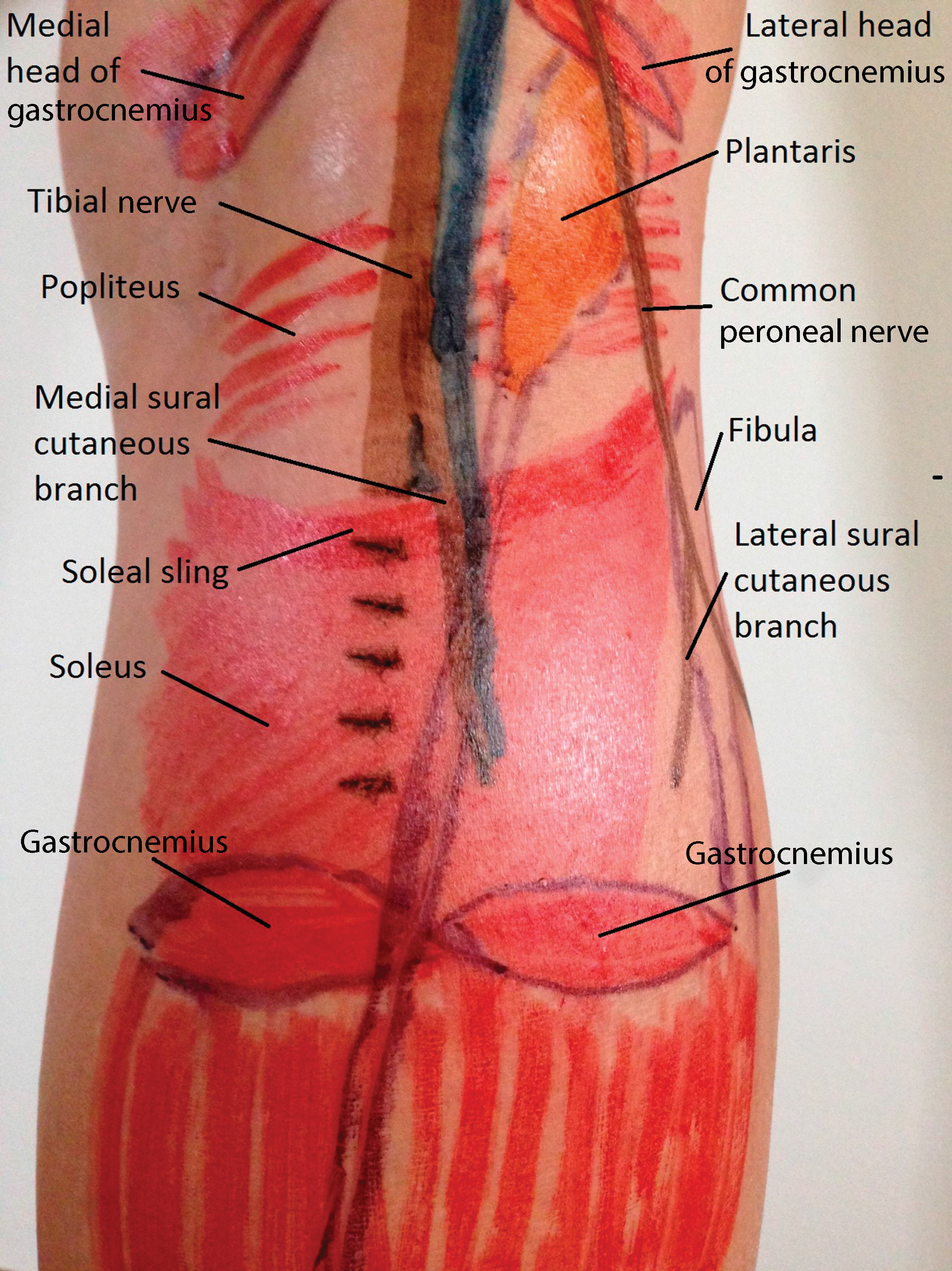
The tibial nerve originates as the popliteal nerve in the popliteal fossa. It courses superficial to the popliteus muscle, and at the inferior portion of the popliteal fossa it dives deep to the soleus muscle. Before diving deep to the soleus, it divides and gives off a small medial sural branch (Fig. 1). The soleus muscle has its origin at the proximal tibia and fibula, forming a sling between the two bones (Fig. 2). This soleal sling has a proximal fibrous margin that has been implicated as a site of compression of the tibial nerve. [9]
Figure 1.
Magnetic resonance image of the tibial nerve giving off a medial sural branch at the superior margin of the soleus muscle.
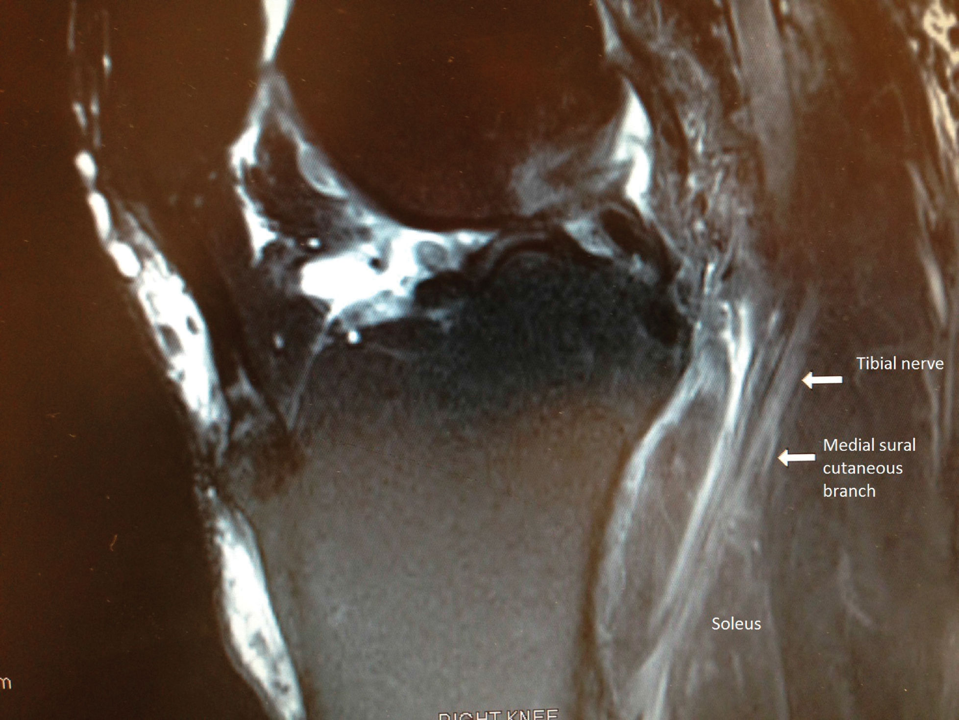
Figure 2.
Anatomical reference points for localization of the soleal sling at the posterior proximal leg.
The tibial nerve then dives deep to the soleus muscle and courses distally deep to the flexor retinaculum of the leg. The distal portion of the flexor retinaculum at the ankle has been termed the laciniate ligament. This is analogous to the distal portion of the arm proximal to the carpal tunnel. As the nerve courses through the laciniate ligament it bifurcates into the medial and lateral plantar nerves. Those nerve branches course deep into the medial and lateral plantar tunnels, respectively.
Existing Diagnostic Literature
Nerve conduction velocity/electromyography technology has been able to localize tarsal tunnel entrapment of the tibial nerve with variable sensitivity and somewhat better specificity. However, there is no specific electrodiagnostic testing for soleal sling entrapment. Magnetic resonance imaging findings of extraneural (n = 4) and intraneural (n = 3) ganglion cysts and idiopathic thickening of the soleal sling (n = 2) have been reported. The sensitivity of this modality is unknown because the only report has a very small sample size. [11] An additional limitation of the magnetic resonance imaging examination is the inability to perform dynamic examinations. Diagnostic ultrasound allows dynamic evaluation of anatomical structures. This modality seems ideally suited for the examination of tibial nerve entrapment at the soleal sling as the foot moves through dorsiflexion and plantarflexion.
Physical examination has been taught by The Association of Extremity Nerve Surgeons but has not been published. That society teaches that compression of the soleal sling by a finger that provokes local pain is a common finding of soleal sling entrapment neuropathy. This is consistent with the Valleix phenomenon. The positive provocative findings are combined with objective symptoms of weak toe flexion and plantar foot paresthesia or sensory impairment to make a clinical diagnosis.
Ultrasound Examination
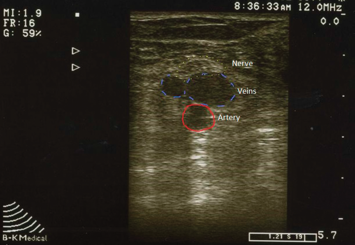
Patient positioning is critical. The patient is placed in a prone position. The leg is flexed approximately 30°. The probe is placed caudal to the popliteal fossa in the transverse plane (Fig. 3). If the leg is not flexed, occlusion of the venae comitans will occur, making identification very difficult. Slightly lateral to the center line, the tibial artery, venae comitans, and tibial nerve will be visible in cross section (Fig. 4). This is the initial reference point.
Figure 3.
Patient positioning for ultrasound examination of the soleal sling.
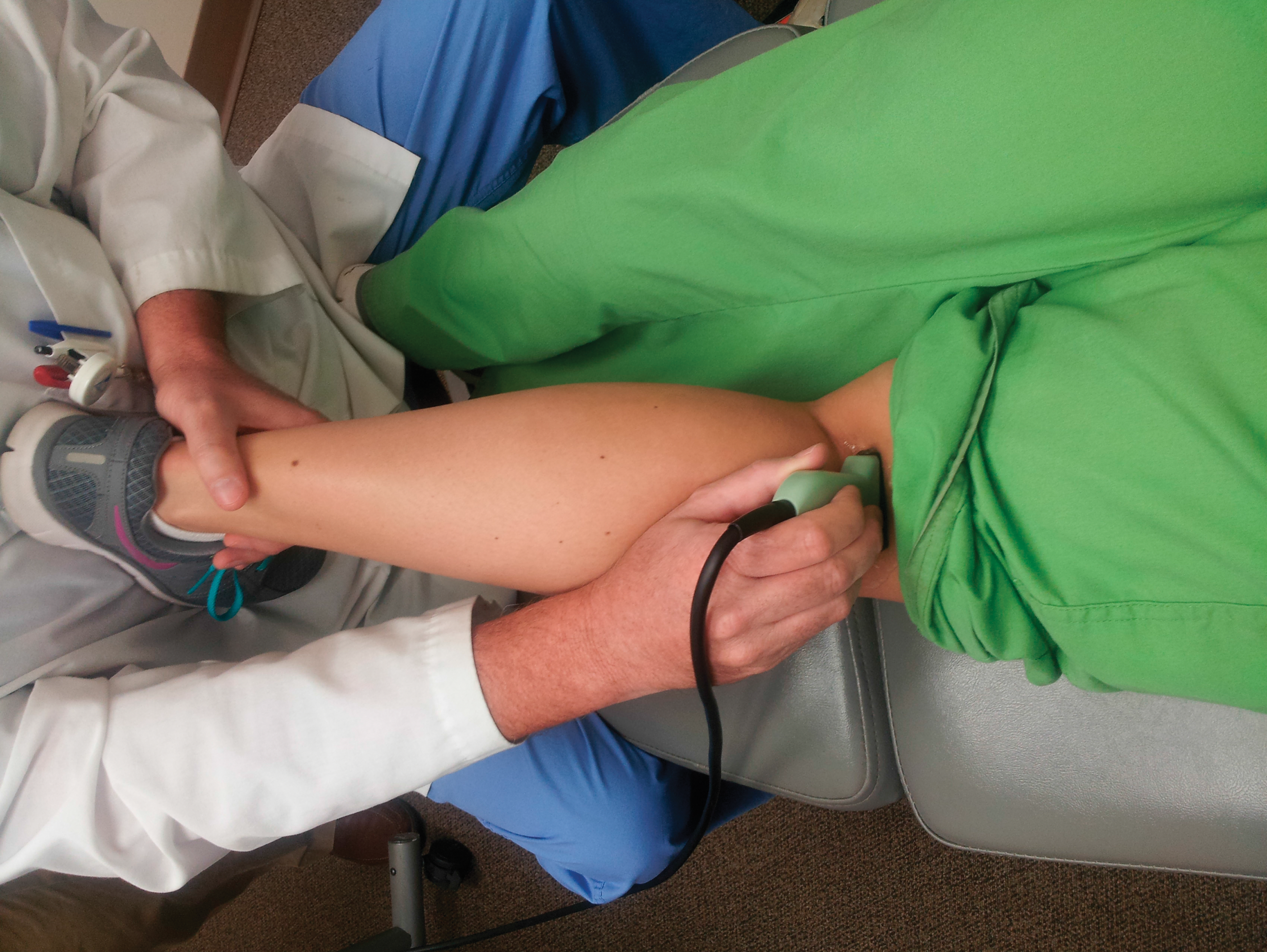
Figure 4.
Short axis (cross-sectional) view of the tibial nerve (superficially) and the venae comitans and tibial artery (deep) at the level of the distal popliteal fossa.
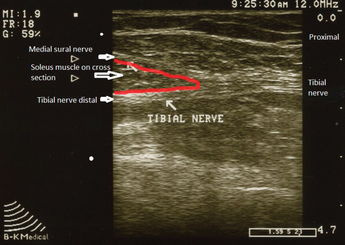
Turning the probe longitudinally allows visualization of the fibrillar echotexture of the tibial nerve fascicles (Fig. 5). A hypoechoic signal along the nerve or loss of normal fibrillar echotexture indicates injury to the nerve. The hypoechoic signal is consistent with an accumulation of axoplasmic fluid often found proximal and distal to sites of compression injury. The loss of normal echotexture is found when there is scarring and nerve damage. Continuing the examination distally, the tibial nerve will bifurcate and give off the medial sural cutaneous branch, which courses in a superficial direction. Just distal to the site of bifurcation, the soleal sling will be visible on cross section (Fig. 6). At that point, dorsiflexion and plantarflexion of the foot will reveal any compression of the nerve on dynamic ultrasound examination.
Figure 5.
Long axis (longitudinal) view of the tibial nerve (superficially) and the tibial vein and artery (deep) at the distal popliteal fossa. Arrows indicate the superficial and deep boundaries of the tibial nerve.
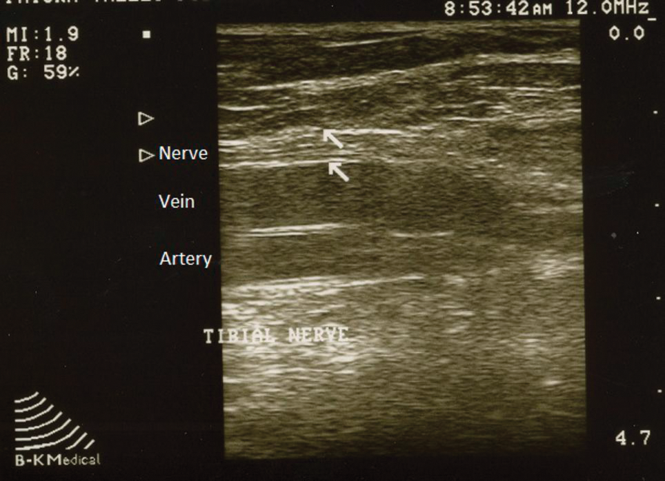
Figure 6.
Long axis view of the tibial nerve bifurcating into the tibial and medial sural nerve branches at the level of the soleal sling.
Cadaveric Study
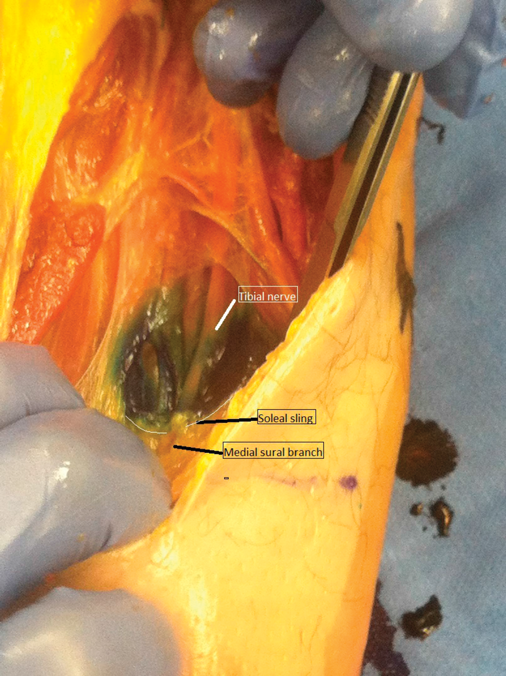
The ultrasound examination technique was subsequently validated on cadaver specimens. Three cadaver above-the-knee–amputated limbs were used. Ultrasound was used to localize the suspected soleal sling–tibial nerve interface. Using the technique described previously herein, methylene blue was then injected under ultrasound guidance into the sling-nerve interface, and follow-up dissection was performed. In all cases, the methylene blue was found directly at the soleal sling, as anticipated (Fig. 7).
Figure 7.
Confirmatory cadaveric dissection of the soleal sling injected with methylene blue using ultrasonography.
Conclusions
The soleal sling is a known entrapment site of the tibial nerve and can produce distal plantar foot pain. Decompressive release of the soleal sling has shown promise for relieving associated foot pain. The difficulty lies in objectively diagnosing this entrapment. Ultrasound examination has the ability to show static and dynamic images of the soleal sling–tibial nerve interaction. At this point, ultrasound is readily available in many physician offices and is an inexpensive reproducible method for examining peripheral nerves. Using the technique described herein can provide the physician with direct visualization of the tibial nerve as the foot moves through dorsiflexion and plantarflexion. With that knowledge, it is anticipated that preoperative examination reliability can be increased and the success of soleal sling decompression improved.
Financial Disclosure
None reported.
Conflicts of Interest
None reported.
References
- Skalley TC, Schon LC, Hinton RY, et al: Clinical results following revision tibial nerve release. Foot Ankle Int 15: 360, 1994.
- Novotny DA, Kay DB, Parker MG: Recurrent tarsal tunnel syndrome and the radial forearm free flap. Foot Ankle Int 17: 641, 1996.
- Womack JW, Richardson DR, Murphy GA, et al: Long-term evaluation of interdigital neuroma treated by surgical excision. Foot Ankle Int 29: 574, 2008.
- Dellon AL: The four medial ankle tunnels: a critical review of perceptions of tarsal tunnel syndrome and neuropathy. Neurosurg Clin North Am 19: 629, 2008.
- Barrett SL, Walsh AS: Endoscopic decompression of intermetatarsal nerve entrapment: a retrospective study. JAPMA 96: 19, 2006.
- Dellon AL: Treatment of symptomatic diabetic neuropathy by surgical decompression of multiple peripheral nerves. Plast Reconstr Surg 89: 689, 1992.
- Rader AJ, Barry TP, Stanley OL: Characteristics of lower extremity pressure sensation impairment in developing diabetic sensory polyneuropathy. Foot Ankle Spec 1: 39, 2008.
- Williams EH, Rosson GD, Hagan RR, et al: Soleal sling syndrome (proximal tibial nerve compression): results of surgical decompression. Plast Reconstr Surg 129: 454, 2012.
- Williams EH, Williams CG, Rosson GD, et al: Anatomic site for proximal tibial nerve compression: a cadaver study. Ann Plastic Surg 62: 322, 2009.
- Upton AR, McComas AJ: The double crush in nerve entrapment syndromes. Lancet 2: 359, 1973.
- Chhabra A, Williams EH, Subhawong TK, et al: MR neurography findings of soleal sling entrapment. AJR Am J Roentgenol 196: W290, 2011.
© 2016 American Podiatric Medical Association