


Abstract
Background: Persons with diabetes have a higher incidence of fractures compared with persons without diabetes. However, there is little published information concerning the deleterious effect of late-stage diabetes on fracture healing. There are no studies using animal models that evaluate the effect of advanced diabetes on fracture healing. The purpose of our study was to evaluate cytokine expression, specifically macrophage inflammatory protein 1 (MIP-1) and vascular endothelial growth factor, in fracture healing in a type 2 diabetes rat model. Methods: We evaluated biomarker expression after femur fracture using a rat model. The two groups consisted of 24 Zucker diabetic rats (study group) and 12 Zucker lean rats (control group). An independent reviewer was used to assess delayed union. We evaluated serum samples 2, 4, 7, and 14 days after surgery for MIP-1, vascular endothelial growth factor, leptin, and other cytokine levels. Results: At 3 weeks, Kaplan-Meier estimates showed that 45.8% of femur fractures in Zucker diabetic rats had healed, whereas 81.8% of those in Zucker lean rats had healed (P = .02). A logistic regression model to predict fast healing that included the three cytokines and diabetes status showed that the only factor achieving significance was MIP-1α. Vascular endothelial growth factor was the only biomarker to show significance compared with delayed healing. Conclusions: These results confirm significant differences in biomarker expression between diabetic and nondiabetic rats during bone healing. The key factors for bone healing may appear early in the healing process, whereas differences in diabetes versus nondiabetes are seen later in the healing process. Increased levels of MIP-1α were associated with the likelihood of delayed healing.
People with diabetes have a higher incidence of fractures compared with persons without diabetes.[1,2,3] However, there is little published information concerning the deleterious effect of late-stage diabetes on fracture healing. Specifically, there are no studies using animal models that evaluate the effect of advanced diabetes and fractures. Most studies document the effects of diabetes on biomechanical properties or fracture healing by observing histologic changes in the fracture callus in type I diabetes animal models. Fracture healing and complications in the general diabetic population may be part of the spectrum of bone pathologic disorders in diabetes in which Charcot's neuroarthropathy represents end-stage disease. Only a few studies in animals with diabetes have evaluated the effect of peripheral neuropathy or peripheral arterial disease on bone repair. Most animal studies evaluate young animals before diabetes-related complications have been shown to develop. The presence and severity of diabetes-related comorbidities (peripheral neuropathy and peripheral arterial disease), rather than diabetes itself, have been shown to increase the risk of complications.[4,5] Most of the clinical reports involving humans with diabetes and fracture complications focus on ankle fractures. There are a few small case series evaluating patients with Charcot's neuroarthropathy.[6,7] For example, the high rate of malunion and nonunion in ankle fractures in diabetes has been attributed to misdiagnosis of Charcot's neuroarthropathy.[8]
Bone healing constitutes a unique, complex process. Understanding the molecular level of bone healing is imperative for the surgeon so that he or she can preserve and augment these endogenous mechanisms. Multiple growth factors work in synchronization during the healing cascade. For example, vascular endothelial growth factor (VEGF) has extensively been studied for its role in angiogenesis.[8,9] Bone is highly vascular, and when fracture occurs, the vasculature is disrupted, and osteoblasts are deprived of oxygen and other materials used for bone formation.
Several cytokines have been implicated in bone repair. Macrophage inflammatory protein 1 (MIP-1) has been reported recently to act indirectly on osteoclastic precursor cells, activating osteoclasts by way of bone marrow stromal cells or osteoblasts, thus leading to bone destruction.[10] The action of leptin on bone seems to be complex, and positive and negative effects have been reported.[11] It seems that its action may depend on current leptin status and the mode of the action (central or peripheral effects). Leptin has been found to be a negative regulator of bone mass in a mouse model.[12] Therefore, leptin, VEGF, and MIP-1α, along with many other cytokines and growth factors, need to be investigated for their role in fracture healing, particularly in diabetes-related complications. The purpose of this study was to evaluate mainly the effect of MIP-1α, leptin, VEGF, and other cytokine expression on fracture healing in a type 2 diabetes rat model.
Materials and Methods
We evaluated protein biomarker expression after femur fracture using the Zucker rat model. We compared two groups: the diabetic group (n = 24) and the control group (n = 12).
Diabetes Model
Male Zucker diabetic fatty rats, age-matched lean littermates (Zucker lean rats), and Zucker rats 14 weeks of age were obtained from Charles River Laboratories International, Inc (Wilmington, Massachusetts). Zucker diabetic fatty animals are an inbred rat model that closely mimics adult-onset diabetes in humans. Zucker diabetic fatty animals develop impaired glucose tolerance, hyperinsulinemia, insulin resistance, obesity, and hyperlipidemia in all males. Glucose intolerance is observed at approximately 7 weeks, and animals are fully diabetic by 8 to 12 weeks. Zucker obese rats are the parent strain and exhibit insulin resistance, hypertension, dyslipidemia, and vascular dysfunction, but they do not become hyperglycemic. All of the animals were fed rat chow (Purina LabDiet Formulab 5008; Purina, Richmond, IN) ad libitum and had free access to water. Glucose checks were performed daily, and fasting blood glucose levels were measured weekly. Metformin was administered by a veterinarian for glucose in the diabetic group. Metformin, 300 mg/kg, was dissolved in drinking water and administered orally. Metformin concentrations were readjusted based on water consumption after an approximately 2-week evaluation period. Body weights were measured at the onset of the study and weekly.
Measures of Neuropathy
Von Frey monofilaments were used to evaluate neuropathy by observing the frequency of mechanical withdrawal using calibrated 4-, 10-, and 26-g monofilaments at 2, 4, and 6 weeks. Animals were placed in a plexiglass cage with holes in the bottom. Each paw was tested three times for 1 sec each until the monofilament bent. Withdrawal was graded from 0 (no withdrawal) to 2 (maximum withdrawal) for each probe.[13,14]
Model to Create Unstable Fracture
All of the rats underwent stabilization of the right femora using the established technique of intramedullary pinning with Kirschner wire. A medial parapatellar incision was used. The femoral canal was reamed by introducing a 0.062 Kirschner wire through the intercondylar notch.[15] This technique provided stabilization during and after fracture. Immediately after stabilization, the right femur was subjected to a deforming force by a guillotine-style blunt-fracture blade. The closed method has been well described and used by others.[15,16] In all of the animals, a 0.062 Kirschner wire was used to ream the femoral canal, and it was replaced by a smaller-diameter, 0.045 smooth Kirschner wire. This encourages instability and, hence, deliberately induces an unstable fracture. Garcia et al[17] reported a high rate (55%) of nonunion due to rotational instability using this type of intramedullary Kirschner wire fixation technique. Radiographs were taken weekly up to 6 weeks. An independent reviewer assessed delayed union. Delayed union was defined as not having complete bridging of two cortices 3 weeks after the fracture. Radiographs were taken weekly until termination at 6 weeks. We evaluated serum samples 2, 4, 7, and 14 days after reduction of the femur fracture and measured levels of VEGF, MIP-1α, and leptin.
Analysis of Biomarkers
We used a Bio-Rad kit (Bio-Rad Laboratories, Hercules, California) combined with our Bio-Plex system to test bone-forming cytokines. Essentially, the Bio-Plex system offers a postgenomic approach to decipher interrelationships among proteins involved in the inflammatory response or signal transduction pathways. All of the concentrations are presented in picograms per milliliter. Serum samples were frozen and stored at −80°C until analysis.
Statistical Analysis
A contingency table was used to compare the control and diabetic groups. A Kaplan-Meier estimate was used to compare time to healing between both groups. We performed t tests comparing all of the cytokines between the fast and slow healers at all of the time points, setting P = 0.05 as a cutoff value for statistical significance. To account for differences between diabetic and nondiabetic rats, we ran a logistic regression model to predict fast healing that included VEGF, MIP-1α, and leptin and diabetes status. We performed t tests to compare cytokine levels between diabetic and nondiabetic rats, setting P = 0.0001 as a cutoff value for statistical significance.
Results
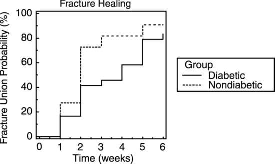
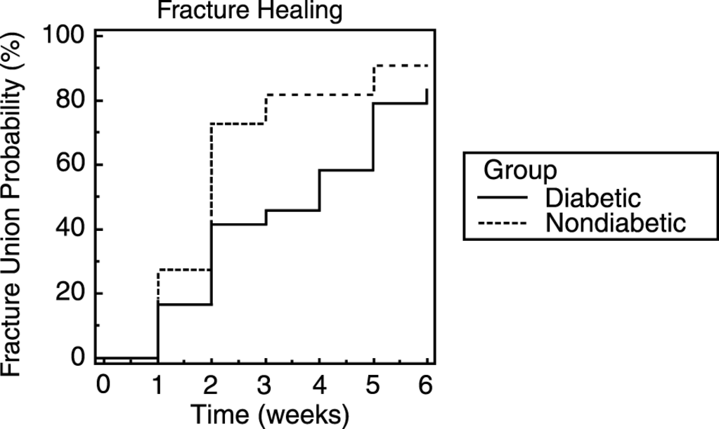
The mean serum glucose level was 276 mg/dL for the diabetic group. Eleven diabetic rats healed quickly (≤3 weeks), and 13 healed slowly (>3 weeks). On the other hand, the control group was more likely to heal within 3 weeks (9 of 11). This subjective trend does not achieve significance, with a Fisher exact test P = .069. At 3 weeks, Kaplan-Meier estimates showed that 45.8% of diabetic rats had healed, whereas 81.8% of control rats had healed (P = .02) (Figure 1). There was no statistically significant difference between rats with and without neuropathy in terms of delayed fracture healing.
Figure 1.
At week 3, Kaplan-Meier estimates found that 45.8% of diabetic rats had healed, whereas 81.8% of nondiabetic rats had healed (P = .02).
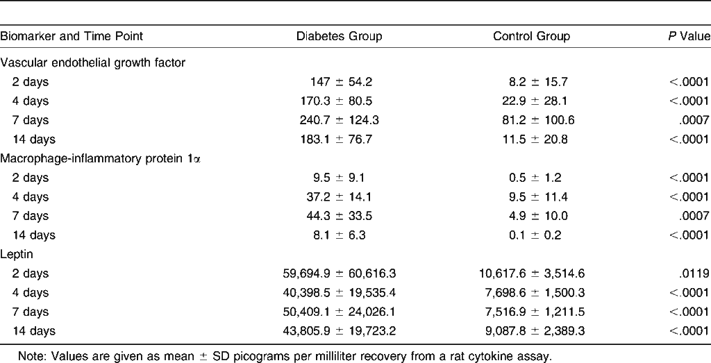
Table 1 demonstrates biomarker values between the diabetic and control groups at different time points during the postoperative period. When we looked at how the levels of cytokines differed between diabetic and nondiabetic rats, we found increased expression of MIP-1α at all four time points (2, 4, 7, and 14 days) and of VEGF at time points 1, 3, and 4 (2, 7, and 14 days) (Table 1). The only biomarker to also show significance in those with and without delayed healing was VEGF. This was noted 2 days postoperatively, with those healing by 3 weeks having a mean ± SD VEGF level of 80.11 ± 76.58 pg/mL. In a logistic regression model, the only factor achieving significance was MIP-1α. This finding may suggest that considering diabetes status fixed, the only cytokine (of those included in the model) that significantly influenced healing was MIP-1α. Increased levels of MIP-1α may be associated with a decreased likelihood of timely healing.
Table 1.
Biomarker Values in the Diabetic and Control Groups at Different Time Points During the Postoperative Period
Discussion
The pathologic process of bone healing in diabetes is not well understood. However, complications related to this process are discussed in the medical literature.[4,5] Diabetic animal models have demonstrated that structural and metabolic changes in diabetes resemble those occurring with advanced age.[18,19] In an unpublished medical record review of ankle fractures, we identified very high rates of wounds, infections, nonunions, and joint pain and the need for long-term bracing in diabetic compared with nondiabetic individuals (Lavery LA, La Fontaine J, Green T, 2009).
A few reports in the literature have indicated that diabetic bones are weaker.[20,21,22,23,24,25] In addition, the fracture repair process in the diabetic state seems to be defective.[14,15] However, no studies have assessed fracture healing rates and characteristics of nonunions while considering the effect of the presence or severity of peripheral neuropathy and peripheral arterial disease in an adult animal model.
In a preliminary study, 20 rat femora were tested using three-point bending. Eight femora were from diabetic rats (mean glucose level, 385 mg/dL) and 12 were from lean rats. The diabetic rats used in these preliminary studies had diabetes induced using streptozotocin 6 weeks before testing. Because the rats used in the preliminary study varied from 280 to 456 g, the structural properties were normalized with respect to the mass of each femur. Statistical analysis indicated that there was a significant difference between the normal and diabetic femora in terms of maximum force tolerated and structural stiffness. Several studies have suggested a deleterious effect of diabetes on wound and fracture healing due to reduced levels of growth factors.[21,22,23]
There are several studies that identify abnormalities of growth factors (platelet-derived growth factor [PDGF], transforming growth factor-β[TGF-β], VEGF, and insulin-like growth factor) in animal and human studies of acute and chronic diabetic wounds.[24,25] Application of exogenous growth factors has been shown to improve healing of diabetic foot ulcers in humans[26,27,28,29] and fractures in animals.[30,31]
Three prospective longitudinal studies have evaluated serum levels of growth factors in long bone fractures and showed reduced cytokine levels in patients (without diabetes) developing nonunions. Abnormalities were identified early in the healing process. There were lower levels of TGF-β at 1 week (P = .052), basic fibroblast growth factor at 2 weeks (P < .05), and PDGF[32] and TGF-β at 4 weeks (P < .05) in patients with nonunions.[23,33,34] However, evaluation of growth factor abnormalities in fractures in diabetes has relied exclusively on induced diabetes animal models. For example, Tyndall et al[23] identified significantly lower levels of PDGF in the early phase of healing in diabetic fracture models in Wistar rats. Abnormalities were observed 2, 4, and 7 days after fracture.[23] In addition, Gandhi et al[35] evaluated a femoral fracture model in Wistar rats and identified significant reductions in levels of PDGF, VEGF, TGF-β, and insulin-like growth factor.
In the present study, we showed that there were significant differences in biomarker expression between diabetic and nondiabetic rats after injury, specifically, VEGF and MIP-1α. However, after adjusting for other covariates, the only factor achieving significance was MIP-1α. This might mean that the only cytokine that significantly independently influenced fracture healing was MIP-1α. Increased levels of MIP-1α were associated with the likelihood of delayed healing. In this study, diabetes status itself did not reach statistical significance for delayed healing. Diabetes may, however, indirectly influence healing through its effect on MIP-1α. Macrophage inflammatory protein-1α has been shown to play a role in osteoclastogenesis directly as well as being a chemotactic factor for mature osteoclasts and osteoclast precursors in a rat model.[36] Increased concentrations may promote excessive and prolonged osteoclastic activity and, thus, cause abnormalities in the normal bone-healing process.[10,37]
Although leptin has been associated with increased bone mass, the increase in leptin levels in the diabetic group may relate to the fact that signaling by this hormone is blocked by the mutation of its receptor. This is a classic regulatory mechanism that relates to the nature of the model, not to diabetes. However, the leptin-deficient animal model has shown bone abnormalities.[38] The ability of leptin to regulate body weight and bone mass has been compared and showed that leptin anti-osteogenic functions are affected by the amount of leptin.[38] However, an increased serum leptin level dramatically reduced bone mass.[12] The present study demonstrated increased leptin levels at 4, 7, and 14 days compared with lean rats. Although these findings were nonsignificant, the leptin pathway may need to be investigated further because obesity is associated with a significant increase in serum leptin levels and diabetes.[39,40]
Increased serum concentrations of VEGF have been shown to play a role in diabetic neuropathy, retinopathy, and nephropathy.[41,42] The present study suggested that VEGF plays a role in every step of the fracture repair cascade from the fracture hematoma to the final remodeling stages of fracture repair. Accumulation of VEGF in a fracture defect attracts osteoprogenitor cells and endothelial cells alike, creating an environment where these distinct cell types secrete trophic factors that mutually promote their proliferation and survival.[9] This establishes a zone rich in growth factors and cells necessary for the repair process. Vascular endothelial growth factor is only one of an unknown number of factors that play an integral role in the process of angiogenesis and bone repair, but the literature suggests that this is most essential for angiogenesis and subsequent bone repair by replenishing the blood supply and the population of bone-forming cells.
Conclusions
There are few clinical reports that address fracture complications in diabetes. We showed a direct correlation between diabetes and an increased risk of delayed fracture union. We also identified biomarkers that show a significant difference in subjects between fast healers and slow healers at different time points. Specifically, the VEGF level was shown to be elevated 2 days after injury in subjects with delayed fracture healing, and increased levels of MIP-1α were associated with a decreased likelihood of fast healing. The key factors for bone healing may appear early in the healing process, whereas differences in diabetes versus nondiabetes are later in the healing process. In the logistic model, MIP-1α is a factor in bone healing, whereas diabetes is not.
Acknowledgments
This study was supported by an institutional research grant from Scott and White Hospital, Temple, Texas.
Financial Disclosure
None reported.
Conflicts of Interest
None reported.
References
- Melton, L.J., III; Leibson, C.L.; Achenbach, S.J.; et al. Fracture risk in type 2 diabetes: update of a population-based study. J Bone Miner Res 2008, 23, 1334. [Google Scholar] [CrossRef] [Scilit] [PubMed]
- Melton, L.J., III; Riggs, B.L.; Leibson, C.L.; et al. A bone structural basis for fracture risk in diabetes. J Clin Endocrinol Metab 2008, 93, 4804. [Google Scholar] [CrossRef] [Scilit] [PubMed]
- Schwartz, A.V.; Sellmeyer, D.E.; Ensrud, K.E.; et al. Older women with diabetes have an increased risk of fracture: a prospective study. J Clin Endocrinol Metab 2001, 86, 32. [Google Scholar] [CrossRef] [PubMed]
- Lavery, L.A.; Peters, E.J.; Williams, J.R.; et al. Reevaluating the way we classify the diabetic foot: restructuring the diabetic foot risk classification system of the International Working Group on the Diabetic Foot. Diabetes Care 2008, 31, 154. [Google Scholar] [CrossRef] [Scilit]
- Wukich, D.K.; Kline, A.J. The management of ankle fractures in patients with diabetes. J Bone Joint Surg Am 2008, 90, 1570. [Google Scholar] [CrossRef] [Scilit]
- Papa, J.; Myerson, M.; Girard, P. Salvage, with arthrodesis, in intractable diabetic neuropathic arthropathy of the foot and ankle. J Bone Joint Surg Am 1993, 75, 1056. [Google Scholar] [CrossRef] [Scilit]
- Tisdel, C.L.; Marcus, R.E.; Heiple, K.G. Triple arthrodesis for diabetic peritalar neuroarthropathy. Foot Ankle Int 1995, 16, 332. [Google Scholar] [CrossRef] [Scilit]
- Glowacki, J. Angiogenesis in fracture repair. Clin Orthop Relat Res 1998, 355 (suppl), S82. [Google Scholar] [CrossRef] [Scilit]
- Beamer, B.; Hettrich, C.; Lane, J. Vascular endothelial growth factor: an essential component of angiogenesis and fracture healing. HSS J 2010, 6, 85. [Google Scholar] [CrossRef] [Scilit]
- Tsubaki, M.; Kato, C.; Manno, M.; et al. Macrophage inflammatory protein-1α (MIP-1α) enhances a receptor activator of nuclear factor κB ligand (RANKL) expression in mouse bone marrow stromal cells and osteoblasts through MAPK and PI3K/Akt pathways. Mol Cell Biochem 2007, 304, 53. [Google Scholar] [CrossRef] [Scilit]
- Cao, J.J. Effects of obesity on bone metabolism. J Orthop Surg Res 2011, 6, 30. [Google Scholar] [CrossRef] [Scilit]
- Elefteriou, F.; Takeda, S.; Ebihara, K.; et al. Serum leptin level is a regulator of bone mass. Proc Natl Acad Sci U S A 2004, 101, 3258. [Google Scholar] [CrossRef] [Scilit] [PubMed]
- Brussee, V.; Guo, G.; Dong, Y.; et al. Distal degenerative sensory neuropathy in a long-term type 2 diabetes rat model. Diabetes 2008, 57, 1664. [Google Scholar] [CrossRef] [Scilit] [PubMed]
- Calcutt, N.A.; Chaplan, S.R. Spinal pharmacology of tactile allodynia in diabetic rats. Br J Pharmacol 1997, 122, 1478. [Google Scholar] [CrossRef] [Scilit] [PubMed]
- Bonnarens, F.; Einhorn, T.A. Production of a standard closed fracture in laboratory animal bone. J Orthop Res 1984, 2, 97. [Google Scholar] [CrossRef] [Scilit]
- Macey, L.R.; Kana, S.M.; Jingushi, S.; et al. Defects of early fracture-healing in experimental diabetes. J Bone Joint Surg Am 1989, 71, 722. [Google Scholar] [CrossRef] [Scilit]
- Garcia, P.; Holstein, J.H.; Histing, T.; et al. A new technique for internal fixation of femoral fractures in mice: impact of stability on fracture healing. J Biomech 2008, 41, 1689. [Google Scholar] [CrossRef] [Scilit]
- Clark, J.B.; Palmer, C.J.; Shaw, W.N. The diabetic Zucker fatty rat. Proc Soc Exp Biol Med 1983, 173, 68. [Google Scholar] [CrossRef] [Scilit]
- Chen, D.; Wang, M.W. Development and application of rodent models for type 2 diabetes. Diabetes Obes Metab 2005, 7, 307. [Google Scholar] [CrossRef] [Scilit]
- Bekker, P.J.; Holloway, D.; Nakanishi, A.; et al. The effect of a single dose of osteoprotegerin in postmenopausal women. J Bone Miner Res 2001, 16, 348. [Google Scholar] [CrossRef] [Scilit]
- Bolander, M.E. Regulation of fracture repair by growth factors. Proc Soc Exp Biol Med 1992, 200, 165. [Google Scholar] [CrossRef] [Scilit]
- Kawaguchi, H.; Kurokawa, T.; Hanada, K.; et al. Stimulation of fracture repair by recombinant human basic fibroblast growth factor in normal and streptozotocin-diabetic rats. Endocrinology 1994, 135, 774. [Google Scholar] [CrossRef] [Scilit]
- Tyndall, W.A.; Beam, H.A.; Zarro, C.; et al. Decreased platelet derived growth factor expression during fracture healing in diabetic animals. Clin Orthop Relat Res 2003, 408, 319. [Google Scholar] [CrossRef] [Scilit] [PubMed]
- Blakytny, R.; Jude, E. The molecular biology of chronic wounds and delayed healing in diabetes. Diabet Med 2006, 23, 594. [Google Scholar] [CrossRef] [Scilit] [PubMed]
- Jude, E.B.; Blakytny, R.; Bulmer, J.; et al. Transforming growth factor-β 1, 2, 3 and receptor type I and II in diabetic foot ulcers. Diabet Med 2002, 19, 440. [Google Scholar] [CrossRef] [Scilit] [PubMed]
- Wieman, T.J. Becaplermin Gel Studies Group: Clinical efficacy of becaplermin (rhPDGF-BB) gel. Am J Surg 1998, 176, 74S. [Google Scholar] [CrossRef] [Scilit]
- Wieman, T.J.; Smiell, J.M.; Su, Y. Efficacy and safety of a topical gel formulation of recombinant human platelet-derived growth factor-BB (becaplermin) in patients with chronic neuropathic diabetic ulcers: a phase III randomized placebo-controlled double-blind study. Diabetes Care 1998, 21, 822. [Google Scholar] [CrossRef] [Scilit]
- Bennett, S.P.; Griffiths, G.D.; Schor, A.M.; et al. Growth factors in the treatment of diabetic foot ulcers. Br J Surg 2003, 90, 133. [Google Scholar] [CrossRef] [Scilit]
- Tsang, M.W.; Wong, W.K.; Hung, C.S.; et al. Human epidermal growth factor enhances healing of diabetic foot ulcers. Diabetes Care 2003, 26, 1856. [Google Scholar] [CrossRef] [Scilit]
- Wildemann, B.; Bamdad, P.; Holmer, C.; et al. Local delivery of growth factors from coated titanium plates increases osteotomy healing in rats. Bone 2004, 34, 862. [Google Scholar] [CrossRef] [Scilit]
- Wildemann, B.; Schmidmaier, G.; Brenner, N.; et al. Quantification, localization, and expression of IGF-I and TGF-β1 during growth factor-stimulated fracture healing. Calcif Tissue Int 2004, 74, 388. [Google Scholar] [CrossRef] [Scilit]
- Weiss, S.; Zimmermann, G.; Pufe, T.; et al. The systemic angiogenic response during bone healing. Arch Orthop Trauma Surg 2009, 129, 989. [Google Scholar] [CrossRef] [Scilit] [PubMed]
- Zimmermann, G.; Henle, P.; Kusswetter, M.; et al. TGF-β1 as a marker of delayed fracture healing. Bone 2005, 36, 779. [Google Scholar] [CrossRef] [Scilit] [PubMed]
- Zimmermann, G.; Moghaddam, A.; Reumann, M.; et al. TGF-β1 as a pathophysiological factor in fracture healing [in German]. Unfallchirurg 2007, 110, 130. [Google Scholar] [CrossRef] [Scilit] [PubMed]
- Gandhi, A.; Doumas, C.; O'Connor, J.P.; et al. The effects of local platelet rich plasma delivery on diabetic fracture healing. Bone 2006, 38, 540. [Google Scholar] [CrossRef] [Scilit]
- Kukita, T.; Nomiyama, H.; Ohmoto, Y.; et al. Macrophage inflammatory protein-1 α (LD78) expressed in human bone marrow: its role in regulation of hematopoiesis and osteoclast recruitment. Lab Invest 1997, 76, 399. [Google Scholar]
- Choi, S.J.; Cruz, J.C.; Craig, F.; et al. Macrophage inflammatory protein 1-α is a potential osteoclast stimulatory factor in multiple myeloma. Blood 2000, 96, 671. [Google Scholar] [CrossRef] [Scilit]
- Hamrick, M.W.; Pennington, C.; Newton, D.; et al. Leptin deficiency produces contrasting phenotypes in bones of the limb and spine. Bone 2004, 34, 376. [Google Scholar] [CrossRef] [Scilit]
- van Dielen, F.M.; van't Veer, C.; Schols, A.M.; et al. Increased leptin concentrations correlate with increased concentrations of inflammatory markers in morbidly obese individuals. Int J Obes Relat Metab Disord 2001, 25, 1759. [Google Scholar] [CrossRef] [Scilit]
- Diaz, V.A.; Mainous, A.G., III; Baker, R.; et al. How does ethnicity affect the association between obesity and diabetes? Diabet Med 2007, 24, 1199. [Google Scholar] [CrossRef] [Scilit]
- Mahdy, R.A.; Nada, W.M.; Hadhoud, K.M.; et al. The role of vascular endothelial growth factor in the progression of diabetic vascular complications. Eye 2010, 24, 1576. [Google Scholar] [CrossRef] [Scilit]
- Tavakkoly-Bazzaz, J.; Amoli, M.M.; Pravica, V.; et al. VEGF gene polymorphism association with diabetic neuropathy. Mol Biol Rep 2010, 37, 3625. [Google Scholar] [CrossRef] [Scilit]

© 2014 American Podiatric Medical Association