


Abstract
Hallux varus is defined as a medial deviation of the phalanx at the first metatarsophalangeal joint and can be congenital or acquired. Brachymetatarsia is defined as shortening of the metatarsal bones. A combination of hallux varus and brachymetatarsia is rare. A 15-year-old girl presented to our outpatient clinic complaining of problems with her feet. A distinctive hallux varus was present bilaterally combined with a brachymetatarsia of the first metatarsals. The patient reported discomfort. She was restricted in her activities and had severe psychological strain owing to the deformity. We decided on surgery. First, a Pennig MiniFixator for callus distraction of the first metatarsal bone was applied. Owing to the increased plantar subluxation of the phalanx during distraction, an extension of the external fixator was administered so that the hallux could be repositioned to a physiologically satisfying position. After sufficient callus formation, the hardware was removed 14 weeks after surgery. Thereafter, the phalanx moved back to the subluxed position. Finally, an arthrodesis of the first metatarsophalangeal joint was performed with a locking plate. Surgery should not be made only for cosmesis and associated psychological aspects; but, discomfort should be the deciding factor. The postoperative clinical and cosmetic results in our case were good, and the patient was quite satisfied. There was no longer any preoperative discomfort and pain. (J Am Podiatr Med Assoc 104(1): 85-89, 2014)
Hallux varus is defined as a medial deviation of the phalanx at the first metatarsophalangeal joint and can be congenital or acquired. A congenital hallux varus may occur with an extended or shortened first metatarsal bone, dysplastic metatarsal heads, or with surplus phalanges and metatarsals [1,2,3]. It also may occur in connection with neuromuscular disorders (eg, infantile cerebral palsy), clubfoot, pes equinovarus, and metatarsus adductus. In contrast, an acquired hallux varus usually represents a complication after hallux valgus surgery [3,4,5]. Treatment of a hallux varus ranges from conservative therapy to complex surgical reconstruction. In less severe cases, soft-tissue procedures may suffice.[4,6]. If deformities are more severe, usually the bony structures must be addressed [2,4,6,7,8]. The patients not only have pain but are also stressed owing to the cosmetic aspects and difficulties of wearing certain footwear.
Brachymetatarsia is defined as shortening of the metatarsal bones and a diagnosis is made when a metatarsal ends more than 5 mm proximal to the row of the metatarsal heads. Most often, metatarsals I and IV are shortened [2,9]. Women are affected more often than men, with a 70% incidence of bilateral involvement [2,10]. The congenital form is associated with complex syndromes such as Turner or Apert syndrome [2,9,11]. The acquired type may occur as a result of trauma, tumors, or juvenile rheumatoid arthritis. It also may occur owing to endocrinopathies, for example, pseudohypoparathy-roidism, achondroplasia, and multiple epiphyseal dysplasia [9,12]. Operative treatment of brachymetatarsia is done by elongation of the concerned ray. Techniques range from spacer implantations and bone grafting to callus distraction by an external fixator [2,13,14,15,16,17,18,19]. Patients are often concerned about (transfer) metatarsalgia owing to pathological bio- mechanical behavior of the foot. Cosmetic aspects are also a concern.
Case Report
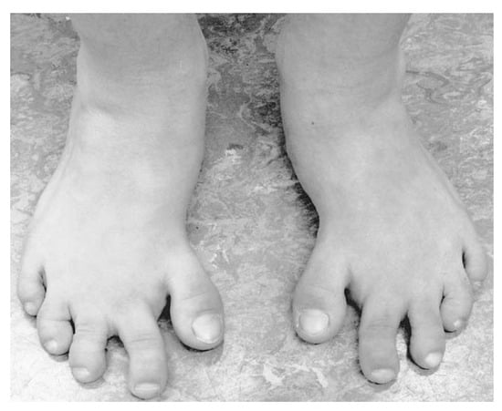
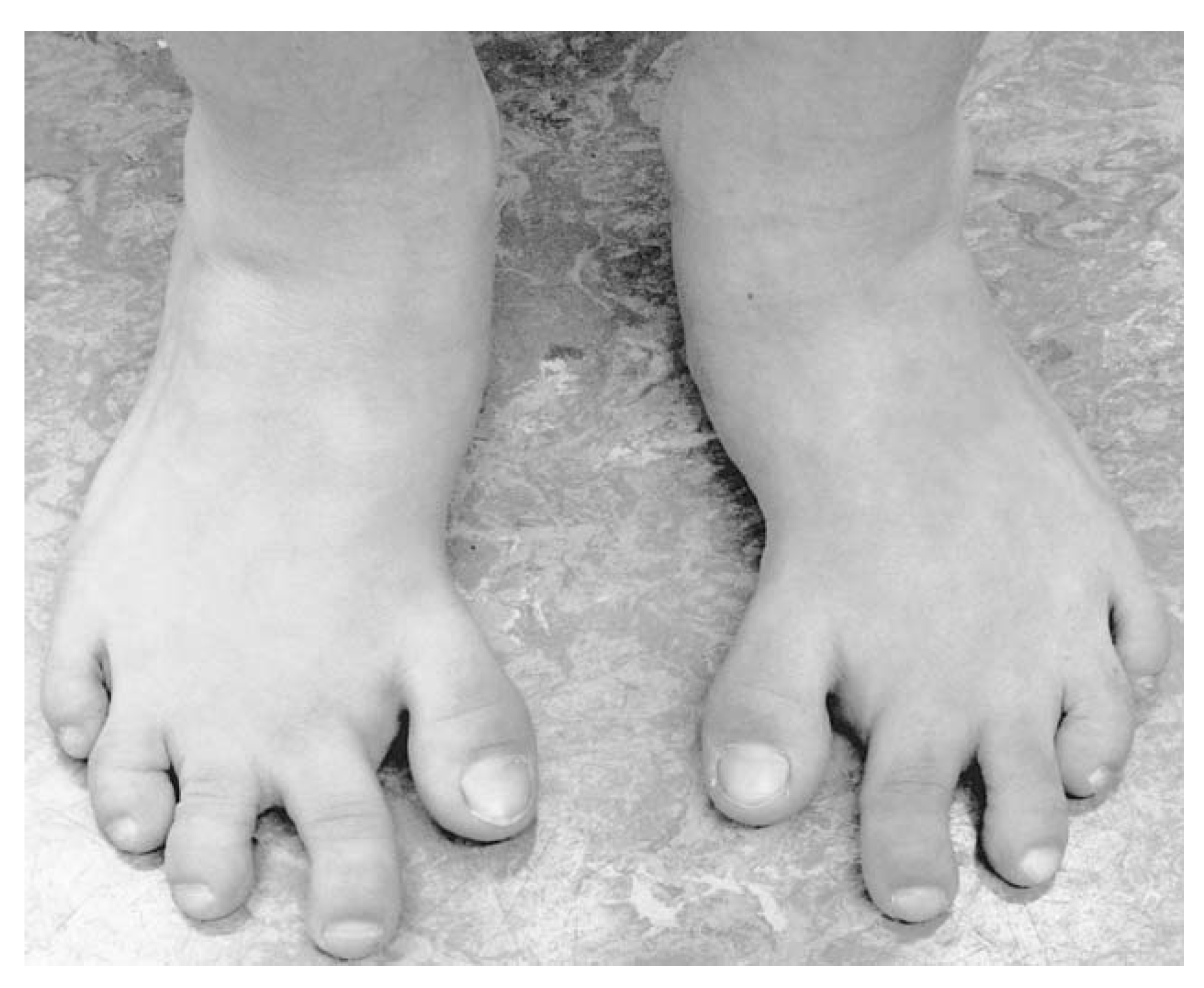
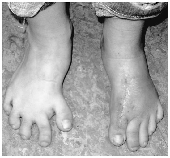
A 15-year-old girl presented to our outpatient clinic with problems with her feet. A distinctive hallux varus was present bilaterally in combination with a brachymetatarsia of the first metatarsals (
Figure 1
). The deformity had been present since birth. Apart from that, the patient was healthy and did not report any familial occurrence of such deformity. There were no complex syndromes or metabolic disorders.
Figure 1.
Preoperative photograph of both feet, showing the bilateral deformity.
The patient reported discomfort and pain not only in the region of the first metatarsophalangeal joint, but also under under the second through fourth metatarsal heads in terms of transfer metatarsalgia. The patient was quite active, and the discomfort and pain of the syndrome significantly restricted her activities. She also had psychological strain owing to the deformity and could not wear normal or modern footwear.
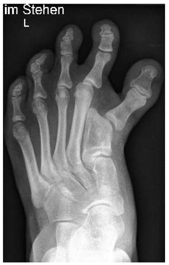
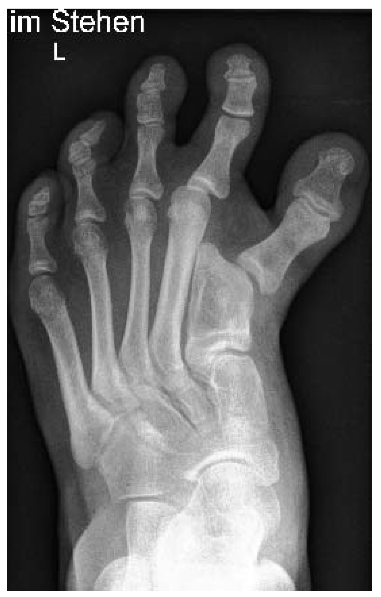
Radiographs showed shortening and bulging of the first metatarsal bilaterally along with a varus- aligned joint surface of the first metatarsal. The epiphyses were closed, and the first metatarsal bone was approximately 24 mm shorter than the second metatarsal (
Figure 2
).
Figure 2.
Preoperative anteroposterior radiograph of the left foot showing the shortened first metatarsal and the deformed first metatarsophalangeal joint with the varus position of the toe.
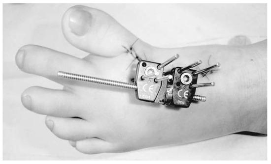
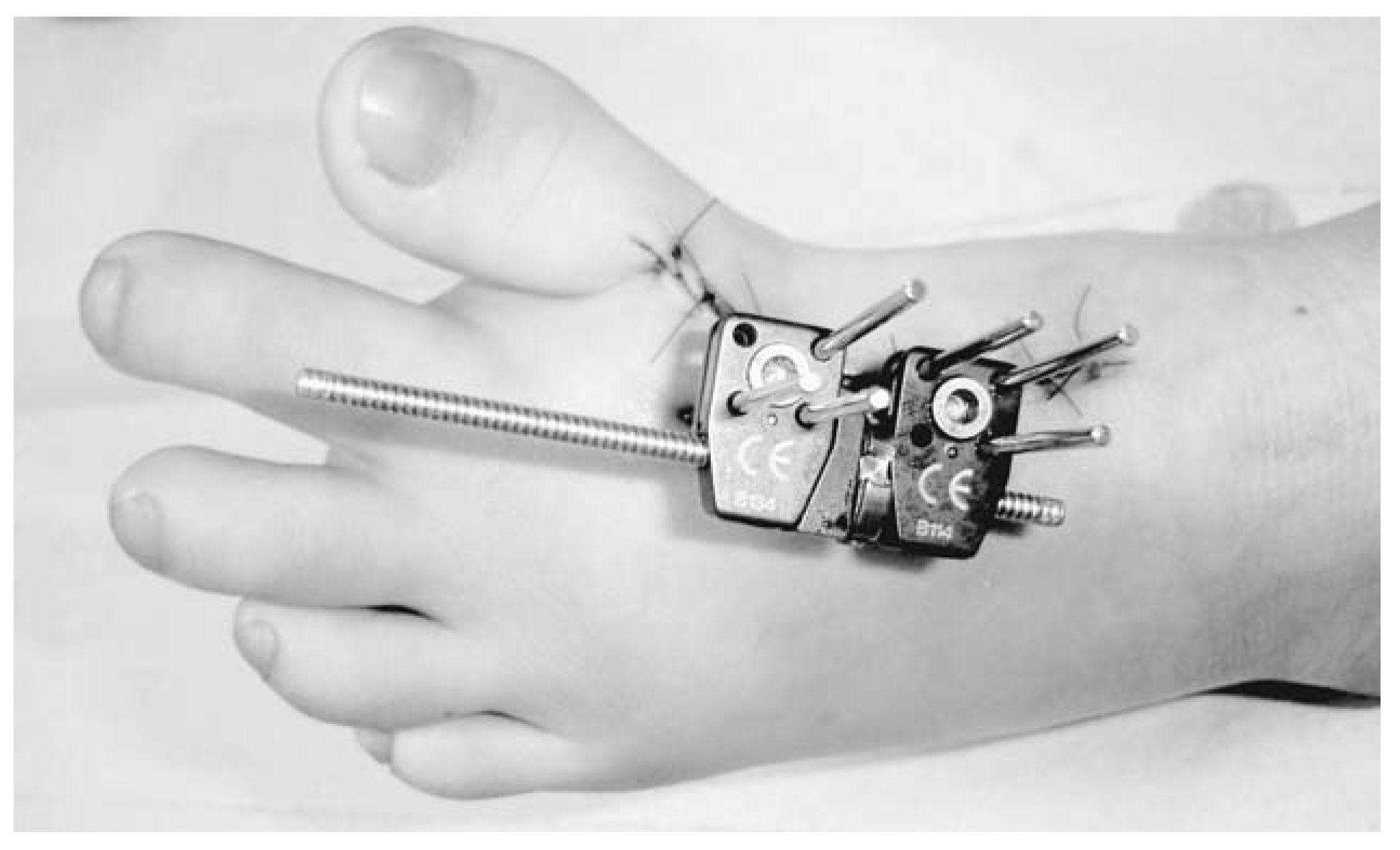
We decided on surgery. A Pennig MiniFixator (Orthofix; Ottobrunn, Germany) for callus distraction of the first metatarsal bone was applied (
Figure 3
). After surgery, the patient was walking with a postoperative shoe. After 1 week, the distraction began by approximately 1 mm daily. Six weeks after surgery, radiographs showed the regular position of the fixator, and the distraction had reached the target-value of 24 mm with a good callus formation in the distraction gap.
Figure 3.
Postoperative photograph of the external fixation device in its original configuration.
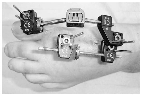
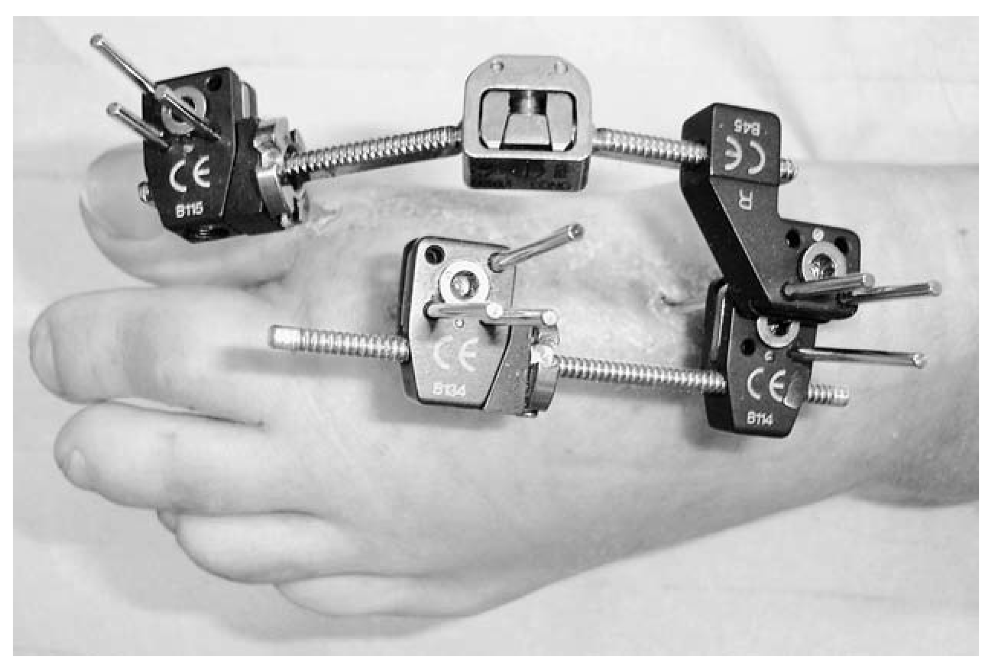
Owing to increased plantar subluxation of the phalanx during distraction, extension of the external fixator was administered so that the hallux could be repositioned to a physiologically satisfying position (
Figure 4
). The soft tissues of the hallux were gradually extended and could finally be correctly positioned. This was done during bone remodeling after distraction was completed; therefore, no treatment time was lost.
Figure 4.
Photograph of the changed external fixator, now involving the proximal phalanx. The integrated joint allows gradual adjustment of position. The calculated length has been achieved, and the hallux has a nice position before hardware removal.
After sufficient callus formation, the hardware was removed 14 weeks after surgery. The phalanx moved back to the subluxed position again, causing pain to the patient. Finally, an arthrodesis of the first metatarsophalangeal joint was done with a locking plate (
Figure 5
). An exclusive soft-tissue procedure would not be effective owing to the highly deformed metatarsophalangeal joint.
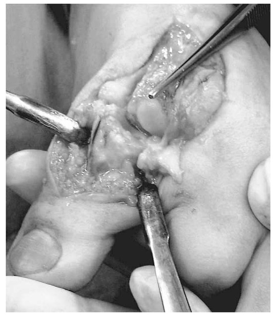
Figure 5.
Intraoperative photograph during arthrodesis. The joint surface of the metatarsal is small with a 458 medial deviation.
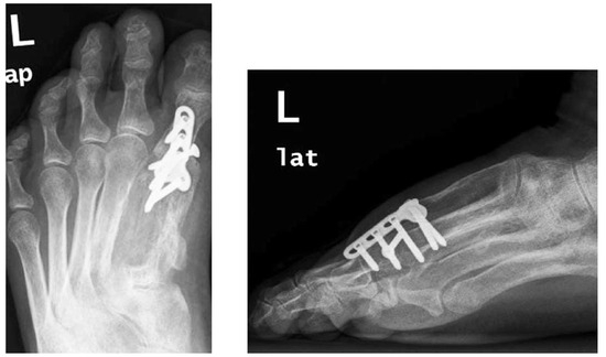
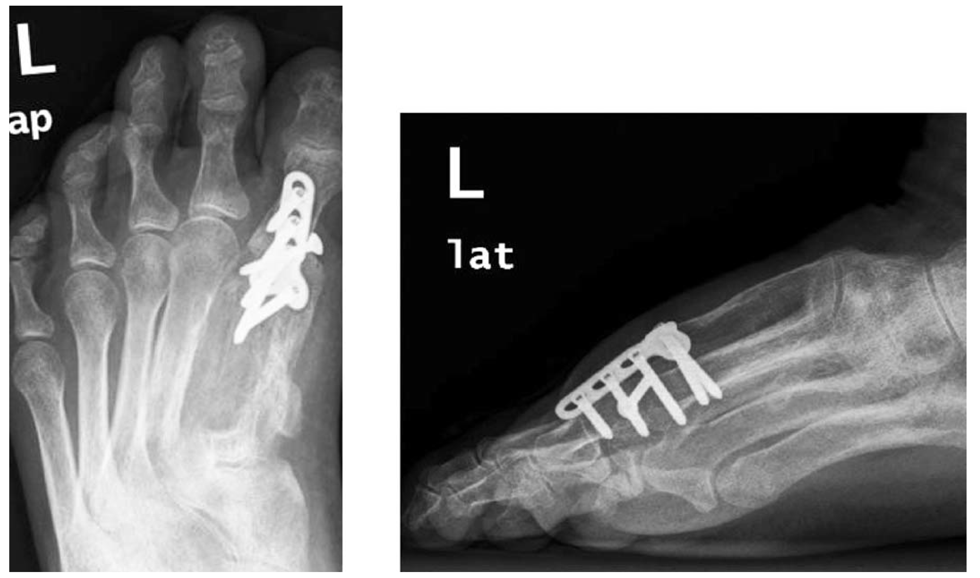
After the arthrodesis, the patient again used a postoperative shoe and was fully weightbearing for 6 weeks. Her postoperative clinical and cosmetic results were good, and the patient was satisfied. She no longer had any discomfort and pain (Figure 6 and Figure 7). A surgical procedure for the opposite side was scheduled.
Figure 6.
End result after final procedure showing the nicely aligned hallux.
Figure 7.
Postoperative anteroposterior and lateral radiographs after consolidated arthrodesis displaying the callus formation and optimal length of the first metatarsal.
Discussion
Apart from age and stage of growth, the extent of the deformity is important for selecting the appropriate surgery. Single-stage intervention or callus distraction is chosen depending on the length to be obtained. Several authors [9,12,20] have determined a maximal extension of approximately 15 mm for single-stage procedures, because neurovascular damage may arise. In the present case, we decided to use callus distraction because our lengthening goal was 24 mm to adapt to the length of the other metatarsals. Glickman and Cornfield [2] also report the deformity of a hallux varus with brachymetatarsia, and surgical treatment of the deformity by bone interposition from the second through fourth metatarsals. This reported single-step procedure included shortening of the second through fourth metatarsals of 1 to 1.5 cm by osteotomy, using the removed bone as an autograft to lengthen the first ray and fixing it with Kirschner wires. With this method, a distance of 17 mm was gained, still leaving an inequality of 7 mm, which is, according to the literature, still defined as brachymetatarsia [12,20,21].
In contrast to Glickman and Cornfield [2], we chose callus distraction using a Pennig MiniFixator to avoid any potential damage to the soft tissue, nerves, and problems with perfusion in gaining a length of 24 mm.
The subsequent arthrodesis, 6 months after the initial intervention, is unusual for an adolescent and was not initially planned. It was necessary owing to the deformed metatarsal joint surface. Although soft-tissue distraction was performed by extending the fixator, the hallux subluxed after we removed the hardware. Reasons for subluxation may have been because of the combination of congenital highly medially deviated joint surface combined with still-elevated tension of the soft tissues, especially the flexor hallucis longus tendon. How- ever, an arthrodesis could be performed only after callus distraction, because by then, enough bone length had been gained.
As described by Gilbody and Nayagam [22], the surgical challenge is to sufficiently fix the pins of the fixator, which is difficult with significantly shortened metatarsal bones. A total length of the metatarsal bone of 3 to 4 cm is critical. The difficulty in setting the pins is that they have to be in the exact plane because the position of the pins, once set, leads the way for lengthening. The preferred site for the osteotomy is the distal metaphysis of the metatarsal, although in most cases, owing to the shortness of the bone, the osteotomy must be performed proximally in the small gap between the pins of the external fixator. Alternatively, joint-bridging pins of the tarsometatarsal joint could be used, although in our opinion, we do not advise involving more joints than needed.
Several authors [2,6,9,13,23] agree that this kind of foot deformity has a major effect on gait and gait pattern, because the hallux varus—apart from not being able to wear normal shoes—causes a loss of hallux purchase, irritates the bursa of the interphalangeal joint, and can involve the lesser toes that also develop an adductus. Brachymetatarsia is highly associated with transfer-metatarsalgia leading to painful calluses and soft-tissue contractures. Apart from the physical symptoms, this disorder also psychologically strains young people, as was seen in our patient.
Because a standardized technique does not exist [9], surgery should be individually planned owing to the complexity, duration of treatment, and possible complications [2,9,14,16]. Surgery should not be made for cosmesis and associated psychological aspects alone; discomfort should be the deciding factor. Surgery was performed only after growth of the patient was complete and discomfort was permanent.
Conclusions
We present the course of surgical treatment of a rare combined deformity of the forefoot by callus distraction of the first metatarsal bone followed by modification of the inserted fixator to reposition the metatarsophalangeal joint. After hardware removal, an arthrodesis of the first metatarsophalangeal joint was performed with the now sufficiently elongated metatarsal. A good clinical and cosmetic result was achieved after surgery.
Financial Disclosure
None reported.
Conflicts of Interest
None reported.
References
- FARMER, A.W. Congenital hallux varus. Am J Surg 1958, 95, 274. [Google Scholar] [CrossRef] [Scilit]
- GLICKMAN, S.H.; CORNFIELD, R.H. Surgical reconstruction of a congenital foot deformity: hallux varus with brachyme- tatarsia of the first metatarsal. J Foot Surg 1990, 29, 499. [Google Scholar]
- JANIS, L.R.; DONICK, I.I. The etiology of hallux varus: A review. J Am Podiatry Assoc 1975, 65, 233. [Google Scholar] [CrossRef] [Scilit]
- BANKS, A.S.; RUCH, J.A.; KALISH, S.R. Surgical repair of hallux varus. JAPMA 1988, 78, 339. [Google Scholar] [CrossRef] [Scilit] [PubMed]
- HAWKINS, F.B. Acquired hallux varus: cause, prevention and correction. Clin Orthop Relat Res 1971, 76, 169. [Google Scholar] [CrossRef] [Scilit] [PubMed]
- MILLS, J.A.; MENELAUS, M.B. Hallux varus. J Bone Joint Surg Br 1989, 71, 437. [Google Scholar] [CrossRef] [Scilit] [PubMed]
- BILOTTI, M.A.; CAPRIOLI, R.; TESTA, J. Reverse Austin osteotomy for correction of hallux varus. J Foot Surg 1987, 26, 51. [Google Scholar]
- STANIFER, E.; HODOR, D.; WERTHEIMER, S. Congenital hallux varus: case presentation and review of the literature. J Foot Surg 1991, 30, 509. [Google Scholar]
- SCHIMIZZI, A.; BRAGE, M. Brachymetatarsia. Foot Ankle Clin 2004, 9, 555, ix. [Google Scholar] [CrossRef] [Scilit]
- URANO, Y.; KOBAYASHI, A. Bone-lengthening for shortness of the fourth toe. J Bone Joint Surg Am 1978, 60, 91. [Google Scholar] [CrossRef] [Scilit]
- NUZZO, J.J.; MUELLER, R.A. Diaphyseal lengthening for brachymetatarsia. J Foot Surg 1987, 26, 332. [Google Scholar] [PubMed]
- DAVIDSON, R.S. Metatarsal lengthening. Foot Ankle Clin 2001, 6, 499. [Google Scholar] [CrossRef] [Scilit] [PubMed]
- GIANNINI, S.; FALDINI, C.; PAGKRATI, S. One-stage metatarsal lengthening by allograft interposition: a novel approach for congenital brachymetatarsia. Clin Orthop Relat Res 2010, 468, 1933. [Google Scholar] [CrossRef] [Scilit]
- LEE, K.B.; YANG, H.K.; CHUNG, J.Y. How to avoid complications of distraction osteogenesis for first brachymetatarsia. Acta Orthop 2009, 80, 220. [Google Scholar] [CrossRef] [Scilit]
- LEE, W.C.; YOO, J.H.; MOON, J.S. Lengthening of fourth brachymetatarsia by three different surgical techniques. J Bone Joint Surg Br 2009, 91, 1472. [Google Scholar] [CrossRef] [Scilit]
- OH, C.W.; SATISH, B.R.; LEE, S.T. Complications of distraction osteogenesis in short first metatarsals. J Pediatr Orthop 2004, 24, 711. [Google Scholar] [CrossRef] [Scilit]
- SHIM, J.S.; PARK, S.J. Treatment of brachymetatarsia by distraction osteogenesis. J Pediatr Orthop 2006, 26, 250. [Google Scholar] [CrossRef] [Scilit]
- STEEDMAN, J.T., JR.; PETERSON, H.A. Brachymetatarsia of the first metatarsal treated by surgical lengthening. J Pediatr Orthop 1992, 12, 780. [Google Scholar] [CrossRef] [Scilit]
- WADA, A.; BENSAHEL, H.; TAKAMURA, K. Metatarsal lengthening by callus distraction for brachymetatarsia. J Pediatr Orthop B 2004, 13, 206. [Google Scholar] [PubMed]
- KIM, H.T.; LEE, S.H.; YOO, C.I. The management of brachymetatarsia. J Bone Joint Surg Br 2003, 85, 683. [Google Scholar] [CrossRef] [Scilit] [PubMed]
- BARTOLOMEI, F.J. Surgical correction of brachymetatarsia. JAPMA 1990, 80, 76. [Google Scholar] [CrossRef] [Scilit] [PubMed]
- GILBODY, J.; NAYAGAM, S. Lengthening of the first metatar- sal through an arthrodesis site for treatment of brachymetatarsia: a case report. J Foot Ankle Surg 2008, 47, 559. [Google Scholar] [CrossRef] [Scilit] [PubMed]
- VISPO-SEARA, J.L.; KRAUSPE, R. [Hallux varus congenitus]. Z Orthop Ihre Grenzgeb 1998, 136, 542. [Google Scholar] [CrossRef] [Scilit] [PubMed]
© 2024 American Podiatric Medical Association