


Abstract
Background: Low arch alignment and metatarsus primus elevatus (MPE) have been postulated to increase dorsal compressive stresses in the joints of the medial column of the foot and to contribute to the development of degenerative changes. The primary purposes of this study were 1) to examine the relationship between radiographic measures of arch alignment and MPE and 2) to assess arch alignment and MPE in individuals with midfoot arthritis and in asymptomatic controls. The secondary aim was to examine the reliability of radiographic measures of arch alignment and MPE. Methods: Radiographic measures of arch height and MPE were quantified on 28 individuals with midfoot arthritis and 22 individuals in a control group. Reliability was assessed using the intraclass correlation coefficient (ICC). The Pearson product moment correlation (r) was used to assess the relationship between arch alignment and MPE. Between-group differences were assessed using a two-sample t test (α = 0.05). Results: Good to excellent reliability was noted for measures of arch height (ICC[2,3] = 0.919–0.994) as well as MPE (ICC[2,3] = 0.891–0.882). A modest positive association was noted between normalized cortical elevation and normalized navicular height (r = 0.274, P = .030) and calcaneal inclination angle (r = 0.263, P = .035). Individuals with midfoot arthritis demonstrated lower arch alignment, reflected in a significantly higher calcaneal–first metatarsal angle (P = .002), lower calcaneal inclination angle (P = .004), and lower normalized navicular height (P < .001) compared with controls. No evidence was found to support between-group differences in lateral intermetatarsal angle (P = .495) and normalized cortical elevation (P = .146). Conclusions: These findings provide objective data establishing the reliability of measures of MPE and arch alignment and their potential clinical significance.
Foot alignment, reflected in the height of the medial longitudinal arch, may influence foot motion[1,2,3] and muscle activity[4] and, consequently, has been linked with the evolution of musculoskeletal symptoms and pathologic abnormalities.[5] Low arch alignment and attendant increases in first metatarsal lowering and forefoot abduction[1,2,3] may potentially contribute to increased articular stress at the transverse-tarsal joints.[6] Similar increases have been postulated to occur at the medial tarsometatarsal and first metatarsophalangeal articulations.[7,8,9] Low arch alignment may increase dorsal compressive stresses in the joints of the medial column of the foot, which, in turn, may lead to the development of focal impingement, degenerative changes, and eventual osteoarthritis.
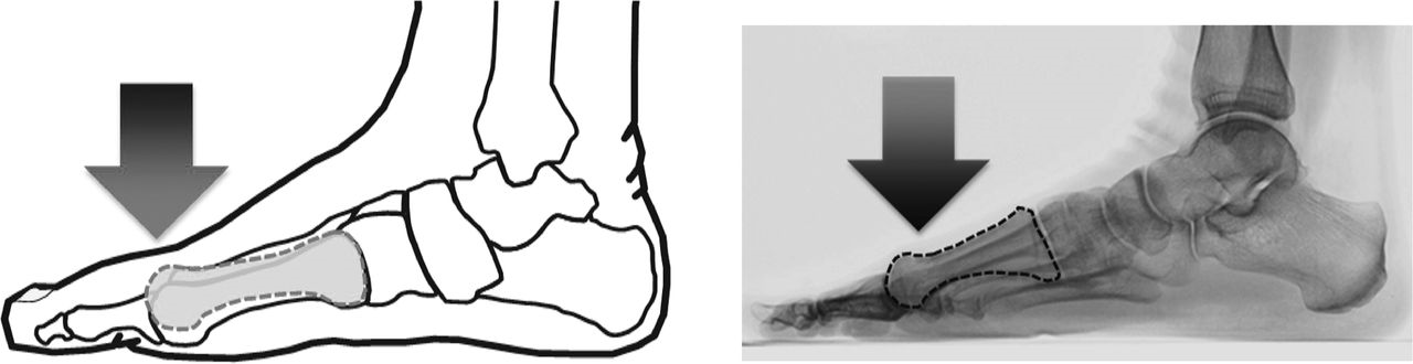
Excessive first metatarsal lowering (dorsiflexion) has been attributed not only to low arch alignment but also to aberrant foot structure reflected as metatarsus primus elevatus (MPE).[10] Metatarsus primus elevatus was first described by Lambrinudi[11] in 1938 as an anatomical variation in which the plane of the first metatarsal lies above the plane of the lesser metatarsals. It has been described as excessive dorsal elevation of the first metatarsal (Fig. 1), and it may be indicative of potential first-ray hypermobility.[12,13] Although clinical reports have suggested that MPE is a key causative factor in the development of arthritic changes at the first metatarsophalangeal joint,[11,13] objective evidence remains equivocal.[12,13,14] To our knowledge, no previous studies have assessed MPE in individuals across a range of arch alignments and degenerative osteoarthritis. Consequently, objective data examining whether MPE is related to first metatarsal lowering that accompanies low arch alignment are not available.
Figure 1.
Schematic representations of metatarsus primus elevatus, ie, dorsal elevation of the first metatarsal and concomitant first metatarsal dorsiflexion.
The gold standard for assessing arch alignment[15,16,17] and MPE[12,13] is weightbearing radiographic measurement. Arch alignment has been quantified on lateral and anteroposterior views to evaluate deviations in the sagittal and transverse planes, respectively. Frequently used measures of arch alignment with well-established reliability include angular measures (such as calcaneal inclination angle, calcaneal–first metatarsal angle, and talonavicular coverage angle)[15,18,19] and linear measures (such as navicular height and dorsum height).[15,16] Metatarsus primus elevatus has been evaluated on lateral weightbearing radiographs using angular[20] and linear measures.[13,14] However, limited objective data are available examining the reliability of radiographic measures used to assess MPE.
Clinical reports have identified arch alignment and MPE as potential contributors to the development of dorsal impingement and consequent degenerative changes at the first metatarsophalangeal joint.[11,13] Low arch alignment has also been identified in individuals with talonavicular osteoarthritis.[5,9] Quantitative data examining the role of arch alignment and MPE in individuals with arthritis at the tarsometatarsal joint (midfoot) are lacking. Midfoot arthritis has emerged as a significant clinical problem owing to an alarming increase in incidence[21,22] combined with a high potential for chronic secondary disability.[23] In vivo kinematic studies indicate that individuals with midfoot arthritis may demonstrate excessive first metatarsal mobility in challenging activities.[24] Assessments of arch alignment based on preoperative radiographs obtained from individuals with midfoot arthritis support low arch alignment in this population with the caveat of wide variability.[25,26] For instance, the preoperative talonavicular coverage angle varied from 5.2° to 21.3° in one cohort of individuals with atraumatic midfoot arthritis.[26] Wide variation in measures of arch alignment and lack of objective data quantifying MPE in patients with midfoot arthritis preclude our ability to evaluate and understand foot alignment in this clinical population.
The primary purposes of this study were 1) to examine the relationship between radiographic measures of arch alignment and MPE and 2) to assess arch alignment and MPE in individuals with midfoot arthritis and in asymptomatic individuals in a control group. The secondary aim was to examine the reliability of radiographic measures of arch alignment and MPE.
Methods
Participants
Radiographic data were obtained retrospectively on 50 participants: 28 individuals with midfoot arthritis (mean ± SD age, 58 ± 8 years; mean ± SD body mass index [calculated as weight in kilograms divided by height in meters squared], 28.7 ± 4.7; 26 women) and 22 asymptomatic individuals in a control group (mean ± SD age, 37 ± 15 years; mean ± SD body mass index, 24.7 ± 6.7; 18 women). Individuals with midfoot arthritis were taking part in a larger study assessing the effect of shoe inserts on foot motion, plantar loading, and self-reported function.[7,27] All of the individuals with midfoot arthritis presented with unilateral pain localized to the medial aspect of the dorsum of their foot, aggravated by weightbearing. The clinical impression of isolated midfoot arthritis was confirmed with weightbearing lateral and anteroposterior radiographs (see the “Procedures” subsection for additional details).
Asymptomatic individuals in the control group were recruited from the community and were screened for lower-extremity pain or symptoms. A single physical therapist (S.R.) screened all of the control participants. The exclusion criteria for both groups included 1) concomitant injury or previous surgery of the index lower extremity; 2) conditions such as stroke, inflammatory arthritis, or diabetes that may influence foot function; and 3) use of an assistive device, such as a walker or a cane. Informed consent was acquired before initiation of the study procedures. All of the procedures were approved by the Institutional Review Boards at the University of Rochester Medical Center and, therefore, have been performed in accordance with the ethical standards of the 1964 Declaration of Helsinki.
Procedures
Weightbearing anteroposterior and lateral digital radiographs were obtained using standard clinical techniques.[15] Participants were instructed to stand in a relaxed bilateral equal weightbearing stance. A focal distance of 40 inches (100 cm) was used, with exposure set to 50 mA at 60 kV. In patients with midfoot arthritis, the involved foot was x-rayed. In control participants, the dominant foot was x-rayed.
Data Analysis
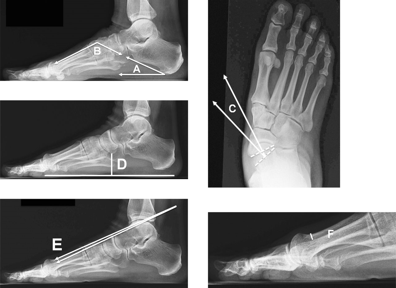
Arch alignment was quantified using the following measures developed in previous studies: 1) calcaneal inclination angle, 2) calcaneal–first metatarsal angle, 3) talonavicular coverage angle, and 4) normalized navicular height (Fig. 2). The calcaneal inclination angle was defined as the angle subtended between the inferior surface of the calcaneus and the supporting surface.[15,28] The calcaneal–first metatarsal angle was defined as the angle subtended by the inferior surface of the calcaneus and a line parallel to the dorsum of the midshaft of the first metatarsal.[15,28] The talonavicular coverage angle was defined as the angle subtended by the bisection of the anteromedial and the anterolateral extremes of the talar head and the bisection of the proximal articular surface of the navicular.[29] Normalized navicular height was defined as the distance between the anteroinferior prominence of the navicular tuberosity and the supporting surface and is expressed normalized to truncated foot length.[30] Truncated foot length was measured from the most posterior portion of the calcaneus to the center of the first metatarsophalangeal joint.[16]
We quantified MPE using the following measures developed in previous studies: 1) lateral intermetatarsal angle and 2) normalized cortical elevation (Fig. 2). The lateral intermetatarsal angle was measured, consistent with previous reports,[13,20] as the angle between the dorsal shafts of the first and second metatarsals. In terms of sign convention, plantarflexion of the first metatarsal relative to the second was assigned negative values, and dorsiflexion of the first metatarsal relative to the second was assigned positive values. First metatarsal head cortical elevation was measured relative to the second metatarsal and was quantified as the perpendicular distance from the dorsal aspect of the second metatarsal shaft to the dorsal aspect of the first metatarsal shaft at the level of the metatarsal necks (ie, the metaphyseal-diaphyseal junction).[13] Truncated foot length was measured from the most posterior portion of the calcaneus to the center of the first metatarsophalangeal joint.[16] Consistent with the convention used for the lateral intermetatarsal angle, plantarflexion of the first metatarsal relative to the second was assigned negative values, and dorsiflexion of the first metatarsal relative to the second was assigned positive values.
All of the readings were obtained by a single podiatric physician with more than 10 years of clinical experience (K.B.) using ImageJ software.[31] Three readings of each measure were obtained. Briefly, the tester obtained readings on all 50 images. Next, the images were randomly reordered. The tester was blinded to the previous readings (by starting a new Excel workbook) and rated all 50 images again. The same procedure was repeated to obtain the third reading.
Statistical Analysis
All of the dependent variables were assessed for normality and variance homogeneity. Reliability was assessed using the intraclass correlation coefficient (ICC[2,3]). The Pearson product moment correlation (r) was used to assess the relationship between arch alignment and MPE. Statistical significance (Ho: ρ = 0) was assessed using approximate tests based on the Fisher Z transformation (α = 0.05). Between-group differences were assessed using a two-sample t test (α = 0.05).
Figure 2.
Representative data illustrating techniques used to measure the calcaneal inclination angle (A), calcaneal first metatarsal angle (B), talonavicular coverage angle (C), normalized navicular height (D), lateral intermetatarsal angle (E), and normalized cortical elevation (F).
Results
Reliability
The ICC[2,3] and 95% confidence interval indicated good to excellent intrarater reliability for lateral intermetatarsal angle (0.891; 0.825–0.935), normalized cortical elevation (0.882; 0.789–0.934), normalized navicular height (0.989; 0.982–0.998), calcaneal inclination angle (0.994; 0.991–0.997), calcaneal–first metatarsal angle (0.991; 0.986–0.995), and talonavicular coverage angle (0.919; 0.870–0.952); standard errors were 0.53°, 0.75%, 0.75%, 1.47°, 2.77°, and 0.79°, respectively.
Associations
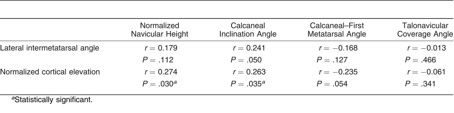
A modest positive association was noted between normalized cortical elevation and normalized navicular height (r = 0.274, P = .030) and calcaneal inclination angle (r = 0.263, P = .035). No other significant relationships were noted between measures of arch alignment and MPE (Table 1).
Between-Group Differences
Individuals with midfoot arthritis demonstrated lower arch alignment reflected in a significantly higher calcaneal–first metatarsal angle (P = .002), lower calcaneal inclination angle (P = .004), and lower normalized navicular height (P < .001) compared with individuals in the control group (Fig. 3 and Table 2). Trends toward a higher talonavicular coverage angle (P = .128) were noted in individuals with midfoot arthritis compared with those in the control group. No evidence was found to support between-group differences in lateral intermetatarsal angle (P = .495) or normalized cortical elevation (P = .146).
We assessed the role of age and body mass index as covariates. We conducted an analysis of covariance including age and body mass index as covariates when assessing the effect of group (midfoot arthritis versus controls). However, neither age nor body mass index played a significant role as covariates (P > .05). This analysis may suggest that age and body mass index did not play a significant role in mediating the between-group differences in arch alignment and MPE noted in this study.
Table 1.
Summary of Correlations Between Measures of Arch Alignment and Metatarsus Primus Elevatus
Discussion
The primary purposes of this study were 1) to examine the relationship between radiographic measures of arch alignment and MPE and 2) to assess arch alignment and MPE in individuals with midfoot arthritis and in asymptomatic individuals in the control group. The secondary aim was to examine the reliability of radiographic measures of arch alignment and MPE. The chief findings of this study indicate that weightbearing radiographs may be used to obtain reliable measures of arch alignment and MPE. We found a modest positive relationship between normalized cortical elevation and normalized navicular height and between normalized cortical elevation and calcaneal inclination angle, suggesting that arch alignment may explain approximately 10% of the variance in MPE. Patients with midfoot arthritis demonstrated lower arch alignment and similar MPE compared with their counterparts in the control group. Taken together, these results provide objective data establishing the reliability of measures of MPE and arch alignment and their possible clinical significance.
These findings of excellent reliability for angular measures of arch alignment (ICC[2,3] > 0.90) are consistent with previous studies[15,18,19,28,32,33,34] that have reported reliability coefficients of 0.89 to 0.99 for calcaneal inclination angle,[15,32,33,34] 0.99 for calcaneal–first metatarsal angle,[15] and 0.79 to 0.99 for talonavicular coverage angle.[18,19,32,33,34] Mean values of arch alignment noted in the control group (Fig. 3) were consistent with previous reports of 19° to 24°, 132°, and 12° to 20° for the calcaneal inclination, calcaneal–first metatarsal, and talonavicular coverage angles, respectively.[15,18,19,32,33,34] The findings of the present study suggest that measures of MPE also demonstrate acceptable reliability. Reliability (ICC[2,3] = 0.89) and mean lateral intermetatarsal angle were comparable with those reported by Bryant et al.[20] Several studies have quantified cortical elevation, but limited data exist examining its reliability, and wide variability in mean values has been reported in the literature.[13,14] In a recent report, Bouaicha et al[12] refined the measurement technique used to quantify MPE and reported reliability of 0.92 and a mean value of 2.6 mm in controls. In contrast, previous studies suggest that cortical elevation of up to 8 mm may be noted in controls.[13,14,35] As a strategy to reduce variability that may be related to foot size, we chose to measure cortical elevation using previously reported techniques,[13,14,35] and we express it relative to truncated foot length.[36] This strategy resulted in reliability of 0.882 and mean normalized cortical elevation of 2.57%. Normalized cortical elevation may provide a reliable alternative strategy for quantifying MPE using standard lateral weight-bearing radiographs.
Having established the reliability of the measures of arch alignment and MPE in the present cohort, we evaluated the relationship between arch alignment and MPE. Previous studies have noted that first metatarsal lowering is a feature that is common to MPE and low arch alignment[12]; therefore, we expected to find a significant relationship between arch alignment and MPE. Contrary to our expectation, these results indicate that arch alignment explains a small (~10%) albeit statistically significant portion of the variance in MPE. Because most variance in MPE is not explained by arch alignment, these results may suggest that arch alignment and MPE reflect different aspects of foot mechanics. These findings are consistent with the work of Coughlin and Kaz,[37] who noted similar magnitudes of MPE in individuals with different arch alignment (patients with adult-onset flatfoot deformity versus controls).
Figure 3.
Summary of mean between-group differences in arch alignment and metatarsus primus elevatus between individuals with midfoot arthritis (MOA) and individuals in the control group (CTRL). A, Lateral intermetatarsal angle. B, Normalized (to truncated foot length) navicular height. C, Calcaneal–first metatarsal angle. D, Normalized (to truncated foot length) cortical elevation. E, Calcaneal inclination angle. F, Talonavicular coverage angle. Error bars represent SD. *Statistically significant differences.
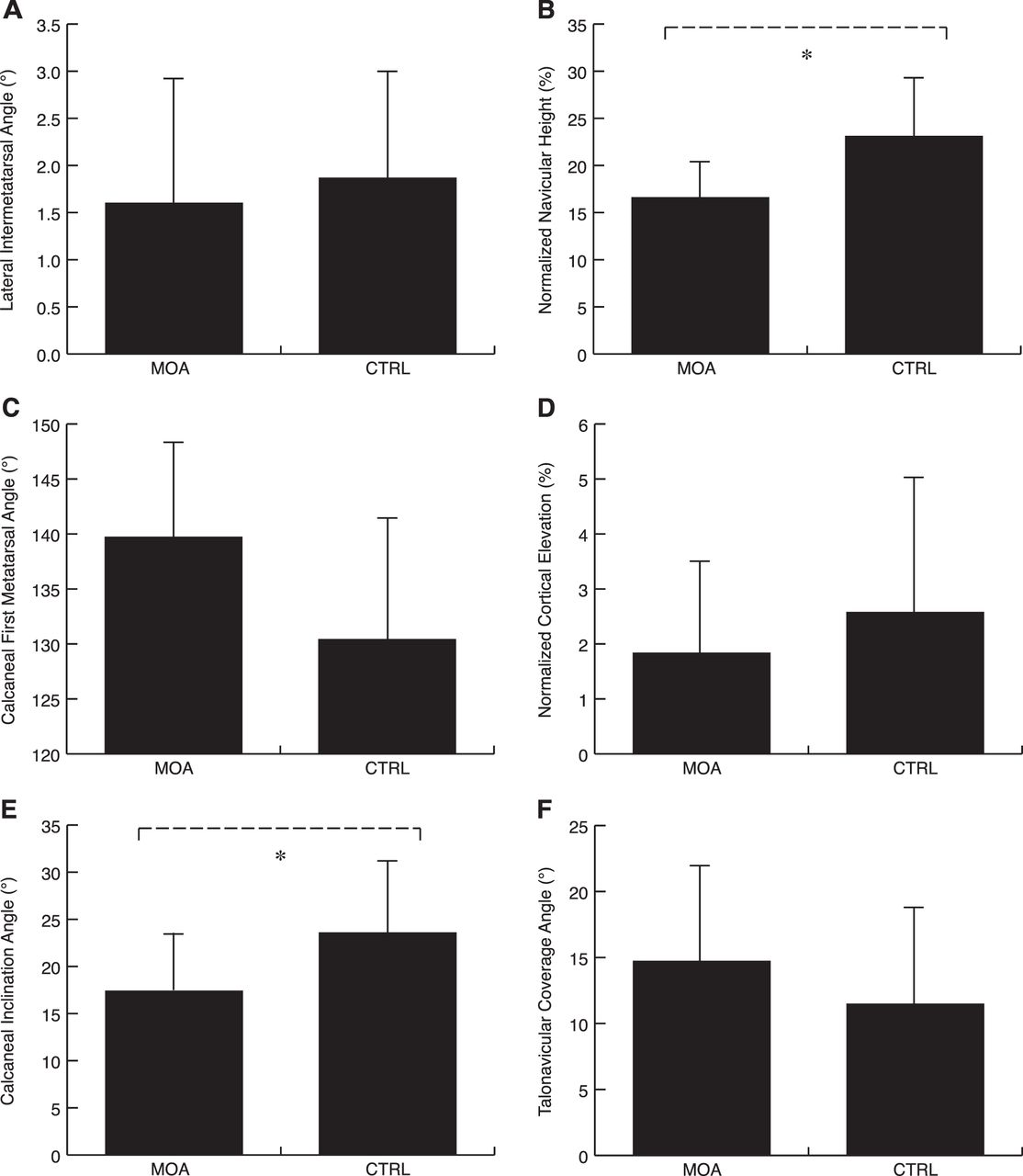
Table 2.
Summary of Between-Group Differences in Arch Alignment and Metatarsus Primus Elevatus Between Individuals with Midfoot Arthritis (MOA) and Those in the Control Group (CTRL)
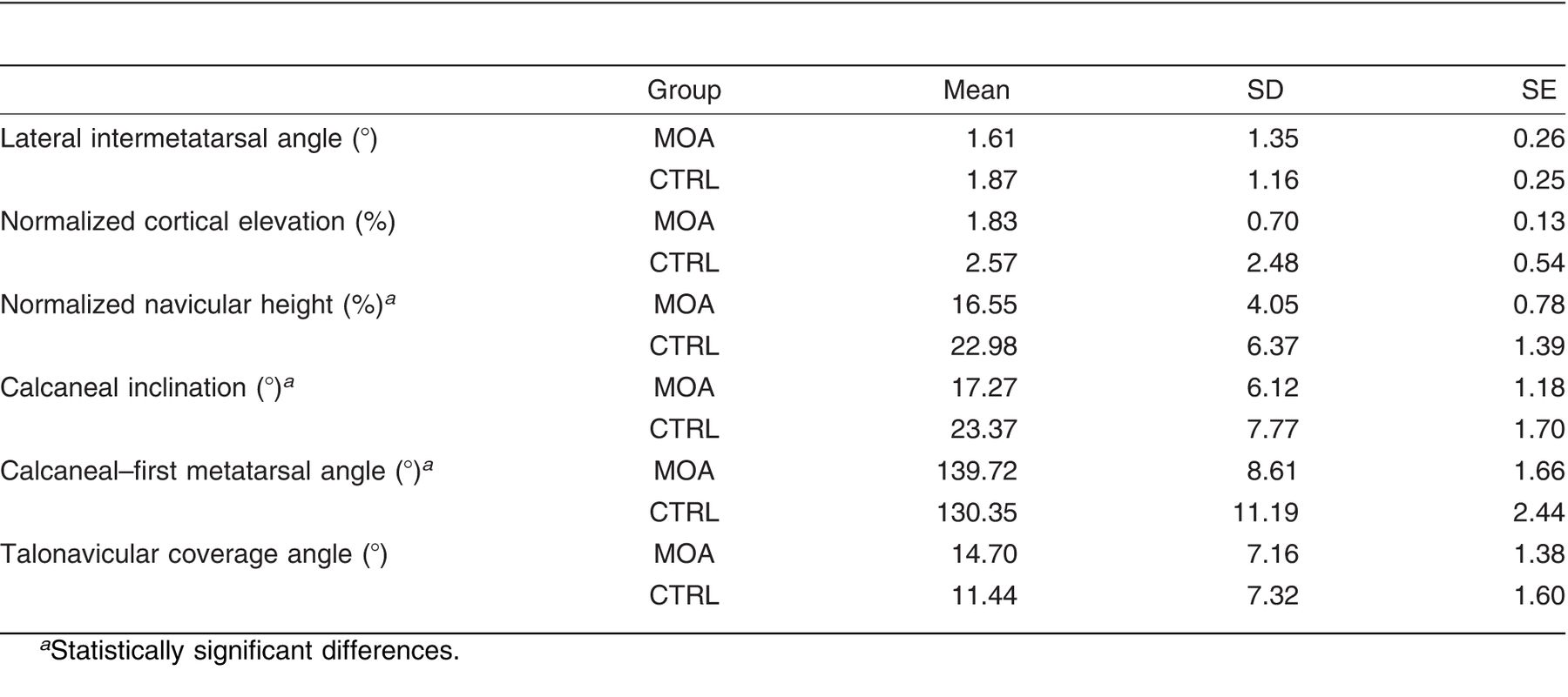
We also compared arch alignment and MPE in patients with midfoot arthritis compared with individuals in a control group. These results indicate that patients with midfoot arthritis show significant sagittal plane deviations in alignment, reflected as a lower calcaneal inclination angle, higher calcaneal–first metatarsal angle, and lower normalized navicular height (Fig. 3). Trends toward transverse plane deviations were noted in terms of a modest increase in talonavicular coverage angle compared with controls. Based on previous work indicating that individuals with midfoot arthritis demonstrate excessive first metatarsal mobility in challenging activities,[24] we expected to see radiographic evidence of significant MPE in individuals with midfoot arthritis. Contrary to our hypothesis, no differences were noted in MPE between patients with midfoot arthritis and their counterparts in the control group. Taken together, these findings underscore the importance of sagittal plane alignment in midfoot arthritis. The reduced variability in these results compared with previous reports[25,26] may be attributed to differences in samples between studies. The present patients represent a cohort with early degenerative changes compared with previous studies that assessed preoperative radiographs in patients with midfoot arthritis who failed conservative management and were seeking operative intervention.
In conclusion, the chief findings of this study indicate that MPE and arch alignment can be reliably measured using standard weightbearing radiographs. We found that arch alignment explains a statistically significant but small (~10%) portion of the variance in MPE, suggesting that arch alignment and MPE may capture different aspects of foot mechanics. Patients with midfoot arthritis demonstrated lower arch alignment, particularly in sagittal plane measures, and similar MPE compared with individuals in the control group. These results may help delineate the nature and extent of aberrant foot alignment in patients with midfoot arthritis.
Financial Disclosure: None reported. | |
Conflict of Interest: None reported. |
References
- Levinger, P, GSMurley, CJBarton, et al. :A comparison of foot kinematics in people with normal- and flat-arched feet using the Oxford.Foot Model. Gait Posture32::519. :2010. .
- Tome, J, DANawoczenski, AFlemister, et al. :Comparison of foot kinematics between subjects with posterior tibialis tendon dysfunction and healthy controls. .J Orthop Sports Phys Ther36::635. :2006. .
- Cobb, SC, LLTis, JTJohnson, et al. :The effect of low-mobile foot posture on multi-segment medial foot model gait kinematics. .Gait Posture30::334. :2009. .
- Murley, GS, HBMenz, and KBLandorf. :Foot posture influences the electromyographic activity of selected lower limb muscles during gait. .J Foot Ankle Res2::35. :2009. .
- Menz, HB, SEMunteanu, GVZammit, et al. :Foot structure and function in older people with radiographic osteoarthritis of the medial midfoot. .Osteoarthritis Cartilage18::317. :2010. .
- Kitaoka, HB, ZPLuo, and KNAn. :Contact features of the talonavicular joint of the foot. .Clin Orthop Relat Res325::290. :1996. .
- Rao, S, JFBaumhauer, JTome, et al. :Orthoses alter in vivo segmental foot kinematics during walking in patients with midfoot arthritis. .Arch Phys Med Rehabil91::608. :2010. .
- Shereff, MJ and JFBaumhauer. :Hallux rigidus and osteoarthrosis of the first metatarsophalangeal joint. .J Bone Joint Surg Am80::898. :1998. .
- Greisberg, J, STHansenJr, and BSangeorzan. :Deformity and degeneration in the hindfoot and midfoot joints of the adult acquired flatfoot. .Foot Ankle Int24::530. :2003. .
- Bryant, A, PTinley, and KSinger. :A comparison of radiographic measurements in normal, hallux valgus, and hallux limitus feet. .J Foot Ankle Surg39::39. :2000. .
- Lambrinudi, C. :Metatarsus primus elevatus. .Proc R Soc Med31::1273. :1938. .
- Bouaicha, S, CEhrmann, BKMoor, et al. :Radiographic analysis of metatarsus primus elevatus and hallux rigidus. .Foot Ankle Int31::807. :2010. .
- Roukis, TS. :Metatarsus primus elevatus in hallux rigidus: fact or fiction?JAPMA95::221. :2005. .
- Horton, GA, YWPark, and MSMyerson. :Role of metatarsus primus elevatus in the pathogenesis of hallux rigidus. .Foot Ankle Int20::777. :1999. .
- Saltzman, CL, DANawoczenski, and KDTalbot. :Measurement of the medial longitudinal arch. .Arch Phys Med Rehabil76::45. :1995. .
- Williams, DS and ISMcClay. :Measurements used to characterize the foot and the medial longitudinal arch: reliability and validity. .Phys Ther80::864. :2000. .
- Bryant, JA. :A comparison of radiographic foot measurements taken in two different positions. .JAPMA91::234. :2001. .
- Schon, LC, SBWeinfeld, GAHorton, et al. :Radiographic and clinical classification of acquired midtarsus deformities. .Foot Ankle Int19::394. :1998. .
- Menz, H and SMunteanu. :Validity of 3 clinical techniques for the measurement of static foot posture in older people. .J Orthop Sports Phys Ther35::479. :2005. .
- Bryant, A, BMahoney, and PTinley. :Lateral intermetatarsal angle: a useful measurement of metatarsus primus elevatus?JAPMA91::251. :2001. .
- Richter, M, BWippermann, CKrettek, et al. :Fractures and fracture dislocations of the midfoot: occurrence, causes and long-term results. .Foot Ankle Int22::392. :2001. .
- Patel, A, SRao, DNawoczenski, et al. :Midfoot arthritis. .J Am Acad Orthop Surg18::417. :2010. .
- Teng, AL, MSPinzur, LLomasney, et al. :Functional outcome following anatomic restoration of tarsal-metatarsal fracture dislocation. .Foot Ankle Int23::922. :2002. .
- Rao, S, JFBaumhauer, JTome, et al. :Comparison of in vivo segmental foot motion during walking and step descent in patients with midfoot arthritis and matched asymptomatic control subjects. .J Biomech42::1054. :2009. .
- Mann, RA, DPrieskorn, and MSobel. :Mid-tarsal and tarsometatarsal arthrodesis for primary degenerative osteoarthrosis or osteoarthrosis after trauma. .J Bone Joint Surg Am78::1376. :1996. .
- Jung, HG, MSMyerson, and LCSchon. :Spectrum of operative treatments and clinical outcomes for atraumatic osteoarthritis of the tarsometatarsal joints. .Foot Ankle Int28::482. :2007. .
- Rao, S, JFBaumhauer, LBecica, et al. :Shoe inserts alter plantar loading and function in patients with midfoot arthritis. .J Orthop Sports Phys Ther39::522. :2009. .
- Murley, G, HMenz, and KLandorf. :A protocol for classifying normal- and flat-arched foot posture for research studies using clinical and radiographic measurements. .J Foot Ankle Res2::22. :2009. .
- Scharfbillig, R, AEvans, ACopper, et al. :Criterion validation of four criteria of the foot posture index. .JAPMA94::31. :2004. .
- Williams, DS and ISMcClay. :Measurements used to characterize the foot and the medial longitudinal arch: reliability and validity. .Phys Ther80::864. :2000. .
- Rasband, WS. :ImageJ[computer program]. ,US National Institutes of Health. ,Bethesda, MD. ,1997–2009. .
- Ellis, SJ, JCYu, BRWilliams, et al. :New radiographic parameters assessing forefoot abduction in the adult acquired flatfoot deformity. .Foot Ankle Int30::1168. :2009. .
- Lee, KM, CYChung, MSPark, et al. :Reliability and validity of radiographic measurements in hindfoot varus and valgus. .J Bone Joint Surg Am92::2319. :2010. .
- Davids, JR, TWGibson, and LIPugh. :Quantitative segmental analysis of weight-bearing radiographs of the foot and ankle for children: normal alignment. .J Pediatr Orthop25::769. :2005. .
- Meyer, JO, LRNishon, LWeiss, et al. :Metatarsus primus elevatus and the etiology of hallux rigidus. .J Foot Surg26::237. :1987. .
- McPoil, TG, MWCornwall, BVicenzino, et al. :Effect of using truncated versus total foot length to calculate the arch height ratio. .Foot (Edinb)18::220. :2008. .
- Coughlin, MJ and AKaz. :Correlation of Harris mats, physical exam, pictures, and radiographic measurements in adult flatfoot deformity. .Foot Ankle Int30::604. :2009. .