


Abstract
Stress fractures are common overuse injuries of the bone that are most often seen in athletes and military personnel. These types of injuries seem to be commonly missed or delayed in diagnosis. Physicians should consider the possibility of such an injury while determining the diagnosis of a long-lasting foot pain. The aim of this paper was to report an isolated stress fracture of the lateral cuneiform bone. A review of the literature failed to find a similar case.
Stress fractures are common overuse injuries of the bone that are most often seen in athletes and military personnel. This type of injury occurs when bone experiences low force that happens repetitively for a long period. Potential causes of stress fractures include numerous intrinsic and extrinsic risk factors. Intrinsic factors include sex; endocrine factors that can cause hormonal imbalance; low bone mineral density; nutritional imbalance; and neuromusculoskeletal factors, such as high arches and a supinated foot type.[1,2] Regarding sex, compared with men, women have shorter and smaller limbs relative to body length, increased joint flexibility, and less muscular thigh development, which may all predispose women to certain overuse injuries.[3] Extrinsic risk factors include an incorrect training regimen, inadequate footwear, and other environmental factors.
Case Report
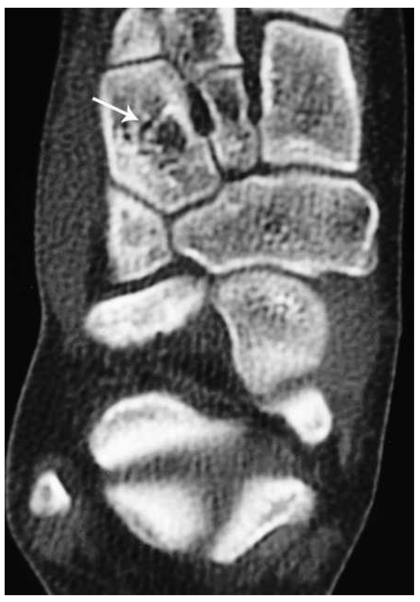
A 19-year-old female competitive soccer player presented to the Department of Orthopaedic Surgery, University Hospital Sveti Duh, Zagreb, Croatia, with a 12-month history of midfoot pain. She reported an insidious onset and a gradual increase in pain during that period. She noted dull, aching pain aggravated by walking and running. She pointed out that the focal spot is on the lateral side of the dorsal midfoot. Kicking a ball with the lateral dorsal side of the foot was especially painful. She did not report any specific incident or acute trauma. She felt pain initially only at the beginning of her soccer practice and later started to feel a constant pain gradually during the entire practice. She also felt moderate pain when performing her normal weightbearing activities, such as walking, but did not feel any pain at rest while not weightbearing. Foot or ankle swelling, skin changes, and anatomical deformity were not found during physical examination. Results of the hop test, as described by Matheson et al,[4] were positive. In the hop test, the patient attempts to hop on the injured leg, and this produces sharp, pinpoint pain in the affected area. Severe tenderness was elicited on direct palpation over the lateral midfoot. A stress fracture of the navicular bone was suspected. Plain radiographs of the foot failed to reveal fracture, and computed tomography (CT) of the foot was suggested. A CT scan of the right foot in the area of the lateral cuneiform bone (11–15 layers) revealed a visible transparent transverse fracture line extending from one cortical area to another, with minor sclerosation surrounding it. This was interpreted as a stress fracture of the lateral cuneiform bone (Fig. 1).
The patient was given nonweightbearing cast immobilization for 6 weeks. Afterward, rehabilitation treatment that consisted of four consecutive stages was initiated. The main treatment for stress fractures is rest from the aggravating activity, and it is usually conducted as a step-by-step treatment algorithm.[1,3] Because there are no algorithms for conservative treatment of an isolated stress fracture of the cuneiform bone, we followed the algorithm for conservative treatment of a partial tarsal navicular stress fracture in athletes.[5] In this algorithm, nonweightbearing short-leg cast immobilization is obliged for 6 to 8 weeks.[5,6] Afterward, rehabilitation treatment is performed in four consecutive stages, and each stage lasts for 2 weeks. We conducted a physical examination after each stage to determine whether the patient could proceed to the next stage of rehabilitation. At every physical examination, we reviewed whether the patient felt severe tenderness with direct palpation over the lateral midfoot and whether the hop test finding was positive. We reviewed whether there was foot or ankle swelling or skin changes. We also reviewed range of motion, dorsiflexion and plantar-flexion, as well as supination and pronation of the ankle joint, which were within normal limits at every physical examination.
Figure 1.
Computed tomographic scan showing a stress fracture of the lateral cuneiform bone (white arrow).
In the first stage, patients are instructed to walk with the help of crutches and not to bear weight on the affected leg. In this stage, patients start with physical therapy exercises, but only for the upper extremities and the unaffected leg. In the second stage, normal walking is allowed and patients continue with physical therapy exercises, but now exercises for both the upper and lower extremities are performed. In the third stage, patients are instructed to run in a straight line every other day and to use small weights while conducting the exercises. The distance they are allowed to run gradually increases. In the last stage, patients are gradually included in athletic training. With time, if there is no recurrence of symptoms, athletes return to their competition level.
At the 14-week follow-up visit, the patient returned to her full training activities equal to those before the injury. At the 6-month and 1-year follow-up visits, the patient did not show any recurrence of pain, and she has efficiently returned to competition.
Discussion
We presented a case of a 19-year-old female competitive soccer player who presented to us with a 12-month history of midfoot pain and was eventually diagnosed as having a stress fracture of the lateral cuneiform bone. In making a final diagnosis, plain radiographs are used as a standard method, but they have been shown to be very unreliable in the detection of stress fractures. In the study by Khan et al,[6] in diagnosing tarsal navicular stress fracture, plain radiographs demonstrated only 33% sensitivity.
Radionuclide bone scanning has predominantly been represented as a reference standard in the evaluation of stress fractures and as a diagnostic method with high sensitivity.[7] On the bone scan, focal cortical uptake can indicate a stress reaction or a real stress fracture.[4] If the present patient had received a bone scan at an early stage, it might have indicated a stress reaction before the real stress fracture developed. In that case, a shorter duration of conservative treatment would have been required. In illustrated case findings from physical examination, the patient’s medical history and duration of symptoms for 12 months were highly suggestive of a navicular stress fracture, so the bone scan was omitted. Although it has been considered that a negative bone scan finding rules out a stress lesion as a potential diagnosis, several cases in the literature where a bone scan failed to identify stress lesions have been described.[8–10] The absence of a significant osteoblastic response in the presence of early stress reactions, namely, osteopenia, may explain the lack of increased uptake of the radionuclide, especially in the diagnosis of early cortical stress fractures.[8] Radionuclide bone scanning has low specificity and, thus, cannot be used as a solid method for making a diagnosis of a stress fracture; in addition, a positive bone scan result can mimic some other, more aggressive bone diseases, such as osteomyelitis, osteoid osteoma, osteogenic sarcoma, Ewing’s sarcoma, and metastasis.
Magnetic resonance imaging (MRI) and CT could be considered diagnostic procedures of choice for stress fractures because of their greater specificity compared with other methods. The CT scan has been pointed out as a great tool for distinguishing differences between a stress reaction and a stress fracture. In the study by Gaeta et al,[8] CT had the best performance in the identification of cortical abnormalities. In the same study, CT and MRI depicted more lesions than did radionuclide bone scanning, but CT depicted more lesions than did MRI.[8] Magnetic resonance imaging is being used increasingly for detecting stress fractures, and, according to some authors, MRI is a method with greater sensitivity than radionuclide bone scanning.[8,11] What is visible on MRI is periosteal and marrow edema and fracture lines.
If there is no response to conservative treatment, operative treatment should be recommended. In their prospective study of athletes with navicular stress fractures, Saxena and Fullem[12] recommended operative treatment: open reduction and internal fixation for more severe fractures, type II and III injuries, which can be assessed by CT.
Conclusions
These types of injuries seem to be commonly missed or delayed in diagnosis. Physicians should consider the possibility of such an injury when determining the diagnosis of long-lasting foot pain. It is important to note that in the case of this patient, the injury progressed for 1 year until the accurate diagnosis was made. By continuing with sporting activities, stress fractures tend to progress with time, with pain becoming severe or occurring at an earlier stage of exercising. In suspected cases with negative radiographic findings, additional imaging techniques, such as CT, MRI, and radionuclide bone scanning, need to be considered. Radionuclide bone scanning is a diagnostic method with high sensitivity, especially at an early stage, although maybe not 100%. False-negative bone scan findings are mainly confined to case reports. Magnetic resonance imaging is a method of great sensitivity and is being used increasingly for detecting stress fractures. Computed tomography is a good diagnostic method for ascertaining the extent of the fracture and for helping to guide treatment, whether conservative or operative treatment is required. When clinically warranted, we recommend MRI or CT in the final diagnosis of stress fracture because of their greater specificity over other methods.
Financial Disclosure
None reported.
Conflict of Interest
None reported.
References
- Ivkovic, A, IBojanic, and MPecina:Stress fractures of the femoral shaft in athletes: a new treatment algorithm. Br J Sports Med 40: 518, 2006.
- Spitz, DJ and AHNewberg:Imaging of stress fractures in the athlete. Radiol Clin North Am 40: 313, 2002.
- Ivkovic, A, MFranic, IBojanic, et al:Overuse injuries in female athletes. Croat Med J 48: 767, 2007.
- Matheson, GO, DBClement, DCMcKenzie, et al:Stress fractures in athletes: a study of 320 cases. Am J Sports Med 15: 46, 1987.
- Bojanic, I and MMPecina:Conservative treatment of stress fractures of the tarsal navicular in athletes. Rev Chir Orthop Reparatrice Appar Mot 83: 133, 1997.
- Khan, KM, PDBrukner, CKearney, et al:Tarsal navicular stress fracture in athletes. Sports Med 17: 65, 1994.
- Roub, LW, LWGumerman, ENHanley, et al:Bone stress: a radionuclide imaging perspective. Radiology 132: 431, 1979.
- Gaeta, M, FMinutoli, EScribano, et al:CT and MR imaging findings in athletes with early tibial stress injuries: comparison with bone scintigraphy findings and emphasis on cortical abnormalities. Radiology 235: 553, 2005.
- Keene, JS and EGLash:Negative bone scan in a femoral neck stress fracture: a case report. Am J Sports Med 20: 234, 1992.
- Milgrom, C, RChisin, MGiladi, et al:Negative bone scans in impending tibial stress fracture: a report of three cases. Am J Sports Med 12: 488, 1984.
- Fredericson, M, AGBergman, KLHoffman, et al:Tibial stress reaction in runners: correlation of clinical symptoms and scintigraphy with a new magnetic resonance imaging grading system. Am J Sports Med 23: 472, 1995.
- Saxena, A and BFullem:Navicular stress fractures: a prospective study on athletes. Foot Ankle Int 27: 917, 2006.
© 2013 American Podiatric Medical Association