


Abstract
Background: Clinical recommendations for the prevention and healing of diabetic foot ulcers (DFUs) are somewhat clear. However, assessment and quantification of the mechanical stress responsible for DFU remain complex. Different pressure variables have been described in the literature to better understand plantar tissue stress exposure. This article reviews the role of pressure and shear forces in the pathogenesis of plantar DFU.
Methods: We performed systematic searches of the PubMed and Embase databases, completed by a manual search of the selected studies. From 535 potentially relevant references, 70 studies were included in the full-text review.
Results: Variables of plantar mechanical stress relate to vertical pressure, shear stress, and temporality of loading. At this time, in-shoe peak plantar pressure (PPP) is the only reliable variable that can be used to prevent DFU. Although it is a poor predictor of in-shoe PPP, barefoot PPP seems complementary and may be more suitable when evaluating patients with diabetes mellitus and peripheral neuropathy who seem noncompliant with footwear. An in-shoe PPP threshold value of 200 kPa has been suggested to prevent DFU. Other variables, such as peak pressure gradient and peak maximal subsurface shear stress and its depth, seem to be of additional utility.
Conclusions: To better assess the at-risk foot and to prevent ulceration, the practitioner should integrate quantitative models of dynamic foot plantar pressures, such as in-shoe and barefoot PPPs, with the regular clinical screening examination. Prospective studies are needed to evaluate causality between other variables of mechanical stress and DFUs.
Diabetes mellitus (DM) is a common chronic condition that has an increasing prevalence in the United States.[1] In 2011, the Centers for Disease Control and Prevention estimated that 25.8 million Americans (8.3%) had diabetes (both types).[2] Approximately 15% of people affected by DM with symmetrical peripheral neuropathy (PN) will develop at least one foot ulcer,[3] mainly secondary to loss of protective sensation, abnormally high foot plantar pressures,[4] and diminished vascular perfusion and skin oxygenation.[5] Prognosis related to diabetic foot ulcer (DFU) is poor because up to 24% of patients with DMPN will eventually require an amputation.[3] Furthermore, DFU recurrence is also of concern because recurrence is greater than 50% after 3 years.[6]
Healing of DFU is considered optimal with sufficient off-loading and effective off-loading modalities.[7] Cavanagh and Bus[7] demonstrated that a large discrepancy exists between current clinical practice and guidelines regarding off-loading DFU. Accordingly, they also highlighted that healing times of uncomplicated plantar ulcers reported in US clinical trials were largely higher than expected (6–8 weeks). Whereas clinical recommendations for the prevention and healing of DFU are somewhat clear,[8] assessment and quantification of the mechanical stress responsible for DFU remain complex. Different pressure variables have been described in the literature to better understand plantar tissue stress exposure.[9–13] The main objective of quantifying mechanical stress is to improve our understanding of its implication on plantar tissue trauma and breakdown leading to DFU. Modeling and monitoring mechanical plantar stress would probably benefit patients with DMPN in the prevention and healing of DFU. Accordingly, this review presents the role of pressure and shear forces in the pathogenesis of diabetic plantar forefoot ulceration, integrating the latest advances in technology for measuring pressure and shear forces under the foot and in the subsurface.
Methods
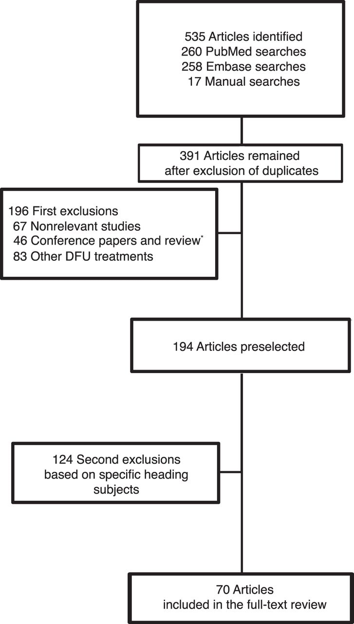
For this review, we performed a literature search in PubMed (the US National Library of Medicine’s database) and in Embase (Elsevier B.V.’s database) of all published articles until July 31, 2011. Figure 1 describes the study selection process. The predetermined keywords used for the initial selection of papers were plantar pressure, ulcer, wound, diabetes, metabolic syndrome, foot, neuropathic foot, diabetic foot, foot ulcer, pressure threshold, time pressure, shear pressure, stress, mechanical stress, tissue, plantar tissue, elder, elderly, aging, skin, skin modification, off-loading and its synonym, ischemia, callus, determinant, weight, waist circumference, obesity, activity, and activity level. Different combinations of the keywords were also used. We extracted 374 English- or French-language unique records. We then performed a second selection based on new specific keywords related to the aim of the review: clinical trial, dynamic plantar pressure, kPa, N/cm, peak plantar pressure, pressure gradient, pressure time, dynamic plantar pressure, static plantar pressure, threshold, vertical pressure, weightbearing, plantar tissue, soft tissue, stiffness, atrophy, shear pressure, shear stress, and off-loading. Seventy articles were reviewed.
Peak Plantar Pressure
Peak plantar pressure (PPP) is referred to as the highest amount of foot pressure under the ball of the foot on a specific area. It has been considered a contributing factor to skin breakdown, especially when repeated at a specific area. The PPP has been the most studied pressure variable for the past two decades, since it has been linked to foot ulceration in patients with DMPN.[4,14]
Figure 1.
Study selection process. DFU indicates diabetic foot ulcer. *Except the review by Cavanagh and Bus published in 2010.[7]
It seems that PPP is higher in patients with DM[4,9,15,16] compared with their healthy counterparts. It also seems that patients with DMPN have greater PPP at the forefoot than those who are diabetic but do not have insensate feet.[17–19] In patients with DMPN and previous foot ulceration, the highest PPP (at the midfoot) has been observed compared with patients with DMPN free of a history of foot ulceration.[20]
The plantar forefoot is where the highest PPPs are recorded.[4,21] Correlations between the location of the DFU and PPP are now well accepted and identify the forefoot as the susceptible area. More precisely, Boulton and coworkers,[22] in 1983, recorded abnormally high dynamic pressures under the metatarsal heads in 51% of patients with DMPN compared with 17% and 7% of patients with DM and control patients, respectively. Note that in the DMPN group, 13 of the 41 patients had previous ulceration, and this may have overestimated the PPP. In addition, in patients with DM and severe neuropathy, forefoot PPPs were 2.3 times higher than rearfoot PPPs, whereas they were 1.3 times higher at the forefoot of patients with DM with mild neuropathy.[9] Thus, this large prospective study showed that the more severe neuropathic group had the highest dynamic PPPs at the forefoot.[9] Furthermore, Mueller et al[19] also demonstrated that forefoot PPP was 34% greater in patients with DMPN than in control patients. Note that these studies, which demonstrate a higher PPP in patients with DMPN, were conducted in the barefoot condition.
Although, the PPP can be measured in the static or dynamic condition, dynamic pressure measurement seems to be more sensitive and reliable for evaluating the at-risk foot.[23] Note that this study was conducted barefoot. Indeed, foot-ground contact pressures and internal foot stresses were considerably higher during gait compared with standing.[24] However, keep in mind that time spent standing could be as much as twice as long as time spent walking in patients with DMPN. Therefore, the cumulative effect of PPP over time during an important standing duration may be very significant and comparable with the cumulative effect of PPP over time during walking (see the “Pressure-Time Integral” subsection later herein).[25]
In addition, in healthy individuals, it was demonstrated that the highest PPPs were found in the dynamic condition at approximately 80% of the stance phase of gait and at the forefoot.[26] It occurs between heel lift and toe lift, where the forefoot experiences relative high forces at the same time as rapidly decreasing contact area between the foot and the ground. Kelly et al[26] demonstrated that there was no difference between the timing of PPP in patients with DM who had a transmetatarsal amputation (both the amputated and the contralateral foot) and the timing of PPP in healthy individuals. This finding suggests that this portion of gait is the most deleterious in the gait cycle, but we cannot extrapolate to DM without amputation.
Is There a Critical Threshold of Plantar Pressure Predicting Forefoot Ulceration?
Considering the role of PPP in the development of diabetic ulcers in insensate feet, many authors were interested in finding a PPP threshold that would cause skin breakdown. The reason to quantify that threshold seemed to be of interest in prevention. However, it seems that no optimal single cutoff point really exists.[27] Although, many barefoot values were suggested for screening patients for at-risk foot ulceration, it is clear that the higher the PPP barefoot, the higher the risk (Table 1).
In addition, prospective studies indicate that patients with diabetes (both types) also seem to be at greater risk for alterations in plantar pressure over time,[31] and patients with type 2 DM are susceptible to elevation of plantar pressure over time.[32]
The difficulty in establishing a standardized or a specific range of PPP thresholds is mainly because DFU is a multifactorial process[5] affected by direct vertical pressure but also by shear stress (the forces that are parallel to the level of soft tissue). More recently, Owings et al[30] evaluated PPP in a group of neuropathic patients with diabetes who had a history of foot ulceration (DMPN+hxU; n = 49). This was the first cohort study (18 months of follow-up) to measure in-shoe PPP and to compare it with barefoot PPP in the same individuals who had previous plantar ulceration that remain healed. Two years earlier, Guldemond et al[33] also evaluated, for the first time, in-shoe PPPs but in patients with DMPN without a history of plantar ulceration (n = 44). They reported similar values as Owings et al.[30] For the DMPN group, mean PPP values ranged from 134 to 275 kPa under the different metatarsal heads (first: 248 kPa, second: 275 kPa, third: 264 kPa, fourth: 187 kPa, and fifth: 134 kPa) when using the same Pedar in-shoe pressure-measurement system (Novel Electronics Inc, St. Paul, Minnesota) at level walking. No threshold values were suggested because it was not the goal of the study.
Owings et al[30] reported (at a previous site of ulceration) a mean barefoot PPP of 556 kPa with great variability (107–1,192 kPa) and a lower mean in-shoe PPP of 207 kPa. As mentioned by the authors, it is important to consider that devices measuring PPP (barefoot and in-shoe) have different sensor sizes, and for this reason, absolute comparisons of pressure are complicated. However, independent of the scaling issue, they showed that barefoot PPP was a poor predictor of peak in-shoe pressure because it explains approximately 35% of the variance in in-shoe PPP. Based on this assumption, they proposed that in-shoe PPP is a key variable that should be examined in a diabetic patient with an at-risk foot.
Table 1.
Suggested Barefoot and In-Shoe Dynamic PPP Thresholds at the Forefoot
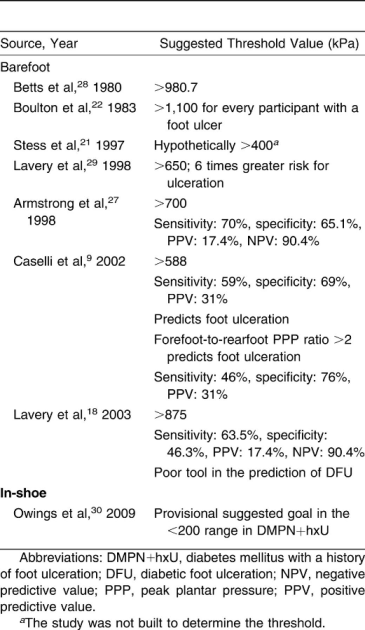
With the Pedar pressure-measurement system, they also proposed for patients with DMPN and a history of ulceration (+hxU) a provisional in-shoe pressure goal of less than 200 kPa to be the aim with appropriate footwear to prevent foot reulceration. It is necessary to emphasize the fact that this provisional goal has been obtained from patients with DMPN+hxU with individual characteristics that may differ from the rest of the DMPN+hxU population. Individual characteristics that have been assessed include the level of compliance and the activity profile. The authors also specified that their subset of patients with DMPN+hxU had a longer ulcer-free survival time than is typical. Note that this threshold was not based on confidence intervals for discrimination issue. For all of these reasons, we have to mention that this in-shoe therapeutic goal may not be extrapolated to cases of noncompliance to footwear.
At this time, in-shoe PPP measurements seem more realistic, but we cannot completely discard barefoot PPP measurements because they could be of added or complementary utility, particularly in patients without compliance to footwear.
Predicting Factors of High PPP
To be able to screen at-risk patients, without having a reliable threshold, authors studied the predicting factors of PPP. Using regression models, in healthy populations, they found several determinants related to forefoot PPP, foot structure, and biomechanical function. Especially, high walking speed,[34,35] abnormal Morton’s index,[36] low soft-tissue thickness,[36,37] abnormal arch index,[36] hallux abducto valgus deformity,[37] forefoot arthropathy,[37] abnormal muscle action, and gait style[38] were highlighted.
However, recently, Guldemond and coworkers[17] contradicted the ability of clinical and radiologic examinations to predict PPP. These authors concluded that the use of a quantitative PPP measurement was, therefore, advocated.[17]
In addition, the intervention of body mass is still debated[5,39–42] and needs to be clarified by prospective study.[40–42] However, in a cross-sectional study of male veterans, Wrobel et al[40] observed an association between several anthropometric measurements and PPP. Whereas body mass did not seem to be a main contributor, weight indices to foot area showed a strong and significant relationship with PPP. Height was also related to PPP in this homogenous population.[40] At the same time, another research team highlighted a correlation between body mass index and foot arch height. Both variables were also associated with PPP.[42]
Because gait speed may interfere with PPP, different methods have been used to alleviate potential errors due to variation in gait speed. Most of the studies reviewed used the patient’s own pace as the set speed for subsequent PPP evaluations. However, this is not ideal, and various researchers have recently developed a technique to objectively assess plantar loading independent of gait speed.[43] This technique, however, has to be tested in larger studies.
In the DM population, PN is obviously a strong predictor of a high PPP.[9,44] Tissue thickness at the metatarsal heads is also a strong predictor because it explains up to 37% of the variance in PPP measured when weightbearing and barefoot.[45] Abouaesha et al[46] have also given tissue thickness values for each metatarsal head below which foot pressures are likely high: 5.05 mm under the fifth metatarsal head, 6.55 mm under the fourth, 6.65 mm under the third, 7.85 mm under the second, and 11.05 mm under the first. Sensitivity and specificity ranged from 72.6% to 96.7% and 52.0% to 83.7%, respectively. Furthermore, an increase in soft-tissue stiffness was also demonstrated and would predict high forefoot PPP.[47–49] Toe deformity was also considered to be a reliable predictor of high PPP in patients with DMPN and in control patients,[37] as previously mentioned.
The presence of plantar callus has been positively correlated with foot ulceration at the forefoot,[16,50,51] and paring down the callus has been described to relieve pressure as much as 26% to 30%.[16,52] Altered blood flow and reduced skin oxygen supply have also been found in the neuropathic foot[53] and have also been positively correlated with the development and healing of DFU.[5] Dryness of the skin caused by peripheral autonomic neuropathy has recently been shown to be involved also,[54] but causality has not been clearly established prospectively.
Walking barefoot on concrete or similar hard surfaces was also associated with an increase in PPP, as wearing shoes decreased PPP under the foot by significantly increasing the contact area under the foot.[55]
Guldemond and coworkers[33] assessed the impact of daily life activities, and level walking was suspected to cause the highest forefoot in-shoe PPP in patients with DM (compared with stairs, ramps, turning when walking, and the Up and Go test). In that study, the DMPN group showed lower PPPs during all of the activities than did patients with DM without PN, but this may have been caused by the significantly lower walking speeds in the DMPN group (because a high walking speed is expected to cause high forefoot PPP, as mentioned earlier). This possible explanation is supported by the fact that in barefoot measurement studies, barefoot PPPs are higher in DMPN than in DM (as mentioned earlier).
Weightbearing activity as a predictor of PPP or plantar ulceration has not been fully explored yet. It is hypothesized that slow increases in weightbearing activity permit the plantar tissue to adapt to the stress and become more resistant.[56,57] It seems that foot ulceration develops significantly more in less active patients with DMPN according to Armstrong et al,[58] who studied a DMPN group that was continuously monitored for 25 weeks. Similarly, a higher weightbearing activity level (>7.5 h/d) is associated with lower reulceration of 80% compared with a weightbearing activity level less than 4.5 h/d in the DMPN+hxU group.[59]
In an observer-blinded, 12-month, randomized controlled trial, Lemaster et al[60] determined that there was no significantly increased risk of foot ulceration or foot lesion in patients with DMPN (the intervention group) who underwent a gradual and modest increase in weightbearing activity. However, note that all of the participants received therapeutic shoes and foot self-care education at baseline and that they were closely supervised and received foot care by providers throughout the study. This conclusion cannot be extrapolated to noncompliant patients or to patients who do not meet these criteria. As mentioned by the authors, this study did not record time spent standing immobile and did not monitor physical activity or cumulative plantar tissue stress continuously throughout the study. Additional studies are required and should monitor participant activity and plantar stress continuously throughout study. Thus, it is impossible at this time to recommend that patients with DMPN increase their weightbearing activities, even if lack of physical activity is not recommended in DM.
In addition, weightbearing time may be divided into different categories, such as duration of walking or standing, or further divided into categories such as average number of steps per day, number of walking episodes, and maximum walking duration.[25]
Pressure Variables Related to PPP
Forefoot-to-Rearfoot Ratio
The forefoot-to-rearfoot ratio is one of the pressure variables related to PPP and is obtained as the maximum PPP at the forefoot divided by the maximum PPP at the rearfoot.[9] In a 30-month prospective study of 248 individuals, Caselli et al[9] determined that rearfoot and forefoot pressures were increased in patients with DMPN, but only the forefoot-to-rearfoot ratio was increased significantly in severe diabetic neuropathy. They described that imbalance in loading or pressure distribution is potentially the source of pressure abnormality that leads to DFU. Moreover, they found that a forefoot-to-rearfoot ratio greater than 2 could predict ulcer development and that a PPP threshold value of 588.42 kPa (6 kg/cm2) was as specific as the forefoot-to-rearfoot ratio when walking barefoot at different walking speeds.
Peak Pressure Gradient
Introduced by Mueller et al[11] in 2005, peak pressure gradient is a pressure variable that gives additional information about mechanical stress and loading. It is defined as the greatest spatial change in plantar pressure around the PPP location. Whereas PPP gives the highest magnitude of pressure, peak pressure gradient indicates the highest change in high pressure between two specific areas around the PPP. More precisely, peak pressure gradient is calculated by subtracting the pressure in each node around the PPP from that in the adjacent node and dividing by the distance between the centers of the nodes.[11] Peak pressure gradient was early on hypothesized to be a good indicator of skin trauma because it was postulated that spatial changes in high plantar pressures would identify high stress concentrations in the soft tissue.[11] When measured in-shoe at different walking speeds in patients with DMPN+hxU, the mean peak pressure gradient was 143% higher in the forefoot than in the rearfoot, whereas the mean PPP was only 36% higher in the forefoot than in the rearfoot. The mean ± SD peak pressure gradient forefoot-to-rearfoot ratio was nearly double that of the PPP forefoot-to-rearfoot ratio (2.84 ± 1.36 versus, 1.48 ± 0.58).
Other studies also indicate that peak pressure gradient seems to be a more discriminating pressure variable than PPP alone[13,19,61] in predicting skin injury. However, it is still unknown whether peak pressure gradient can be used to predict ulceration risk, and further studies are required.
Pressure-Time Integral
Pressure-time integral gives additional information about the cumulative effect of pressure over time at a specific point under the foot. It is believed to be of additive relevance because it gives the summation of pressure and time spent on a specific area of high pressure in a single footstep.[12,20] Although PPP, forefoot-to-rearfoot ratio, and peak pressure gradient are interesting for predicting tissue injury, it is relevant to remember that they do not give information about cumulative pressure or total load exposure.[12] Although a few studies evaluated the use of pressure-time integral, data suggest that it would be complementary to the previous variables and could be used to assess the at-risk foot.[12,21] More recently, Zimny et al[62] found that patients with DMPN presented with significantly limited joint mobility at the ankle and at the first metatarsophalangeal joint that was positively correlated with significantly higher pressure-time integrals of patients with DMPN. Because of the cross-sectional study design, it is impossible to attribute the limited joint mobility to plantar ulceration. However, the higher pressure-time integral values in the DMPN group compared with the control group show us that limited joint mobility leads to abnormal loading of the foot and would, therefore, be a useful screening indicator to assess the at-risk foot in DMPN.[62]
Pressure-time integral seems to be strongly correlated with PPP at the midfoot and the forefoot based on a study comparing in-shoe pressure in five different types of footwear in patients with DMPN (n = 69) (using the Pedar-X system; Novel Electronics Inc).[63] However, this high correlation may be because up to now, calculation of pressure-time integral was based on PPP values.[10] In fact, the Novel software used to evaluate pressure-time integral may not correctly measure cumulative loading because it calculates only the summation of the PPPs recorded per time sample. Accordingly, Melai et al[10] demonstrated that this particular way of estimating pressure-time integral (or pressure-time integral_N) does not take into account the submaximal pressures experienced by the sole of the foot. Consequently, they postulated a new formula to evaluate pressure-time integral (or pressure-time integral_F) based on the integral of force over time divided by the contact area of the foot sole area. Pressure-time integral_F is, therefore, a more valid and accurate approach to determining the actual pressure-time integral because the mean cumulative load per square centimeters is obtained (including the PPP and the submaximal pressures as well) instead of pressure-time integral_N, which is the cumulative pressure and time spent only at the PPP point.[10] From the same study, different correlation variables were obtained for PPP and pressure-time integral_N and for PPP and pressure-time integral_F at different locations. It was found that most of the correlation variables were lower with pressure-time integral_F PPP than the pressure-time integral_N PPP except for the first metatarsal head (0.56 versus 0.83, respectively), the hallux (0.52 versus 0.83, respectively), and the heel (0.36 versus 0.64, respectively). With these results in mind, and because pressure-time integral_F is not calculated exclusively on PPP, it is suggested that pressure-time integral_F is a better model. However, at this time, it cannot be used as an indicator of plantar tissue damage because, to our knowledge, no study has explored the relationship between pressure-time integral_F and plantar tissue damage or ulceration risk.
Shear Stress and Related Variables
In addition to PPP, shear stress and its strain were postulated to be a significant source of mechanical stress.[27] Only a few studies have examined the role of shear stress because of the difficulty of finding a suitable device able to measure these horizontal forces.[64]
In the current literature, there are two major types of shear evaluated: shear stress applied to the surface of the skin and subsurface shear stress, which are the shear forces in the soft tissue.
Two major ways of measuring shear forces near the surface of the foot exist: with a barefoot force plate[65] or with in-shoe transducers incorporated into a customized insole,[64,66–69] the latter being more accurate regarding the real shear forces at the surface of the foot in contact with the shoe. Shear stress at the surface of the skin has two components: one oriented in a longitudinal or anteroposterior way and the other oriented in a transverse or mediolateral way.[68]
Pollard et al[68] were the first to test vertical and shear force components in healthy individuals (n = 9) with in-shoe transducers and controlled walking speed. Their work tells us that shear stress is greater at the surface of the foot under the first metatarsal head when barefoot compared with other footwear.[68] They also demonstrated that use of a plaster cast greatly reduces the three components, with longitudinal shear reduction of 71% to 81%, transverse shear reduction of 45% to 57%, and vertical pressure reduction of 35% to 62%.
To our knowledge, only one study has evaluated the differences in in-shoe shear pressure at the surface of the foot between patients with DMPN and control patients.[69] According to Yavuz et al,[69] patients with DM have higher peak-to-peak anteroposterior and mediolateral shear magnitudes and an important increase in shear-time integral values compared with healthy individuals. Shear-time integral is a temporal parameter similar to pressure-time integral that confers information about the length of time shear stress was applied to the foot.[69] As for pressure-time integral, it gives the length of the mechanical stress and is considered somewhat complementary.[69] Note, however, that this study was conducted barefoot, and we must be careful about the values obtained because they are probably not representative of shear forces at the interface between the skin of the foot and the shoe. Adding that groups were not similar in terms of age (a mean difference of 14.2 years between the control group and the diabetic group), more study is needed in evaluating shear stress between diabetic patients and healthy individuals, particularly at the foot-shoe interface.
Yavuz et al[69] also suggested that when evaluating shear stress with a single transducer, it is important to consider that skin can experience shear stress in two opposite directions, such as braking and propulsive forces. So to not underestimate shear forces, the frequency of shear forces (anteroposterior and mediolateral) is twice that of pressure.
To our knowledge, no predicting factor of shod shear has been determined in patients with DMPN or in the healthy population. However, shoe design and fit have been postulated to be critical factors of shod shear measurement.[66]
That being said, prospective study is still missing, and we cannot make causal association between shear stress and DFU. However, it is suggested that frictional shear stress is responsible for callus formation. At this time, we may question only whether tissue breakdown occurs earlier under shear forces regarding an animal study suggesting that tissue breakdown occurs earlier when shear forces are increased.[70] Lord and Hosein[67] studied a small population of DMPN+hxU (n = 6) and found that maximal in-shoe shear stress corresponds to the areas of higher prevalence of plantar ulceration. Although fragmentary and insufficient, results suggested that in DMPN+hxU, a medial shift in maximal shear stress occurs under the forefoot, and there is also increased step-to-step variability in patients with DM compared with their previous study in healthy individuals (n =9).[66] The maximum shear stresses for both groups (DMPN+hxU and control participants) did not differ significantly (73 and 87 kPa, respectively), but the DMPN+hxU group had higher magnitudes of shear forces for the first/second metatarsal heads (average: 73/64 versus 35/31 kPa, respectively) and lower ones for the third/fourth metatarsal heads (average: 51/39 versus 86.5/71, respectively). Overall, few data exist on the differences in shear stresses between patients with DM and healthy individuals.
Subsurface Shear Stress
Conversely to shear stress, which is measured at the surface of the skin, subsurface shear stress is the stress inside the overall plantar tissue generated by surface skin pressure. There are two major ways to determine subsurface shear stress: with a finite model analysis of the foot, which estimates stress in the soft tissue based on a three-dimensional modeling of the foot,[71,72] or with a mathematical function that estimates stress based on pressure distribution.[13] Both methods are based on a few assumptions and give an estimation of subsurface shear stress because it is impossible to measure it. One of the major assumptions is that plantar soft tissue is homogeneous, isotropic, linearly elastic, and bounded by an infinite plane.[13] Because it plays a key role in the mechanical failure criteria, it is reported that maximum shear stress (MSS) may be used to predict tissue damage and skin breakdown.[73]
Additional parameters were recently described,[13,61] such as peak MSS (PMSS) and the depth of the PMSS below the skin surface (D_PMSS). The PMSS is obtained after comparing the MSS values of each subsurface level. The PMSS is, accordingly, the greatest magnitude of the MSS compared across all of the subsurface levels.
At first, in the DMPN+hxU group, the magnitude of PMSS is much higher than in healthy individuals (controls).[61] However, no statistically significant difference was found between the DMPN group and controls (at the forefoot).[61] The PMSS values are also much higher in the forefoot compared with the rearfoot in patients with DMPN+hxU and in those with DMPN with an active plantar ulcer (DMPN+U).[13,19] This difference between the forefoot and rearfoot PMSS values has also been seen in healthy individuals, but the magnitude of these stress variables seems to be lower than in the DMPN+U group.[19] In DMPN+U, forefoot PMSS values were found to be significantly different from those of healthy individuals, but forefoot PMSS values in patients with DMPN were not found to differ from those found in their healthy counterparts. Although fragmentary, this finding suggests that significantly different high subsurface shear stresses are found in patients with DMPN+hxU/U, particularly at the forefoot, typically where most ulcer and tissue breakdown occurs. It is still unknown whether a particular subsurface shear stress threshold exists and what value would represent an abnormal PMSS value depending on the anatomical site.
Whereas PMSS values are higher in the neuropathic diabetic foot with ulceration or before ulceration at the forefoot, D_PMSS values were found to be lower in the DMPN+hxU[13,61] and DMPN+U[19] groups, still at the forefoot. This means that the highest or peak subsurface shear stresses are located under the forefoot more superficially near the surface of the skin. When examining and comparing the D_PMSS at the forefoot among the three groups (control, DMPN, and DMPN+hxU), the depths were significantly different between the control and DMPN groups and between the control and DMPN+hxU groups.[61] This finding suggests that D_PMSS values in the neuropathic diabetic foot are diminished at the forefoot.
Also, Mueller et al[19] highlighted the fact that except for D_PMSS, all of the pressure variables at the forefoot (PPP-forefoot, peak pressure gradient–forefoot, and PMSS-forefoot) were higher in their study performed barefoot than in in-shoe studies.[13,61] According to Lott et al[61] and Mueller et al,[19] mean ± SD forefoot-PMSS values in healthy individuals were 70 ± 18 kPa in-shoe and 170 ± 59 kPa barefoot. Although the patient groups were different in the two studies, a similar difference was also found: mean ± SD forefoot-PMSS values in DMPN+hxU were 90 ± 30 kPa in-shoe and in DMPN+U were 230 ± 52 kPa barefoot. These preliminary data suggest that therapeutic shoes alone can significantly reduce plantar and subsurface shear stresses compared with walking barefoot.[19]
Some of these authors provided correlations between stress and pressure variables.[13,19,61] As a result, correlations were relatively high in the forefoot and lower in the rearfoot.[19] From that, they concluded that PPP and peak pressure gradient seem to be important variables in identifying the location of PMSSs in the subsurface tissues.[13] Peak pressure gradient and PMSS seem to be more discriminating than PPP alone in individuals at risk for ulceration.[61] Peak pressure gradient, PMSS, and D_PMSS seem to have additional value over PPP in predicting plantar tissue breakdown, with peak pressure gradient being the more discriminating variable between locations and participant groups.[19] However, at this point, it is still unknown whether these variables can predict ulcer risk, so prospective studies are needed.
Discussion
Altogether, research gathered in this literature review highlighted different pressure and force variables that should be considered to better assess the mechanical stress involved in the pathogenesis of DFUs.
Essentially, these variables are based on direct pressure (or vertical forces), shear stress (or parallel forces), and temporality of loading. Although it has been demonstrated that high plantar pressures can predict foot ulceration in diabetic patients, suggested pressure/force variables have not been thoroughly studied prospectively. To our knowledge, besides PPP, causality with tissue breakdown has not been clearly demonstrated with any other pressure/force variables. The forefoot-to-rearfoot ratio has, nevertheless, been studied prospectively and is associated with the severity of PN. Consequently, we can extrapolate only that the forefoot-to-rearfoot ratio is probably a reliable pressure variable because severity of PN has been demonstrated to predict plantar ulceration. Based on preliminary data, most of the pressure/force variables described seem to be of additional utility.
Although predicting factors of high PPP (and plantar tissue damage by extrapolation) have been explored, we did not find data that support predicting factors of high shear forces (except for shoe fit), of high subsurface shear stress, or about temporal parameters (except a few studies for pressure-time integral). In the same way, dynamic PPP thresholds (barefoot and in-shoe) have been suggested, but no specific thresholds of shear stress, subsurface shear stress, and temporal variables have been suggested. Because shear forces may be as important as vertical forces in the pathogenesis of DFU, further research on predicting factors and on shear threshold would be advocated. Difficulties having a suitable device that measures shear forces in good and normative quality explain the scant data.
At this time, only the reliable in-shoe PPP threshold value of 200 kPa suggested by Owing et al[30] can be used as a reference to prevent foot ulceration. Because this value was obtained from the previous ulcer sites of patients with DMPN who have remained healed, we believe that this threshold is safe for all diabetic patients with and without a history of previous DFU. Barefoot PPP threshold values may be used to prevent foot ulceration, but they are less representative of in-shoe PPP. Therefore, we believe that barefoot PPP threshold values should not be used alone for the prevention of DFU because of their inability to discriminate rigorously the at-risk foot prone to ulceration and because of the increased variability in barefoot pressures. Those thresholds may be more suitable for patients noncompliant with footwear. Note that several potential confounding parameters may affect plantar pressure measurements. The presence of a plantar callus, the type of shoe (eg, extra-depth versus regular), the size of the sensor, the walking speed, and the patient’s body mass index should be addressed.
Nevertheless, we believe that different in-shoe PPP threshold values may exist. We believe that the tissue repair threshold might not be the same as the plantar tissue breakdown threshold in DMPN. We also think that the plantar tissue breakdown in-shoe threshold is lower in DMPN+hxU patients than in DMPN patients because the healed site might be more prone to subsequent reulceration. With that being said, we postulate that DFU could be seen as a continuum: DM, DMPN, DMPN+U, and DMPN+hxU would represent the different stages of the plantar tissue ulceration continuum. Accordingly, PPP thresholds may appear in a descending order from DM, DMPN, DMPN+hxU to DMPN+U. In the same way, it is possible that the other pressure/force variables mentioned earlier may act similarly depending on the foot condition. However, these hypotheses still need to be proved.
To better assess the at-risk foot and to prevent DFUs, the practitioner should be aware of integrating a quantitative measurement of dynamic foot plantar pressures, such as in-shoe and barefoot PPP, in conjunction with the regular clinical foot examination. In addition, the cost-benefit ratio favors comprehensive quantitative pressure screening, considering the cost of hospitalization, management, surgery, and rehabilitation for DFU. At this time, PPP is the only in-shoe pressure/force variable that has been highlighted to be good for predicting foot ulceration. The forefoot-to-rearfoot ratio, peak pressure gradient, shear stress, PMSS, and D_PMSS are reported to be variables of great interest. Temporal variables seem promising because they represent cumulative loading. Because causality has not been made between DFU occurrence and the magnitude of pressure and force variables (except for PPP), we urge the need for prospective studies.
Financial Disclosure
None reported.
Conflict of Interest
None reported.
References
- Centers for Disease Control and Prevention .CDC’s National Surveillance Data. ,Centers for Disease Control and Prevention. ,Atlanta. ,2011. .
- Centers for Disease Control and Prevention .National Diabetes Fact Sheet. ,Centers for Disease Control and Prevention. ,Atlanta. ,2011. .
- American Diabetes Association .Consensus Development Conference on Diabetic Foot Wound Care:7–8 April 1999,Boston, Massachusetts.JAPMA89::475. ,1999. .
- Veves, A, HJMurray, MJYoung, et al. :The risk of foot ulceration in diabetic patients with high foot pressure: a prospective study. .Diabetologia35::660. ,1992. .
- Boyko, EJ, JHAhroni, VStensel, et al. :A prospective study of risk factors for diabetic foot ulcer: the Seattle Diabetic Foot Study. .Diabetes Care22::1036. ,1999. .
- Boulton, AJ, LVileikyte, GRagnarson-Tennvall, et al. :The global burden of diabetic foot disease. .Lancet366::1719. ,2005. .
- Cavanagh, PR and SABus. :Off-loading the diabetic foot for ulcer prevention and healing. .JAPMA100::360. ,2010. .
- Boulton, AJ, DGArmstrong, SFAlbert, et al. :Comprehensive foot examination and risk assessment: a report of the task force of the foot care interest group of the American Diabetes Association, with endorsement by the American Association of Clinical Endocrinologists. .Diabetes Care31::1679. ;2008. .
- Caselli, A, HPham, JMGiurini, et al. :The forefoot-to-rearfoot plantar pressure ratio is increased in severe diabetic neuropathy and can predict foot ulceration. .Diabetes Care25::1066. ,2002. .
- Melai, T, THIjzerman, NCSchaper, et al. :Calculation of plantar pressure time integral: an alternative approach. .Gait Posture34::379. ,2011. .
- Mueller, MJ, DZou, and DJLott. :“Pressure gradient” as an indicator of plantar skin injury. .Diabetes Care28::2908. ,2005. .
- Sauseng, S, TKastenbauer, GSokol, et al. :Estimation of risk for plantar foot ulceration in diabetic patients with neuropathy. .Diabetes Nutr Metab12::189. ,1999. .
- Zou, D, MJMueller, and DJLott. :Effect of peak pressure and pressure gradient on subsurface shear stresses in the neuropathic foot. .J Biomech40::883. ,2007. .
- Frykberg, RG, LALavery, HPham, et al. :Role of neuropathy and high foot pressures in diabetic foot ulceration. .Diabetes Care21::1714. ,1998. .
- Pataky, Z, JPAssal, PConne, et al. :Plantar pressure distribution in Type 2 diabetic patients without peripheral neuropathy and peripheral vascular disease. .Diabet Med22::762. ,2005. .
- Pitei, DL, AFoster, and MEdmonds. :The effect of regular callus removal on foot pressures. .J Foot Ankle Surg38::251. ,1999. .
- Guldemond, NA, PLeffers, GHIMWalenkamp, et al. :Prediction of peak pressure from clinical and radiological measurements in patients with diabetes. .BMC Endocr Disord8::16. ,2008. .
- Lavery, LA, DGArmstrong, RPWunderlich, et al. :Predictive value of foot pressure assessment as part of a population based diabetes disease management program. .Diabetes Care26::1069. ,2003. .
- Mueller, MJ, DZou, KLBohnert, et al. :Plantar stresses on the neuropathic foot during barefoot walking. .Phys Ther88::1375. ,2008. .
- Bacarin, TA, ICNSacco, and EMHennig. :Plantar pressure distribution patterns during gait in diabetic neuropathy patients with a history of foot ulcers. .Clinics (Sao Paulo)64::113. ,2009. .
- Stess, RM, SRJensen, and RMirmiran. :The role of dynamic plantar pressures in diabetic foot ulcers. .Diabetes Care20::855. ,1997. .
- Boulton, AJ, CAHardisty, RPBetts, et al. :Dynamic foot pressure and other studies as diagnostic and management aids in diabetic neuropathy. .Diabetes Care6::26. ,1983. .
- Duckworth, T, AJMBoulton, and RPBetts. :Plantar pressure measurements and the prevention of ulceration in the diabetic foot. .J Bone Joint Surg Br67::79. ,1985. .
- Sammarco, GJ and RTHochenbury. : “Biomechanics of the Foot and Ankle. ,” inBasic Biomechanics of the Musculoskeletal System, , edited byNordin, M and VHFrankel. ,3rd Ed. , p222. ,Lea & Febiger. ,Philadelphia. ,2001. .
- Najafi, B, RTCrews, and JSWrobel. :Importance of time spent standing for those at risk of diabetic foot ulceration. .Diabetes Care33::2448. ,2010. .
- Kelly, VE, MJMueller, and DRSinacore. :Timing of peak plantar pressure during the stance phase of walking: a study of patients with diabetes mellitus and transmetatarsal amputation. .JAPMA90::18. ,2000. .
- Armstrong, DG, EJGPeters, KAAthanasiou, et al. :Is there a critical level of plantar foot pressure to identify patients at risk for neuropathic foot ulceration?J Foot Ankle Surg37::303. ,1998. .
- Betts, RP, TDuckworth, IGAustin, et al. :Critical light reflection at a plastic/glass interface and its application to foot pressure measurements. .J Med Eng Technol4::136. ,1980. .
- Lavery, LA, DGArmstrong, SAVela, et al. :Practical criteria for screening patients at high risk for diabetic foot ulceration. .Arch Intern Med158::157. ,1998. .
- Owings, TM, JApelqvist, AStenstrom, et al. :Plantar pressures in diabetic patients with foot ulcers which have remained healed. .Diabet Med26::1141. ,2009. .
- Stess, RM, RMirmiran, JRArmstrong, et al. :A prospective analysis on dynamic pressures and diabetic ulcers. .Dis Manage Clin Outcomes1::142. ,1998. .
- Grimm, A, TKastenbauer, SSauseng, et al. :Progression and distribution of plantar pressure in type 2 diabetic patients. .Diabetes Nutr Metab17::108. ,2004. .
- Guldemond, NA, PLeffers, APSanders, et al. :Daily-life activities and in-shoe forefoot plantar pressure in patients with diabetes. .Diabetes Res Clin Pract77::203. ,2007. .
- Rosenbaum, D, SHautmann, MGold, and LClaes. :Effects of walking speed on plantar pressure patterns and hindfoot angular motion. .Gait Posture2::191. ,1994. .
- Warren, GL, RMMaher, and EJHigbie. :Temporal patterns of plantar pressures and lower-leg muscle activity during walking: effect of speed. .Gait Posture19::91. ,2004. .
- Cavanagh, PR, EMorag, AJBoulton, et al. :The relationship of static foot structure to dynamic foot function. .J Biomech30::243. ,1997. .
- Mueller, MJ, MHastings, PKCommean, et al. :Forefoot structural predictors of plantar pressures during walking in people with diabetes and peripheral neuropathy. .J Biomech36::1009. ,2003. .
- Morag, E and PRCavanagh. :Structural and functional predictors of regional peak pressures under the foot during walking. .J Biomech32::359. ,1999. .
- Cavanagh, PR, DSSimsJr, and LJSanders. :Body mass is a poor predictor of peak plantar pressure in diabetic men. .Diabetes Care14::750. ,1991. .
- Wrobel, JS, NJBirkmeyer, JLDercoli, et al. :Do clinical examination variables predict high plantar pressures in the diabetic foot?JAPMA93::367. ,2003. .
- Ahroni, JH, EJBoyko, and RCForsberg. :Clinical correlates of plantar pressure among diabetic veterans. .Diabetes Care22::965. ,1999. .
- Van Schie, C and AJBoulton. :The effect of arch height and body mass on plantar pressure. .Wounds12::88. ,2000. .
- Najafi, B, RCrews, DGArmstrong, et al. :Can we predict outcome of surgical reconstruction of Charcot neuroarthropathy by dynamic plantar pressure assessment? a proof of concept study. .Gait Posture31::87. ,2010. .
- Payne, C, DTurner, and KMiller. :Determinants of plantar pressures in the diabetic foot. .J Diabetes Complications16::277. ,2002. .
- Abouaesha, F, CHvan Schie, GDGriffths, et al. :Plantar tissue thickness is related to peak plantar pressure in the high-risk diabetic foot. .Diabetes Care24::1270. ,2001. .
- Abouaesha, F, CHvan Schie, DGArmstrong, et al. :Plantar soft-tissue thickness predicts high peak plantar pressure in the diabetic foot. .JAPMA94::39. ,2004. .
- Klaesner, JW, MKHastings, DZou, et al. :Plantar tissue stiffness in patients with diabetes mellitus and peripheral neuropathy. .Arch Phys Med Rehabil83::1796. ;2002. .
- Pai, S and WRLedoux. :The compressive mechanical properties of diabetic and non-diabetic plantar soft tissue. .J Biomech43::1754. ,2010. .
- Zheng, YP, YKChoi, KWong, et al. :Biomechanical assessment of plantar foot tissue in diabetic patients using an ultrasound indentation system. .Ultrasound Med Biol26::451. ,2000. .
- Murray, HJ, MJYoung, SHollis, et al. :The association between callus formation, high pressures and neuropathy in diabetic foot ulceration. .Diabet Med13::979. ,1996. .
- Pataky, Z, AGolay, LFaravel, et al. :The impact of callosities on the magnitude and duration of plantar pressure in patients with diabetes mellitus: a callus may cause 18,600 kilograms of excess plantar pressure per day. .Diabetes Metab28::356. ,2002. .
- Young, MJ, PRCavanagh, GThomas, et al. :The effect of callus removal on dynamic plantar foot pressures in diabetic patients. .Diabet Med9::55. ,1992. .
- Zimny, S, FDessel, MEhren, et al. :Early detection of microcirculatory impairment in diabetic patients with foot at risk. .Diabetes Care24.:1810. ,2001. .
- Tentolouris, N, CVoulgari, SLiatis, et al. :Moisture status of the skin of the feet assessed by the visual test neuropad correlates with foot ulceration in diabetes. .Diabetes Care33::1112. ,2010. .
- Mohamed, O, KCerny, WJones, et al. :The effect of terrain on foot pressures during walking. .Foot Ankle Int26::859. ,2005. .
- Maluf, KS and MJMueller. :Novel Award 2002: comparison of physical activity and cumulative plantar tissue stress among subjects with and without diabetes mellitus and a history of recurrent plantar ulcers. .Clin Biomech (Bristol, Avon)18::567. ,2003. .
- Mueller, MJ and KSMaluf. :Tissue adaptation to physical stress: a proposed “Physical Stress Theory” to guide physical therapist practice, education, and research. .Phys Ther82::383. ,2002. .
- Armstrong, DG, LALavery, KHoltz-Neiderer, et al. :Variability in activity may precede diabetic foot ulceration. .Diabetes Care27.:1980. ,2004. .
- Lemaster, JW, GEReiber, DGSmith, et al. :Daily weight-bearing activity does not increase the risk of diabetic foot ulcers. .Med Sci Sports Exerc35::1093. ,2003. .
- Lemaster, JW, MJMueller, GEReiber, et al. :Effect of weight-bearing activity on foot ulcer incidence in people with diabetic peripheral neuropathy: feet first randomized controlled trial. .Phys Ther88::1385. ,2008. .
- Lott, DJ, DZou, and MJMueller. :Pressure gradient and subsurface shear stress on the neuropathic forefoot. .Clin Biomech23::342. ,2008. .
- Zimny, S, HSchatz, and MPfohl. :The role of limited joint mobility in diabetic patients with an at-risk foot. .Diabetes Care27::942. ,2004. .
- Waaijman, R and SABus. :The interdependency of peak pressure and pressure-time integral in pressure studies on diabetic footwear: no need to report both parameters. .Gait Posture35::1. ,2012. .
- Perry, JE, JOHall, and BLDavis. :Simultaneous measurement of plantar pressure and shear forces in diabetic individuals. .Gait Posture15::101. ,2002. .
- Curryer, M and EDLemaire. :Effectiveness of various materials in reducing plantar shear forces: a pilot study. .JAPMA90::346. ,2000. .
- Hosein, R and MLord. :A study of in-shoe plantar shear in normals. .Clin Biomech15::46. ,2000. .
- Lord, M and RHosein. :A study of in-shoe plantar shear in patients with diabetic neuropathy. .Clini Biomech15::278. ,2000. .
- Pollard, JP, LPLe Quesne, and JWTappin. :Forces under the foot. .J Biomed Eng5::37. ,1983. .
- Yavuz, M, ATajaddini, GBotek, et al. :Temporal characteristics of plantar shear distribution: relevance to diabetic patients. .J Biomech41::556. ,2008. .
- Goldstein, B and JSanders. :Skin response to repetitive mechanical stress: a new experimental model in pig. .Arch Phys Med Rehabil79.:265.,1998. .
- Gefen, A. :Plantar soft tissue loading under the medial metatarsals in the standing diabetic foot. .Med Eng Phys25::491. ,2003. .
- Chen, WM, TLee, PVSLee, et al. :Effects of internal stress concentrations in plantar soft-tissue: a preliminary three-dimensional finite element analysis. .Med Eng Phys32::324. ,2010. .
- Hills, DA, DNowell, and ASackfield. :Mechanics of Elastic Contacts. ,Butterworth-Heinemann Ltd. ,Boston. ,1993. .