


Abstract
Background: Split-thickness skin grafts can be used for foot wound closure in diabetic and nondiabetic patients. It is unknown whether this procedure is reliable for all diabetic patients, with or without comorbidities of diabetes, including cardiovascular disease, neuropathy, retinopathy, and nephropathy. Methods: We retrospectively reviewed 203 patients who underwent this procedure to determine significant differences in healing time, postoperative infection, and need for revisional surgery and to create a predictive model to identify diabetic patients who are likely to have a successful outcome. Results: Overall, compared with nondiabetic patients, diabetic patients experienced a significantly higher risk of delayed healing time and postoperative complication/infection and, hence, are more likely to require revisional surgery after undergoing the initial split-thickness skin graft procedure. These differences seemed to be related more to the presence of comorbidities than to diabetic status itself. Diabetic patients with preexisting comorbidities experienced a significantly increased risk of delayed healing time and postoperative infection and a higher need for revisional surgery compared with nondiabetic patients or diabetic patients without comorbidities. However, there were no significant differences in outcome between diabetic patients without comorbidities and nondiabetic patients. Conclusions: For individuals with diabetes but without exclusionary comorbidities, split-thickness skin grafting may be considered an effective surgical alternative to other prolonged treatment options currently used in this patient population.
Along with the dramatic rise in prevalence of diabetes mellitus in recent years, the rates of complications of this multisystem disease have steadily increased. The lifetime risk of developing a foot ulcer for a person with diabetes is estimated to be 25% [1,2]. Diabetic foot wounds can often be challenging to treat owing to risk of infection and compromised healing, with the resultant threat of lower-extremity amputation. Split-thickness skin grafts (STSGs) have become a popular option for closure of foot wounds because of the relative simplicity of the procedure as well as shorter healing times and lower cost compared with other wound-healing modalities [3]. Despite the widespread use of this technique, previous studies regarding STSG outcomes in diabetic patients have been limited and have offered mainly brief reports with anecdotal evidence of delayed healing [4–12]. Usually, the number of diabetic patients has been very small in relation to the remainder of patients in these studies, and this has limited the power to analyze outcomes in this population. The primary goal of this study was to compare STSG outcomes in foot wounds in diabetic versus nondiabetic patients. We hypothesized that the increased healing time and complication rates observed in diabetic patients undergoing this procedure occur primarily in those with preexisting comorbidities, namely, peripheral vascular disease, retinopathy, nephropathy, and cardiovascular disease. The goal was to provide surgeons the ability to predict in advance which patients were likely to have successful STSG outcomes and, therefore, to decrease the risk of complications from this procedure.
Patients and Methods
With approval from the institutional review board of the University of Texas Health Science Center at San Antonio (San Antonio, Texas), a retrospective review of 205 electronic medical records of patients who had undergone STSG for the foot between January 1, 2005, and December 31, 2010, was performed. This study included only patients with available medical record documentation of postoperative follow-up of at least 6 months after the date of STSG. Medical records were excluded from the review if follow-up was less than 6 months. Patients found to have clinically significant arterial occlusive disease of the lower extremity were not considered for STSG because adequate perfusion to the surgical site is a preoperative requirement for this procedure [3]. The sample size was calculated according to a previous study regarding STSG for foot wounds, which had indicated that 79% of nondiabetic patients and 60% of diabetic patients had complete healing within 7 days [4]. Assuming that 70% of the patient sample was diabetic, a total sample of 200 patients would be sufficient to detect this healing rate difference by the log-rank test at α = 0.05 with 91% power. The power analysis was performed using PASS 6.0 software (Dynalab Test Systems, Reynoldsburg, Ohio).
The diagnoses of diabetes mellitus and its comorbidities—peripheral vascular disease, retinopathy, nephropathy, and cardiovascular disease—were identified by the presence of the corresponding International Classification of Diseases codes for these conditions in the patients’ medical records. The following three groups were identified: nondiabetic patients (group A; n = 69), diabetic patients without comorbidities (group B; n = 44), and diabetic patients with comorbidities (group C; n = 90). A potential fourth group of nondiabetic patients with comorbidities could be identified in the general population; however, this group was not included in the study because there were not enough patients that fell into this group from the initially reviewed medical records to maintain power for analysis. Only 11 nondiabetic patients with foot STSG in this period were identified with comorbidities, which was too small to include as a separate group. The remaining nondiabetic patients were generally healthy individuals with no mention of comorbidities in medical record documentation.
From the medical records, the following baseline characteristics were recorded: age, sex, ethnicity, date of STSG application, date of healing, presence of diabetes, duration of diabetes, body mass index (the weight in kilograms divided by the square of the height in meters), smoking history, history of amputation on the ipsilateral foot (toes, partial ray, transmetatarsal, or midfoot), history of chronic kidney disease, history of renal dialysis, and history of revascularization (lower-extremity bypass, angioplasty, stent, or atherectomy). The types of wounds included in the study were as follows: neuropathic ulcerations, wounds after amputation/debridement, traumatic wounds, and dehisced surgical wounds. Locations for STSG were at nonweightbearing surfaces, most commonly at the dorsal aspect of the foot and, to a lesser extent, at the medial or lateral aspects of the foot when dealing with wounds after amputation. The STSG was also used for covering donor defects of plantar flaps and in coverage of certain muscle flaps. All STSGs were performed in wounds with a healthy granular base without exposed bone or tendon, without infection, and in patients without arterial disease that could preclude wound healing. All stand-alone STSG procedures were performed under a regional anesthetic block with intravenous sedation. Spinal or general anesthesia was used if other more involved surgical procedures were performed concomitantly.
The following preoperative laboratory values were collected for each patient: hemoglobin A1c, serum albumin, erythrocyte sedimentation rate, C-reactive protein, blood urea nitrogen, and creatinine. Noninvasive preoperative vascular test results, including ankle-brachial index, toe-brachial index, and Doppler waveforms, were abstracted if they were present in the medical record. Intraoperative wound culture results were recorded in the following categories: staphylococcus/streptococcus only, gram-negative organisms only, polymicrobial (more than two different bacterial organisms), anaerobic organisms only, and no growth. Follow-up examination findings were scrutinized for the development of the following complications: wound dehiscence, infection, and need for revisional surgery.
Surgical Technique
Surgical management consisted of debridement of the foot wound to healthy, bleeding tissue before STSG. Deep soft-tissue cultures of the recipient site were obtained, and the wound was cleansed via pulsatile lavage with sterile normal saline. Local hemostasis was achieved by application of topical thrombin if required, and the recipient site was measured. The donor site for the STSG at the ipsilateral side of the leg or thigh was prepared by local subcutaneous infiltration of 1% lidocaine with epinephrine, and the skin was prepared topically with mineral oil. A 2-inch-wide STSG with 0.018-inch thickness was harvested using a power dermatome in the length previously measured for the recipient site. In the case of larger-width recipient sites, an additional STSG was harvested appropriately. The donor site was dressed with povidone-iodine solution and nonadherent gauze. The graft was meshed in a 1:1.5 ratio using a commercially available mesher and then was secured to the recipient site by skin staples under minimal tension. A bolster dressing consisting of nonadherent petrolatum gauze with sterile plain sponges moistened in saline was applied to firmly secure the graft in place. A well-padded short-leg cast or posterior splint was applied to the affected lower extremity. Postoperatively, patients were maintained nonweightbearing to the affected lower extremity for approximately 3 to 4 weeks or for longer if other adjunctive osseous reconstructive procedures were performed. A minimum 2-week course of oral antibiotic therapy was completed by all of the patients; the exact antibiotic regimen was adjusted based on positive culture and sensitivity results. After removal of any immobilization devices, patients were placed in nonweightbearing short-leg casts or surgical boots until clinically healed, which was defined as complete epithelialization of the wound.
Statistical Methods
For testing the effect of diabetes and comorbidities on the healing time from STSG, the outcome measure of interest was the time to heal from STSG, and, hence, the Cox regression method was used to test for significant differences in healing time, adjusted for postoperative complications, grouped age, and sex for the following comparisons:
- Nondiabetic patients (group A) versus diabetic patients (groups B and C)
- Diabetic patients without comorbidities (group B) versus diabetic patients with comorbidities (group C)
- Nondiabetic patients (group A) versus diabetic patients without comorbidities (group B)
- Nondiabetic patients (group A) versus diabetic patients with comorbidities (group C)
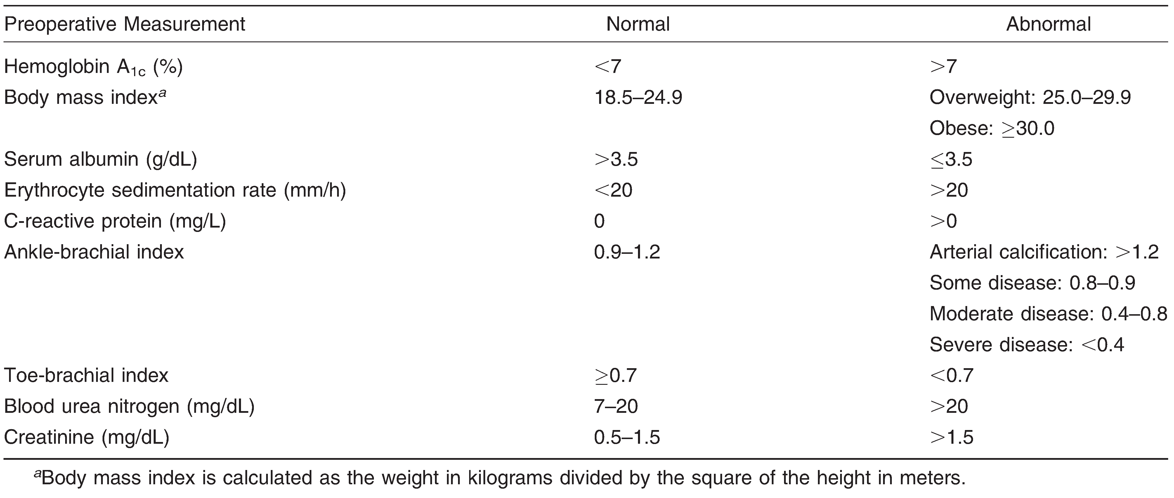
For testing the effect of diabetes and comorbidities on the risk of infection after STSG, the occurrence of postoperative infection was analyzed using contingency tables and logistic regression methods to test for significant differences in infection risks for the intergroup comparisons listed previously herein. The need for revisional surgery after STSG was recorded as a dichotomous variable, and intergroup differences were assessed using contingency tables and logistic regression methods. To screen predictors for the successful outcomes of the STSG application on diabetic patients, multivariable logistic regression methods were used to develop a predictive model based on patient demographics and factors drawn from the parameters obtained from the medical record review. For this purpose, specific laboratory values or categories were established for each of the parameters. Table 1 demonstrates the basic laboratory and vascular noninvasive study values that were used in the analysis. Where appropriate, observations were grouped or combined to create nominal or ordinal categorical variables. All of the statistical analyses were performed using the SAS JMP 8 (SAS Institute, Inc, Cary, North Carolina) and CRAN R, version 2.13, software programs.
Table 1.
Laboratory and Vascular Noninvasive Study Values Used for this Analysis
Results
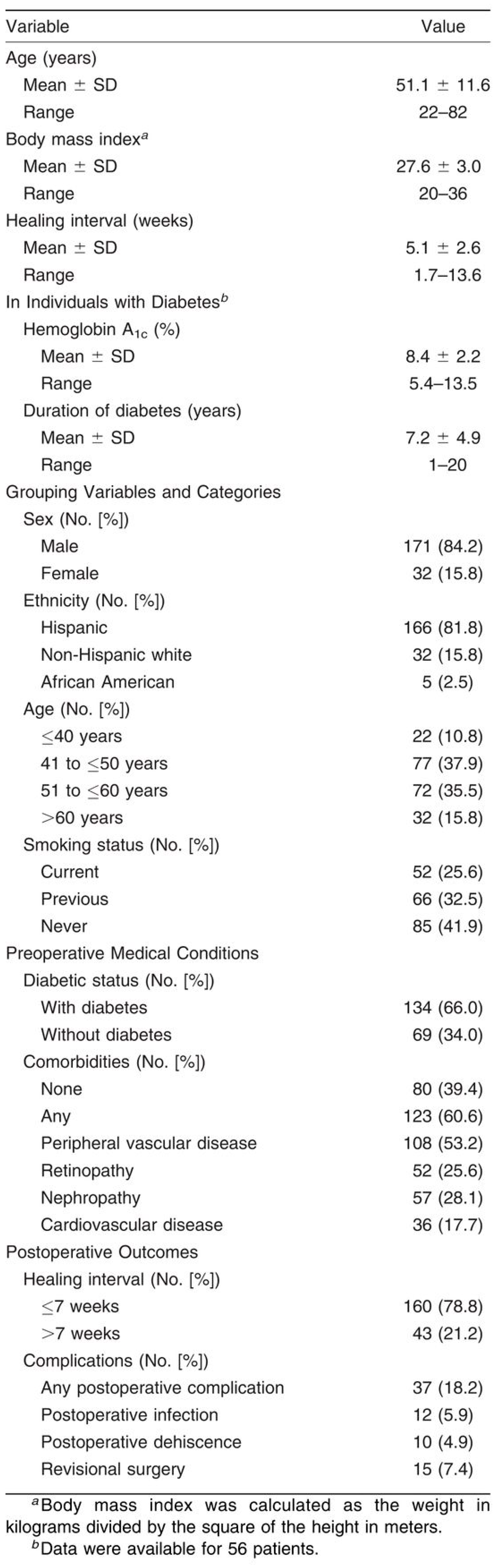
Based on the inclusion and exclusion criteria, 203 of the original 205 patients were identified for further analysis. Two patients were excluded owing to less than 6 months of documented follow-up after STSG. The distribution of each variable presented in the data set is summarized in Table 2. Most of the 203 patients observed were male (n = 171; 84.2%) and Hispanic (n = 166; 81.8%). The age distribution was unimodal and approximately symmetrical. The number of diabetic patients was approximately twice that of nondiabetic patients. More than half of the patients (n = 123; 60.6%) had at least one type of comorbidity, including diabetic and nondiabetic patients. Nearly half of the study patients had clinically diagnosed peripheral vascular disease, whereas the presence of other types of comorbidities (eg, retinopathy, nephropathy, and cardiovascular disease) was much smaller. For 63.4% of diabetic patients (n = 85), the duration of diabetes was longer than 5 years, and most diabetic patients were overweight (body mass index > 25). Most patients had normal erythrocyte sedimentation rate, C-reactive protein, toe-brachial index, blood urea nitrogen, and creatinine values, and most had no revascularization procedures, renal dialysis, chronic kidney disease, or amputation on the same foot as the graft. Most wounds were not traumatically induced, and most patients had an acceptable or normal ankle-brachial index (n = 134; 66.0%) and no bacterial growth on intraoperative wound cultures (n = 112; 55.2%). The distribution of the number of weeks to heal from STSG was positively skewed, but in most patients (n = 160; 78.8%) it was less than 7 weeks. The total cases of postoperative complication (18.2%, 37 patients), which included infection (5.9%, 12 patients), dehiscence (4.9%, 10 patients), and revisional surgery (7.4%, 15 patients), were all small.
Table 2.
Distribution Summary of Variables with Grouping Categories in the 203 Study Patients
Impact of Diabetes and Comorbidities on Healing Time
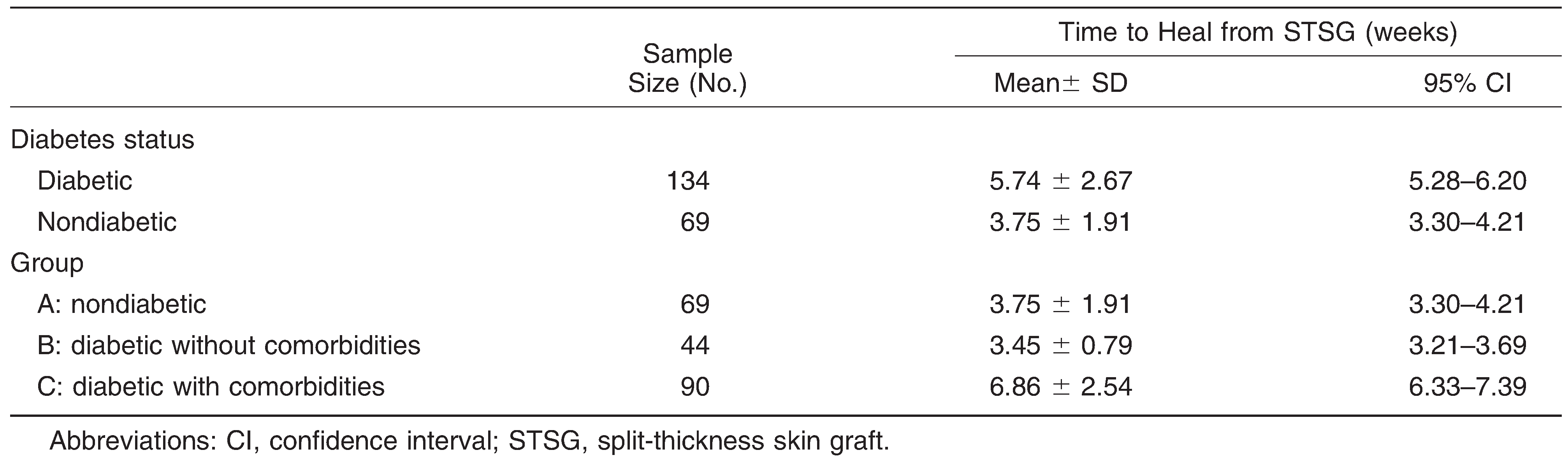
The number of weeks to heal from STSG was significantly correlated with or dependent on all of the other variables (P < .04 for all) except sex and smoking status. Table 3 shows the summary of the number of weeks to heal from STSG for nondiabetic and diabetic patients. Overall, healing time was 1.99 weeks longer for diabetic patients than for nondiabetic patients, and this difference was significant (P < .0001). Further stratification of these patients based on the three study groups revealed more striking differences in healing times between those with comorbidities and those without (Table 3). However, in those without comorbidities, there was no significant difference in healing times between diabetic and nondiabetic patients. The average difference between groups B and C was 3.41 weeks, a difference that nonparametric and t tests confirmed to be a significant difference (P < .0001). The same was true for the average difference of 3.11 weeks between groups A and C. In contrast, the average difference of 0.30 weeks between groups A and B was not significant (P = .443). This finding demonstrated that the overall difference between diabetic and nondiabetic patients was likely due to the presence of comorbidities and, therefore, warranted further investigation.
Table 3.
Time to Heal from STSG Stratified by Diabetes Status and Group (N = 203)
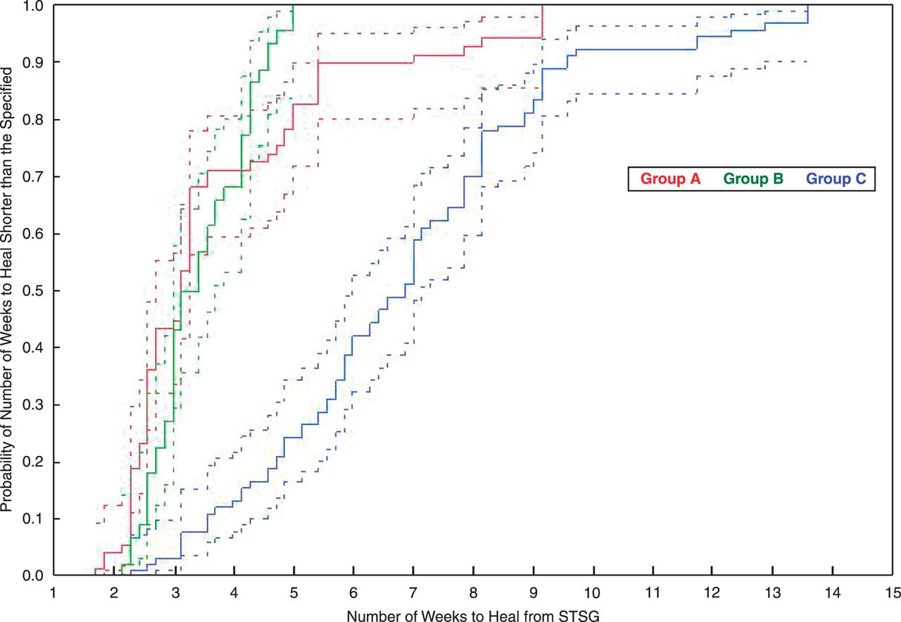
A Cox proportional hazard model (P < .001) was used to fit the healing time from STSG with the group as covariate adjusted for postoperative complication, grouped age, and sex. There was no significant effect of other preoperative variables in the model. The log-rank test and the Wilcoxon test confirmed an overall significant difference in healing times among the three groups (P < .001). Compared with patients without diabetes, diabetic patients overall experienced a 5.15 times higher risk of postoperative complications after STSG compared with patients without diabetes (95% confidence interval, 1.24–21.39; P = .009). As expected, patients without postoperative complications had a much higher rate of healing from STSG compared with those with complications (risk ratio, 7.40). Diabetic patients with comorbidities were at a significantly higher risk for delayed healing time from STSG compared with diabetic patients without comorbidities and nondiabetic patients (risk ratios, 5.05 and 3.14, respectively). The risk difference was not significant between nondiabetic patients and diabetic patients without comorbidities (risk ratio, 1.61). The Kaplan-Meier probability plot shown in Figure 1 illustrates these distinctive observations with 95% confidence intervals (dotted lines).
Figure 1.
Kaplan-Meier probability plot of shorter healing time per group.
To explain the relationship between diabetes and healing time from STSG, another Cox regression model (P < .001) was fitted but now with diabetes status and comorbidities as covariates, adjusted for postoperative complications, grouped age, and sex. The estimate of each effect confirmed that diabetic patients were at a significantly higher risk for delayed healing time from STSG compared with nondiabetic patients (risk ratio, 2.31).
Impact of Diabetes and Comorbidities on Risk of Infection
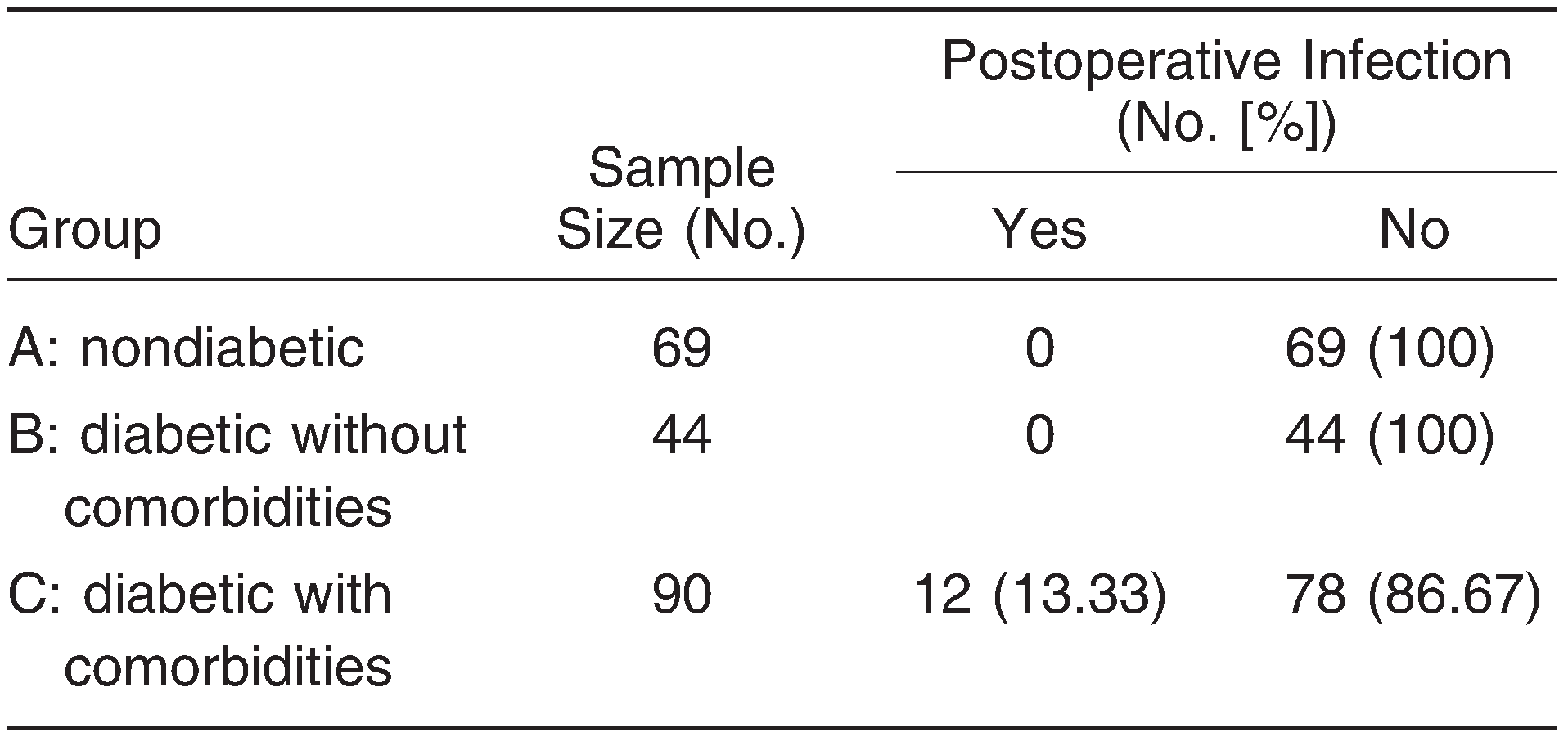
Postoperative infection from STSG was assessed in each of the three study groups, and an overall significant difference in the risk of infection was found among the groups (P = .0003). Further analysis revealed that the difference was attributed to group C alone (Table 4). From the contingency analysis using a logistic regression model, it was found that there was a higher risk of postoperative infection in patients with diabetes compared with those without diabetes (the Fisher exact test P = .006). Also, there was an increased risk of postoperative infection in group C compared with group A (the Fisher exact test P = .0008) and group B (the Fisher exact test P = .007). No statistically significant difference of the infection risk was present between nondiabetic patients and diabetic patients without comorbidities.
Table 4.
Occurrence of Postoperative Infection from Split-Thickness Skin Graft Stratified by Group (N = 203)
Impact of Diabetes and Comorbidities on Need for Revisional Surgery
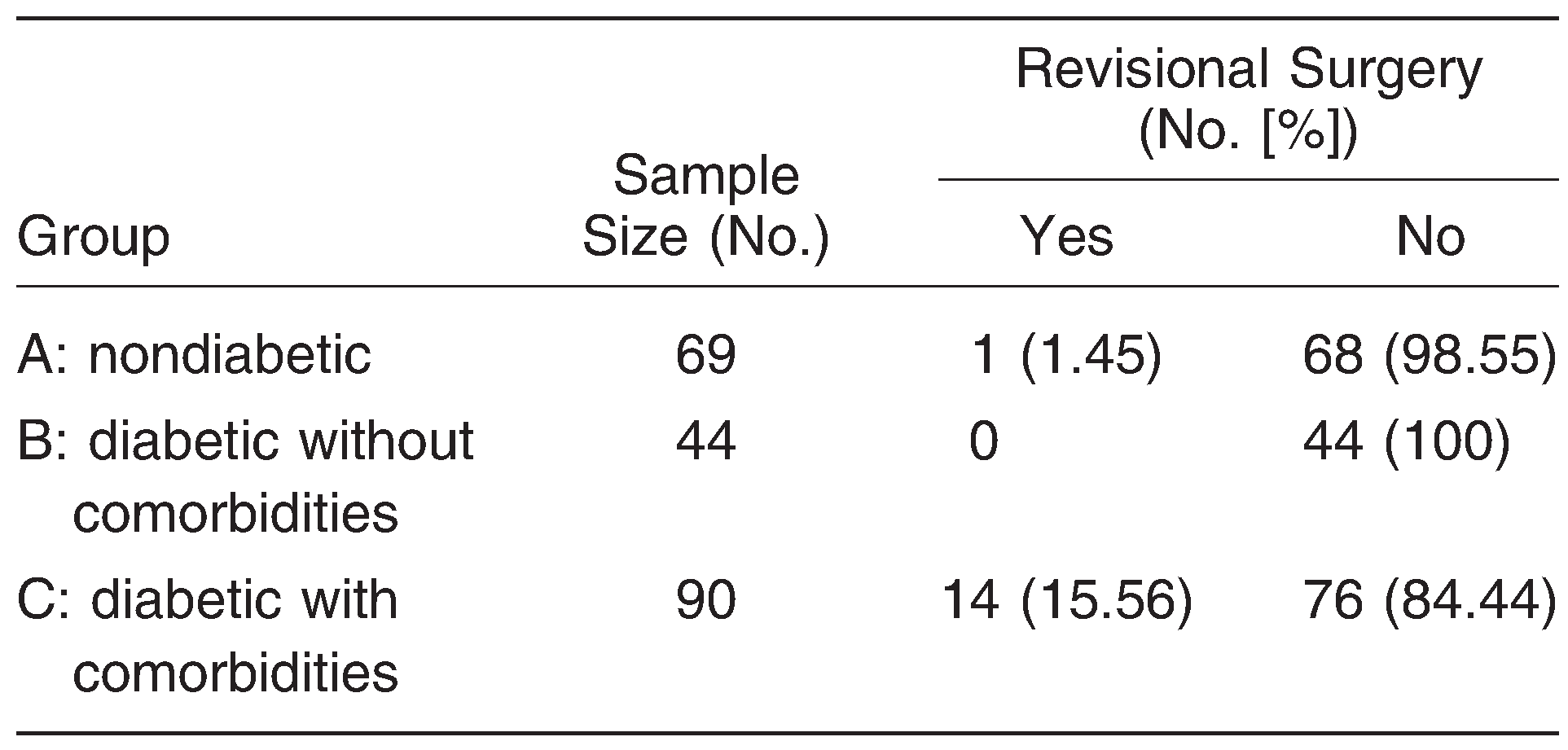
The need for revisional surgery after the STSG application was stratified by the three study groups, and an overall significant difference was found among the groups (P < .001). Further analysis revealed that the difference was again mainly attributed to group C alone (Table 5). From the contingency analysis using a logistic regression model, it was found that patients with diabetes were more likely to require revisional surgery after STSG compared with patients without diabetes (the Fisher exact test P =.014). Also, there was a higher need for revisional surgery from STSG in group C compared with group A (the Fisher exact test P = .002) and group B (the Fisher exact test P = .003). There was no significant difference in the need for revisional surgery between groups A and B (P = .423).
Table 5.
Need for Revisional Surgery from Split-Thickness Skin Graft Stratified by Group (N = 203)
Selective Predictors for Successful Outcomes of STSG
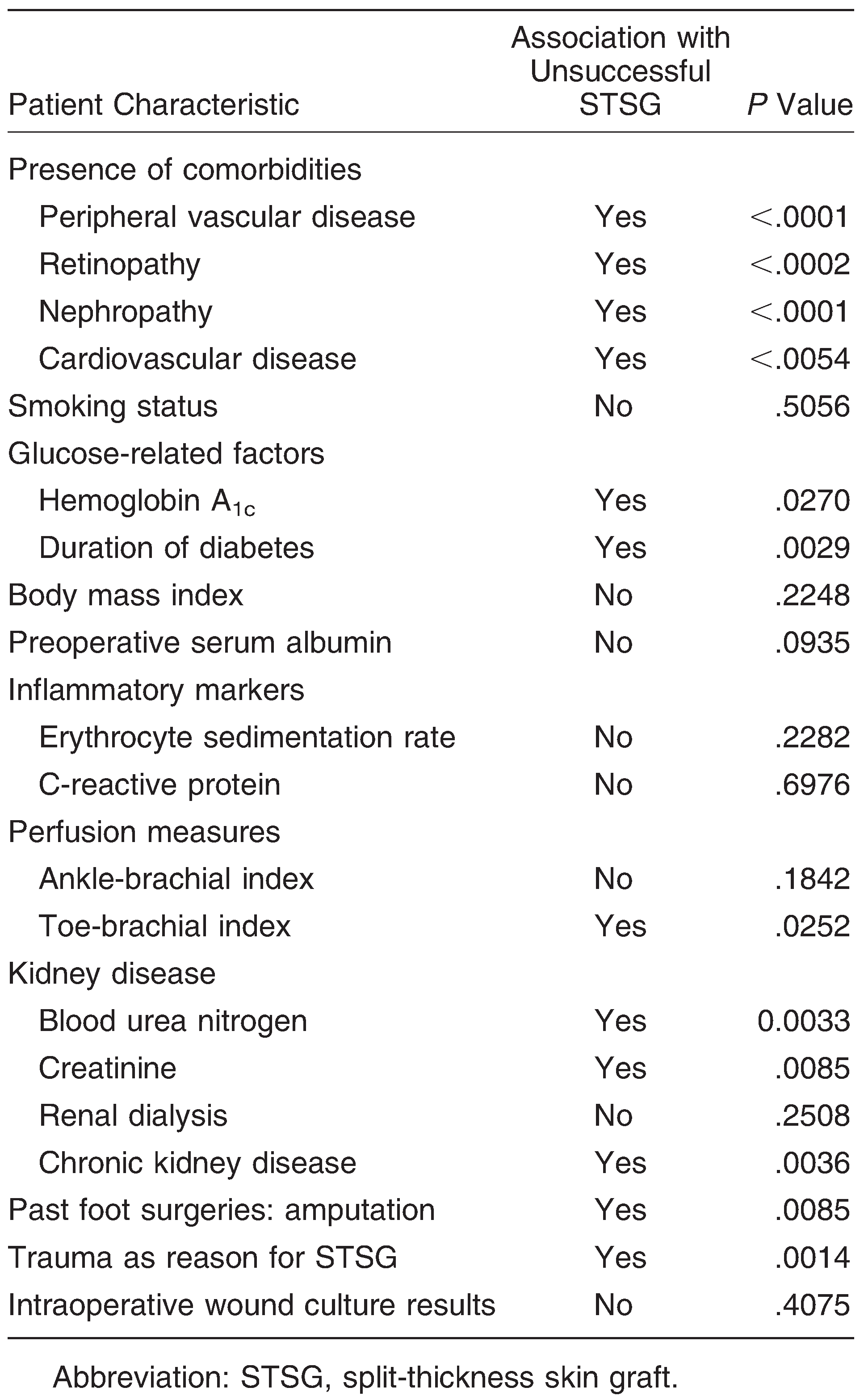
The variables in Table 6 were examined in patients with diabetes to predict which patients were more likely to have a successful outcome of STSG. The notable patient characteristics found to negatively impact outcomes after STSG were the presence of any of the comorbidities studied; glucose-related factors, particularly duration of diabetes and hemoglobin A1c level; some of the kidney disease indicators, namely, blood urea nitrogen level, creatinine level, and history of chronic kidney disease; history of amputation; and whether the reason for surgery was trauma. We used multiple logistic regression modeling and discriminant analyses in an attempt to develop a comprehensive predictive model to identify specific patient characteristics predictive of successful STSG application. After a thorough investigation of numerous combinations of these factors, however, a model of selective predictors could not be established because they did not exhibit a significant effect on predicting a successful STSG when adjusted for other factors.
Table 6.
Patient Characteristics and Their Associations with Outcome of STSG
Discussion
Since the establishment of the procedure, STSG has played a pivotal role in soft-tissue reconstruction. In recent years, STSG has entertained increasing popularity for use in diabetic foot wound coverage, yet no firm guidelines exist to guide reconstructive surgeons on the proper selection of this procedure in this patient population. The literature is replete with reports on the use of this technique for burn wounds; however, these previous studies did not focus on foot wounds and reported only brief results regarding diabetic patients [4–6,9,11]. A study by Jewell et al [4] on outcomes for 52 consecutive patients undergoing STSG for burn wounds reported slightly prolonged healing times in patients with diabetes; however, there were only three patients with diabetes in this series. Thourani et al [5] reported that the presence of diabetes had an adverse affect on the healing of STSG in burn wounds throughout the body when combined with increased age and increased total body surface area of burn, but their analyses did not distinguish between patients with and without the comorbidities described in the present study. Likewise, Mowlavi et al [6] reported decreased graft take in individuals with hyperglycemia; however, the authors used only preoperative serum glucose level, rather than a diagnosis of diabetes, in their analyses and did not distinguish between patients with and without the comorbidities associated with diabetes. The present results demonstrate interesting findings relating to the differential contributions of diabetes and its comorbidities to outcomes for STSG in foot wounds.
Most patients in the present study were male, Hispanic, diabetic, and with a high body mass index. All of the patients in the present study demonstrated complete healing at their last follow-up evaluation. Most study patients (n = 160; 78.8%) healed their STSG in less than 7 weeks, a healing interval consistent with other studies. Mahmoud et al [7] showed that 62% of 50 skin-grafted foot wounds in diabetic patients healed by postoperative week 8. Likewise, Younes et al [8] found the same healing time in their study of 16 patients treated with STSG for diabetic foot wounds. Most of the nondiabetic patients in the present study healed in less than 4 weeks, a period similar to that in other studies of STSG in nondiabetic populations, which reported healing times of 2 to 4 weeks [4].
Smoking status did not have a significant effect on healing times in this patient sample. Smoking has generally been shown to negatively affect wound healing through decreased tissue oxygenation and a subsequent decrease in collagen synthesis [12]. The number of postoperative complications found in the present patients was very small, demonstrating the overall reliability of STSG procedures. We observed only partial wound dehiscence and infection as postoperative complications in 10.8% of patients. Infection has been reported as one of the most common causes of STSG failure in several studies, regardless of the presence of diabetes [10,12,13].
A close examination of age in the present patients showed that with increasing age, the odds of successful STSG decrease substantially. Previous studies have shown prolonged wound healing and decreased wound tensile strength with increasing age [5,6]. It is well known that the prevalence of diabetes in the general population increases with age [14]. Advancing age also means that diabetic patients are more likely to have multiple progressive complications of their diabetes [11]. Older diabetic patients are at higher risk for retinopathy, nephropathy, amputation, and cardiovascular disease, findings that were apparent in the present study population [5,6,14].
Overall, the present study showed a statistically significant difference in healing time between diabetic and nondiabetic patients. Furthermore, as we hypothesized, there was also a statistically significant increase in healing time in diabetic patients with comorbidities compared with those without comorbidities. A previous study by Jones et al [15] regarding ankle fractures in diabetic patients demonstrated similar findings. Delayed healing times in diabetic patients can be attributed to several factors, including the presence of microvascular or macrovascular disease, hyperglycemia, and advanced glycosylation end products [16–19]. Diabetic patients with comorbidities had statistically significant increases in postoperative infection and need for revisional surgery after STSG. As expected, patients who experienced a postoperative complication had longer healing times. Note that very few patients in the present study had postoperative complications. In addition, most of the cases of postoperative dehiscence and infection were resolved through local wound care and oral antibiotic therapy. Other studies report substantially larger complication rates after STSG [4,7,8]. Factors contributing to this finding may include wound size and etiology because most other studies have dealt with larger surface areas requiring coverage as a result of burns, which are more prone to postoperative complications. Only a small number of patients in this study had traumatically induced foot wounds, whereas most were due to previous surgical debridement for the management of infection. Moreover, environments of acute versus chronic wounds often differ in cellular activity, leading to altered inflammatory mechanisms that often impact healing times [20].
Each comorbidity described in this study has the potential to directly impact wound-healing outcomes. Diabetic retinopathy is the leading cause of blindness in people of working age in industrialized countries [21]. A study by Kohner et al [22] in the United Kingdom showed that up to 40% of people with newly diagnosed diabetes have some form of retinopathy. Impaired vision often leads to decreased patient awareness, reporting, and care of foot wounds. We speculate that the patients with diabetic retinopathy described herein were at a more advanced diabetic stage with more pronounced immunologic defects, which contributed to the delayed healing observed in this group.
The most common comorbidity present in the diabetic patients in this study was a history of peripheral vascular disease, as reflected by International Classification of Diseases coding found in the medical record. Patients were screened preoperatively by noninvasive vascular testing and were evaluated by a vascular surgeon if needed. Previous studies, including that by Wattanakit et al [23], have shown a strong association between peripheral vascular disease and chronic kidney disease, and increased risk of foot complications has been demonstrated in even the earliest states of diabetic nephropathy. Renal impairment can have a variety of manifestations, ranging from low-stage chronic kidney disease to end-stage renal disease with hemodialysis, with the latter being associated with the highest risk of peripheral vascular disease [24]. The fact that most patients had only very mild peripheral vascular disease may also be linked to the lower number of patients with end-stage renal disease undergoing hemodialysis found in the present study. Microalbuminuria, the hallmark of diabetic nephropathy, is known to be an independent risk factor for foot ulceration [25]. However, this relationship was not analyzed in this study because data on the albumin to creatinine ratio was not available in the medical records of all of the diabetic patients.
Cardiovascular disease, as reflected by the presence of hypertension, coronary artery disease, myocardial infarction, or cerebral vascular accident, has also been associated with risk of delayed healing in diabetic foot wounds. Löndahl et al [26] suggested that chronic foot ulcers may be considered a sign of cardiac disease because most patients in their study demonstrated cardiac dysfunction on echocardiography. Cardiovascular disease was the least-mentioned comorbidity in the present study, comprising only 36 patients (17.7%); therefore, its specific contribution to STSG healing in this population was not established.
Peripheral neuropathy, often reported as a common complication of diabetes mellitus, has been shown to reduce healing time in diabetic foot ulcers. Its specific contribution to STSG healing is unknown, however [27]. This diagnosis was not taken into account for the present analyses on diabetic comorbidities because there were no consistent objective measures of this condition recorded in the medical records; when listed at all, the diagnosis was often based on unreliable subjective measurements.
As the final part of this analysis, we had hoped to define a comprehensive multivariable model to identify specific patient characteristics that predict a successful outcome of STSG. We could not formulate such a model based on this patient sample. Despite the fact that this study may represent one of the largest in the literature pertaining to STSG in diabetic versus nondiabetic foot wounds, the sample size remains small relative to the number of relevant comorbidities involved. We were, therefore, unable to fit a comprehensive predictive model to this particular study group. Furthermore, larger studies may be undertaken to accomplish such a task. For diabetic patients, the presence of any preexisting comorbidity, history of amputation, and traumatic reason for STSG were negatively associated with the successful outcome of STSG. Furthermore, duration of diabetes, hemoglobin A1c level, chronic kidney disease, blood urea nitrogen level, and creatinine concentration represent modifiable characteristics that may need to be addressed when selecting patients for STSG in diabetic foot wounds. As suggested by several previous studies [28–30], a comprehensive medical and surgical approach is imperative to improve the outcomes of these patients.
A major limitation of this study is its retrospective design. The definitions of each of the diabetic comorbidities depended entirely on previously recorded information available from the medical record. Using this method can lead to missing or misapplied diagnoses. Retinopathy could more accurately have been defined using international criteria, for example [31]. For nephropathy, the American Diabetes Association criteria could have been used based on microalbuminuria and creatinine clearance [32]. Peripheral vascular disease could have been defined as the absence of pedal pulses and an ankle-brachial index less than 0.9 [33]. The present patient population is not necessarily representative of the diabetic population as a whole; consequently, it is possible that these diabetic patients experienced more comorbidities than those at other facilities. The basic laboratory and noninvasive vascular study values used in these analyses to designate normal or abnormal were based on those of University Hospital (San Antonio, Texas); thus, the results may vary if using another institution’s accepted values. In addition, the surgical parameters for performing this procedure were carefully selected, and outcomes may have been masked by the exclusion of patients with the most serious preexisting comorbidities.
Conclusions
Among these patients undergoing STSG for foot wounds, the diagnosis of diabetes alone was not associated with significantly prolonged healing times or increased complication rates compared with nondiabetic patients. The presence of comorbidities, however, such as peripheral vascular disease, retinopathy, nephropathy, and cardiovascular disease, conferred significantly increased risk of delayed healing and postoperative complications leading to revisional surgery. This study reinforces the importance of prevention and early treatment of diabetic comorbidities in efforts to decrease lower-extremity complications in this expanding patient population.
Financial Disclosure
None reported.
Conflict of Interest
None reported.
References
- Singh, N, DGArmstrong, and BALipsky: Preventing foot ulcers in patients with diabetes. JAMA293:217, 2005.
- ClaytonJr, W and TAElasy: A review of the pathophysiology, classification, and treatment of foot ulcers in diabetic patients. Clin Diabetes27:52, 2009.
- Roukis, TS: “Skin Grafting Techniques for Open Diabetic Foot Wounds,” inSurgical Reconstruction of the Diabetic Foot and Ankle, edited byZgonis, T, p129, Lippincott Williams & Wilkins, Philadelphia, 2009.
- Jewell, L, RGuerrero, ARQuesada, et al: Rate of healing in skin-grafted burn wounds. Plast Reconstr Surg120:451, 2007.
- Thourani, VH, WLIngram, and DVFeliciano: Factors affecting success of split thickness skin grafts in the modern burn unit. J Trauma54:562, 2003.
- Mowlavi, A, KAndrews, SMilner, et al: The effects of hyperglycemia on skin graft survival in the burn patient. Ann Plast Surg45:629, 2000.
- Mahmoud, SM, AAMohamed, SEMahdi, et al: Split-skin graft management of diabetic foot ulcers. J Wound Care17:303, 2008.
- Younes, N, AAlbsoul, DBadran, et al: Wound bed preparation with 10-percent phenytoin ointment increases the take of split-thickness skin graft in large diabetic ulcers. Dermatol Online J12:5, 2006.
- Johnson, TM, DRatner, and BRNelson: Soft tissue reconstruction with skin grafting. J Am Acad Dermatol27:151, 1992.
- Browne, EZ: Complications of skin grafts and pedicle flaps. Hand Clin2:353, 1986.
- McCampbell, B, NWasif, ARabbitts, et al: Diabetes and burns: retrospective cohort study. J Burn Care Rehabil23:157, 2002.
- Manchio, JV, CRLitchfield, SSati, et al: Duration of smoking cessation and its impact on skin flap survival. Plast Reconstr Surg124:1105, 2009.
- Reiber, GE: The epidemiology of diabetic foot problems. Diabet Med13 (suppl 1):S6, 1996.
- Fagot-Campagna, A, IBourdel-Marchasson, and DSimon: Burden of diabetes in an aging population: prevalence, incidence, mortality, characteristics and quality of care. Diabetes Metab31(Spec No. 2):5S35, 2005.
- Jones, KB, KAMaiers-Yelden, JLMarsh, et al: Ankle fractures in patients with diabetes mellitus. J Bone Joint Surg Br87:489, 2005.
- Peppa, M, PStavroulakis, and SARaptis: Advanced glycoxidation products and impaired diabetic wound healing. Wound Repair Regen17:461, 2009.
- Blakytny, R and EBJude: Altered molecular mechanisms of diabetic foot ulcers. Int J Low Extrem Wounds8:95, 2009.
- Krishnan, ST, CQuattrini, MJeziorska, et al: Neurovascular factors in wound healing in the foot skin of type 2 diabetic subjects. Diabetes Care30:3058, 2007.
- Liu, ZJ and OCVelazquez: Hyperoxia, endothelial progenitor cell mobilization, and diabetic wound healing. Antioxid Redox Signal10:1869. ,2008.
- Trengove, NJ, MCStacey, SMacAuley, et al: Analysis of the acute and chronic wound environments: the role of proteases and their inhibitors. Wound Repair Regen7:442, 1999.
- Williams, AS: Recommendations for desirable features of adaptive diabetes self-care equipment for visually impaired persons: Task Force on Adaptive Diabetes for Visually Impaired Persons. Diabetes Care17:451, 1994.
- Kohner, EM, SJAldington, IMStratton, et al: United Kingdom Prospective Diabetes Study, 30: diabetic retinopathy at diagnosis of non-insulin-dependent diabetes mellitus and associated risk factors. Arch Opthalmol116:297, 1998.
- Wattanakit, K, ARFolsom, ESelvin, et al: Kidney function and risk of peripheral arterial disease: results from the Atherosclerosis Risk in Communities (ARIC) Study. J Am Soc Nephrol18:629, 2007.
- Ndip, A, LALavery, and AJBoulton: Diabetic foot disease in people with advanced nephropathy and those on renal dialysis. Curr Diab Rep10:283, 2010.
- Guerrero-Romeo, F and MRodriguez-Moran: Relationship of microalbuminuria with the diabetic foot ulcers in type II diabetes. J Diabetes Complications12:193, 1998.
- Löndahl, M, PKatzman, OFredholm, et al: Is chronic diabetic foot ulcer an indicator of cardiac disease?J Wound Care17:12, 2008.
- Marston, WA: Risk factors associated with healing chronic diabetic foot ulcers: the importance of hyperglycemia. Ostomy Wound Manage52:26, 2006.
- Driver, VR, RAGoodman, MFabbi, et al: The impact of a podiatric lead limb preservation team on disease outcomes and risk prediction in the diabetic lower extremity: a retrospective cohort study. JAPMA100:235, 2010.
- Ramanujam, CL, JJStapleton, KLKilpadi, et al: Split-thickness skin grafts for closure of diabetic foot and ankle wounds: a retrospective review of 83 patients. Foot Ankle Spec3:231, 2010.
- Armstrong, DG and BALipsky: Diabetic foot infections: stepwise medical and surgical management. Int Wound J1:123, 2004.
- Massin, P, KAngioi-Duprez, FBacin, et al: Detection, monitoring and treatment of diabetic retinopathy: recommendations of ALFEDIAM. Diabetes Metab22:203, 1996.
- American Diabetes Association :Nephropathy in diabetes. Diabetes Care27:579, 2004.
- American Diabetes Association :Peripheral arterial disease in people with diabetes. Diabetes Care26:3333, 2003.
© 2013 American Podiatric Medical Association