


Abstract
Background: Injections, punctures, and aspirations of the first metatarsophalangeal joint are common interventions. Accurate intra-articular placement of the needle is a prerequisite for the achievement of desirable results and the avoidance of complications. We evaluated the rate of successful intra-articular injections and the influence of the degree of operator experience in achieving this success. Methods: A total of 106 cadaveric metatarsophalangeal joints were injected with a methylene blue–containing solution and subsequently dissected to distinguish intra-articular from periarticular injections. To evaluate the importance of experience, 38 injections were performed by a student, 38 by a trained resident, and 30 by an experienced surgeon. In the second part of the study, we examined the relation of pathologic findings of the metatarsophalangeal joint and the accuracy of intra-articular injection. Results: The overall rate of unintentional periarticular injections remained low (9.4%; 10 of 106 joints). The student achieved a successful intra-articular injection in 86.8% of joints (33 of 38), the resident in 92.1% (35 of 38), and the specialist in 93.3% (28 of 30). The number of extra-articular injections increased significantly with the presence of deformity (hallux valgus) and arthritis of the first metatarsophalangeal joint. Conclusions: The presence of pathologic changes reduces the rate of successful intra-articular joint puncture. However, the overall frequency of successful intra-articular injections can be improved through experience and the use of imaging.
Injections and aspirations of the first metatarsophalangeal joint are common interventions for diagnosis and therapeutic intervention in patients with acute injuries or chronic conditions affecting their metatarsophalangeal joint.[1,2,3,4,5] The aspiration of an effusion or hemarthrosis aids in the confirmation of a diagnosis, and the injection of therapeutic agents, such as corticosteroids, hyaluronic acids, and local anaesthetic agents, forms part of the treatment of these patients. Several complications due to unintentional periarticular placements of injections have been described.[6,7,8,9,10,11] Accurate placement of intra-articular injection avoids complications and improves the therapeutic value of the intervention.[12] The aims of this study were 1) to estimate the frequency of successful intra-articular injections into the first metatarsophalangeal joint without the aid of imaging and 2) to investigate the influence of operator experience and pathologic changes in the first metatarsophalangeal joint on success using a cadaver model.
Materials and Methods
This study was in two main parts. We first compared the rate of successful joint puncture and its relation to operator experience. For this part, joints with signs of pathologic abnormality were excluded, and the rate of successful joint puncture of the student was compared with that of the resident. In the second part of the study, we observed how the presence of pathologic abnormalities and deformities of the metatarsophalangeal joint influenced the rate of success of intra-articular puncture by an experienced surgeon.
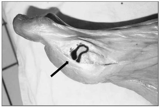
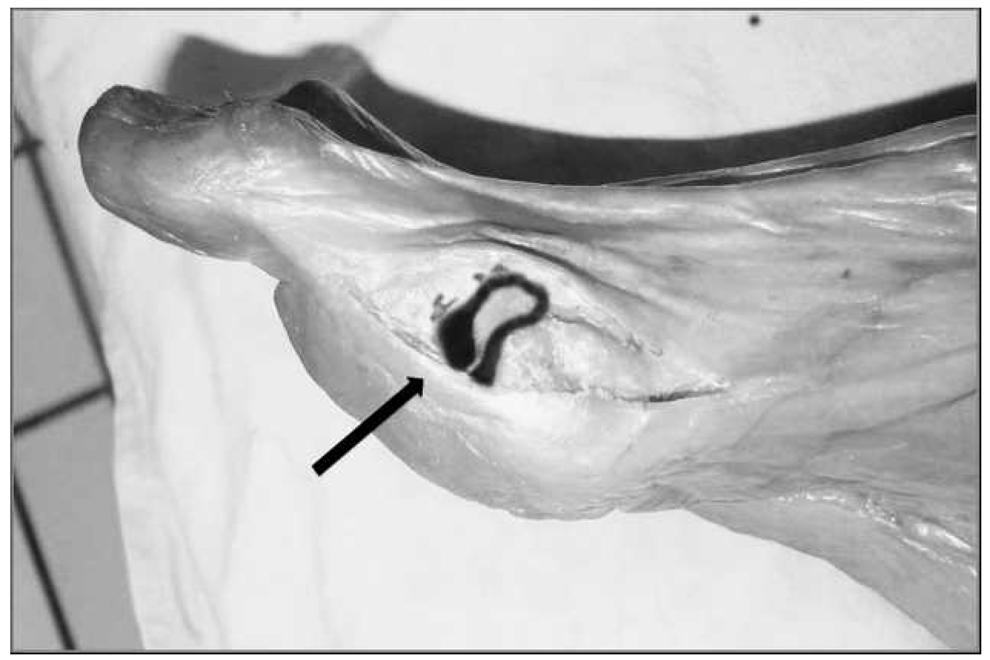
This study used 106 first metatarsophalangeal joints from 53 cadavers (28 men and 25 women) with a mean age of 74.8 years (range, 59–98 years) preserved by the method of Thiel.[13] This special embalming technique, which was developed over a 30-year period, provides a close-to-life model through the preservation of the original tissue color, consistency, and degree of transparency and full range of passive motion of the articular joints. Pathologic skeletal changes were detected by plain radiographs and recorded. A medical student (S.F.), a resident (N.H.), and a skilled specialist (T.K.) performed the injections. The student and resident punctured 38 metatarsophalangeal joints each (19 left and 19 right), and the surgeon punctured 30 (15 left and 15 right). The injections were performed with the cadavers in a supine position. The joint line was located by flexion and extension of the great toe. Gentle distal traction allowed better visualization of the joint line. The intra-articular puncture was then performed with a 14-gauge needle connected to a 1-mL syringe and filled with methylene blue. The needle was angled 60° to 70° to the plane of the foot and pointed distally to match the slope of the joint. Approximately 0.2 mL of methylene blue dye was injected into each joint. An arthrotomy was performed in all of the joints to define the location of the injected methylene blue (Figure 1). The injection was deemed to be a failure if the dye was located outside of the synovial joint.
Figure 1.
Metatarsophalangeal joint after arthrotomy: successful intra-articular puncture with intra-articular–visible methylene blue (arrow).
All of the results were entered into a computerized database and analyzed using Microsoft Excel 2003 (Microsoft Corp, Redmond, Washington). The χ2 test was used to assess correlations. All P values less than 0.05 and χ2 values greater than 3.84 were deemed statistically significant.
Results
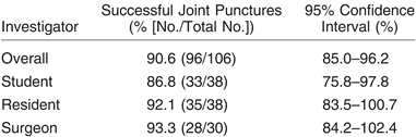
Overall, 90.6% (95% confidence interval [CI], 85.0%–96.2%) of the injections (96 of 106) were within the joint (Table 1). The resident’s rate of intended intra-articular injections was 92.1% (95% CI, 83.5%–100.7%; 35 of 38), and the student was successful in 86.8% (95% CI, 75.8%–97.8%; 33 of 38). No significant correlation between experience and the rate of successful punctures was detected (P =.45, χ2 = 0.56). The specialist had a success rate of 93.3% (95% CI, 84.2%–102.4%; 28 of 30).
Table 1.
Comparison of the Success of Different Investigators.
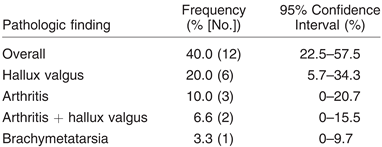
We found that 40.0% (95% CI, 22.5%–57.5%) of the joints (12 of 30) that the specialist injected had evidence of pathologic changes (Table 2). Arthritic changes were present in 10.0% (95 CI, 0%–20.7%) of the joints (3 of 30), hallux valgus in 20.0% (95% CI, 5.7%–34.3%; 6 of 30), and brachymetatarsia in 3.3% (95% CI, 0%–9.7%; 1 of 30). The presence of pathologic abnormality alters the success rate substantially. The combination of arthritis and hallux valgus occurred in 6.6% (95% CI, 0%–15.5%) of the joints (2 of 30), and the injections in both of these cases were extra-articular.
Table 2.
Frequency of Pathologic Findings in 30 Metatarsophalangeal Joints.
Discussion
Accurate intra-articular placement of the needle is a prerequisite for the achievement of desirable results and the avoidance of complication. In this study, we evaluated the rate of successful intra-articular injections and the influence of the degree of operator experience in achieving this success. Although such data are available for other synovial joints, such as the ankle,[14] acromioclavicular,[15] and sternoclavicular[16] joints, this is the first study, to our knowledge, to investigate the rate of successful joint puncture of the first metatarsophalangeal joint using an objective method in a cadaver model. These results are in accordance with those of other authors with a high rate of success in intra-articular punctures (97%), confirming this by visual analog scale for pain, edema, and morning stiffness.[12] The rate of successful joint puncture is high in the first metatarsophalangeal joint, even in relatively inexperienced hands, as evidenced by the present results. Although we did not demonstrate a statistically significant correlation between experience and successful joint puncture, there is certainly a trend toward this. In the second part of the study, pathologic changes akin to those seen in a conventional clinical setting were investigated in relation to a successful joint puncture. The presence of pathologic changes made for a more difficult procedure, as demonstrated by the increased rate of periarticular injections. This finding is of considerable importance because it is patients with pathologic changes who are offered these injections. Of the six joints in this study that had combined hallux valgus and arthritis, two were not successfully punctured. Note that in a study by Solan et al[4] it was these arthritic and deformed joints that had the least benefit from intra-articular injections. It is of questionable wisdom to offer these patients an injection in light of this evidence. Intra-articular punctures are associated with the risk of complications. The unintended introduction of microorganisms into the joint can cause disabling infections; therefore, great care must be taken to ensure a sterile procedure.[6] Other complications include possible adverse effects of local corticosteroid injection, such as tendon rupture, increased ligament stiffness, skin depigmentation, and periarticular soft-tissue calcification.[7,8,9,10] Intra-articular corticosteroid injection is a common therapeutic intervention, and its benefits are well documented. In this study, punctures were not image guided, as in routine clinical practice.
In the clinical setting, joint injections and aspirations are performed in patients with pathologic changes in their joints. It is well known that this makes periarticular injections more likely. In such cases, aspiration of synovial fluid is a clear indicator of correct needle placement, but this is not possible in ‘‘dry’’ joints. In these cases, insufflation of the joint with physiologic saline and subsequent aspiration may confirm needle placement. This method, however, is not foolproof. The position of the needle can also be confirmed with fluoroscopy, but this requires the injection of contrast media and the use of ionizing radiation.[1] An alternative is the use of ultrasound with air as contrast medium to allow real-time visualization of intra-articular placement of the needle.[17] A study by Reach et al[18] demonstrated 100% accuracy of intra-articular injection using ultrasound guidance; this avoids the use of ionizing radiation but is technically demanding, especially in smaller joints. In the metatarsophalangeal joint, the joint space is rather superficial and can be easily identified by gentle traction of the toe.
To our knowledge, this is the first study to explore nonguided intra-articular needle placement of the first metatarsophalangeal joint in an objective manner by using a cadaver model. Even inexperienced physicians have a high frequency of success. However, accurate needle placement in joints with high-graded pathologic findings, such as hallux valgus combined with arthritis, seems difficult, and the therapeutic benefit has to be questioned.
Funding
None reported.
Conflicts of Interest
None reported.
References
- BLIDDAL H: Placement of intra-articular injections verified by mini air-arthrography. Ann Rheum Dis 1999, 58, 641. [CrossRef] [Scilit] [PubMed]
- PONS, M.; ALVAREZ, F.; SOLANA, J. ET AL: Sodium hyaluronate in the treatment of hallux rigidus: a single-blind, randomized study. Foot Ankle Int 2007, 28, 38. [Google Scholar] [CrossRef] [Scilit] [PubMed]
- SARKIN TL: Indications for intra-articular steroid in osteoarthritis of the ankle and big toe joints. S Afr Med J 1974, 48, 2067.
- SOLAN, M.C.; CALDER, J.D.; BENDALL, S.P. Manipulation and injection for hallux rigidus: is it worthwhile? J Bone Joint Surg Br 2001, 83, 706. [Google Scholar] [CrossRef] [PubMed]
- TREPMAN, E.; YEO, S.J. Nonoperative treatment of metatarsophalangeal joint synovitis. Foot Ankle Int 1995, 16, 771. [Google Scholar] [CrossRef] [Scilit] [PubMed]
- BERNAU, A.; HEEG, P. Intra-articular punctures and injections: indications–prevention of infection–technique– complications [in German]. Orthopade 2003, 32, 548. [Google Scholar] [CrossRef] [Scilit] [PubMed]
- DALINKA, M.K.; STEWART, V.; BOMALASKI, J.S. ET AL: Periarticular calcifications in association with intra-articular corticosteroid injections. Radiology 1984, 153, 615. [Google Scholar] [CrossRef] [Scilit] [PubMed]
- GOTTLIEB, N.L. RISKIN WG: Complications of local corticosteroid injections. JAMA 1980, 243, 1547. [Google Scholar] [CrossRef] [PubMed]
- LEMONT H, HETMAN J: Cutaneous foot depigmentation following an intra-articular steroid injection. JAPMA 1991, 81, 606. [CrossRef] [Scilit] [PubMed]
- OXLUND H: The influence of a local injection of cortisol on the mechanical properties of tendons and ligaments and the indirect effect on skin. Acta Orthop Scand 1980, 51, 231. [CrossRef] [Scilit] [PubMed]
- OXLUND H: Long term local cortisol treatment of tendons and the indirect effect on skin: an experimental study in rats. Scand J Plast Reconstr Surg 1982, 16, 61.
- LOPES, R.V.; FURTADO, R.N.; PARMIGIANI, L.; et al. Accuracy of intra-articular injections in peripheral joints performed blindly in patients with rheumatoid arthritis. Rheumatology (Oxford) 2008, 47, 1792. [Google Scholar] [CrossRef] [Scilit] [PubMed]
- THIEL W: The preservation of the whole corpse with natural color [in German]. Ann Anat 1992, 174, 185.
- HEIDARI, N.; PICHLER, W.; GRECHENIG, S. ET AL: Does the anteromedial or anterolateral approach alter the rate of joint puncture in injection of the ankle? a cadaver study. J Bone Joint Surg Br 2010, 92, 176. [Google Scholar] [CrossRef] [Scilit] [PubMed]
- PICHLER, W.; WEINBERG, A.M.; GRECHENIG, S. ET AL: Intra-articular injection of the acromioclavicular joint. J Bone Joint Surg Br 2009, 91, 1638. [Google Scholar] [CrossRef] [Scilit] [PubMed]
- WEINBERG, A.M.; PICHLER, W.; GRECHENIG, S. ET AL: Frequency of successful intra-articular puncture of the sternoclavicular joint: a cadaver study. Scand J Rheumatol 2009, 38, 396. [Google Scholar] [CrossRef] [Scilit] [PubMed]
- FREDBERG, U.; VAN OVEREEM HANSEN, G.; BOLVIG, L. Placement of intra-articular injections verified by ultrasonography and injected air as contrast medium. Ann Rheum Dis 2001, 60, 542. [Google Scholar] [CrossRef] [Scilit] [PubMed]
- REACH, J.S.; EASLEY, M.E.; CHUCKPAIWONG, B. ET AL: Accuracy of ultrasound guided injections in the foot and ankle. Foot Ankle Int 2009, 30, 239. [Google Scholar] [CrossRef] [Scilit] [PubMed]

© 2023 American Podiatric Medical Association