


Abstract
Background: Malalignment and dysfunction of the foot have been associated with an increased propensity for overuse and traumatic injury in athletes. Several multisegment foot models have been developed to investigate motions in the foot. However, it remains unknown whether the kinematics measured by different multisegment foot models are equivocal. The purpose of the present study is to examine the efficacy of two multisegment foot models in tracking aberrant foot function. Methods: Ten high-arched and ten low-arched female athletes walked and ran while ground reaction forces and three-dimensional kinematics were tracked using the Leardini and Oxford multisegment foot models. Ground reaction forces and joint angles were calculated with Visual 3D (C-Motion Inc, Germantown, MD). Repeated-measures analyses of variance were used to analyze peak eversion, time to peak eversion, and eversion excursions. Results: The Leardini model was more sensitive to differences in peak eversion angles than the Oxford model. However, the Oxford model detected differences in eversion excursion values that the Leardini model did not detect. Conclusions: Although both models found differences in frontal plane motion between high- and low-arched athletes, the Leardini multisegment foot model is suggested to be more appropriate as it directly tracks frontal plane midfoot motion during dynamic motion.
Aberrant foot structures have been associated with unique injury patterns in the athletic population.[1,2] Individuals with abnormal foot structures exhibit a greater propensity for injury, with high-arched individuals experiencing bony injuries to the lateral aspect of the lower extremity and low-arched individuals experiencing soft-tissue injuries to the medial aspect of the lower extremity.[1,2] These different injury patterns have been attributed to differences in lower-extremity kinematic and kinetic patterns, including decreased knee flexion excursion, an increased vertical loading rate, and greater leg stiffness during running.[3] Although lower-extremity kinematic and kinetic patterns contribute to lower-extremity injury patterns exhibited by high-and low-arched athletes, several in vivo multisegment foot studies have shown that the specific mechanics of the foot should not be ignored.[4,5,6,7,8,9]
The foot is most often modeled as a single segment.[10] However, this is a limitation of previous investigations into mechanisms of lower-extremity injury as the interactions of the midtarsal and subtalar joints were not considered despite a wealth of research suggesting that limited or excess motion in these joints may result in injury. The use of multisegment foot models in kinematic investigations of foot function in healthy and pathologic movement has become more common in recent years.[6,11,12,13,14,15] Although multisegment foot models provide better insight into the kinematics in the foot, there are difficulties with many of these models in collecting data in shod conditions. Furthermore, the novelty of multisegment foot models has resulted in several different methods for establishing these models.[4,5,6,11,13,14,15,16]
Although two models have been used regularly in recent literature,[11,13] many multisegment foot models have been created, ranging in complexity from three to nine segments.[4,5,11,12,14,17,18] The Leardini multisegment foot model[13] is a relatively simple multisegment foot model that is composed of five segments: the shank, rearfoot, midfoot, forefoot (first ray), and hallux. It has been implemented using skin-mounted[7] and wand-mounted markers.[13] The Oxford multisegment foot model[11] is another relatively simple multisegment foot model composed of the shank, rearfoot, forefoot, and hallux. The Oxford model has been used to investigate multisegment foot mechanics in healthy[11] and patient populations.[19] The central difference between the Leardini and Oxford models is the definition and tracking of a midfoot segment. The Oxford model does not define a midfoot segment, whereas the Leardini model includes a midfoot segment. Previous research has used bone pins to track individual bones in the foot and ankle complex and reported frontal plane ranges of motion near 15°.[20] A separate bone pin study identified several functional units in the foot and ankle, including the proximal medial array consisting of the talus, navicular, and medial cuneiform. Although these studies have provided a platform from which further research may be conducted, the invasive method is a limitation to further study, especially in vivo studies investigating large populations. Other research using a multisegment foot has directly investigated the effect of foot type on motions in the foot[8] and has revealed that individuals with high-arched feet exhibited smaller peak eversion angles in the multisegment foot and at the ankle. However, the greatest differences between the two groups occurred at the midfoot-forefoot joint. The Leardini and Oxford models have been shown to produce reliable data.[11,13] Repeatability analyses have revealed that the rearfoot-midfoot and midfoot-forefoot joints of the Leardini model have coefficients of multiple correlation of 0.67 and 0.75, respectively. Furthermore, the Oxford model has been shown to have intertrial standard deviations of less than 1°.
Although these multisegment foot models have been developed for use as a tool to investigate the mechanics of the foot and its effect on lower-extremity kinematics, little research has focused on the difference between the multisegment foot models and their ability to measure foot function in individuals with differing structure.[4,5,7] Furthermore, few studies have investigated the effect of including the midfoot as an individual segment.[9] Therefore, the purpose of the present study was to compare kinematics measured by two multisegment foot models in high- and low-arched individuals. As frontal plane motion has been identified as a key contributor to the differences in injury patterns observed between high- and low-arched feet, the scope of the present study was limited to the frontal plane. Furthermore, to avoid the confounding factor of sex, only female athletes participated in the present study. It was hypothesized that owing to the presence of a defined midfoot segment, the Leardini model would be more sensitive than the Oxford model in detecting differences in frontal plane motion in the multisegment foot between the high-and low-arched groups.
Methods
Participants
Ten high-arched and ten low-arched women were included in this study from a larger study of healthy recreational athletes. Participants were 18 to 28 years old. Arch type was defined based on arch index measurements[21] taken during bilateral standing and were 1.5 SD above and below a previously reported mean ± SD value established from a sample of 604 feet (0.330 ± 0.031).[22] Participants placed into the high- and low-arched groups had arch index values greater than 0.377 or less than 0.283, respectively. All of the participants were free of major lower-extremity injury for the 6 months before participation in the present study. Each participant read, understood, and signed an informed consent form approved by the University of Texas of the Permian Basin institutional review board before participation in the study.
Experimental Protocol
Each participant was involved in two data collection sessions. The first session was used for inclusion by collecting anthropometric data, including height, mass, total foot length, truncated foot length, and dorsum of foot height.[21] In the second session, each participant performed five trials of barefoot walking and running at a self-selected speed determined during three practice trials before data collection. Three-dimensional kinematic and ground reaction force data were collected simultaneously.
Instrumentation
A seven-camera motion analysis system (240 Hz; Vicon Motion Systems Ltd, Oxford, England) was used to collect three-dimensional kinematic data on the right foot and shank. A force platform (1,200 Hz; OR6-7; AMTI, Watertown, Massachusetts) was synchronized with the motion analysis system and was used to measure ground reaction force data. A standing calibration was taken during quiet standing. During the standing calibration, the arms were placed across the chest and the feet were pointed forward in line with the global coordinate system. Two pairs of photocells (63501 IR; Lafayette Instruments Inc, Lafayette, Indiana) were used to determine and monitor the walking and running velocities of each participant.
Multisegment Foot Models
Two multisegment foot models were used in the present study. All of the segments used in both multisegment foot models were defined and tracked using skin-mounted, retroreflective markers. In the Leardini model, axis definition and orientation was previously described by Leardini et al.[13] The rearfoot was defined and tracked by two 9.5-mm retroreflective markers placed on the superior and inferior calcaneus in line with the Achilles tendon, the peroneal tubercle, and the sustentaculum tali. The midfoot was defined and tracked using 9.5-mm retroreflective markers placed on the lateral aspect of the tuberosity of the cuboid, the superior aspect of the lateral cuneiform, the superior aspect of the medial cuneiform, and the navicular tuberosity. The first metatarsal was defined by 9.5-mm retroreflective markers placed on the base of the first metatarsal, the shaft of the first metatarsal, and the mediolateral and superolateral sides of the first metatarsophalangeal joint. The Oxford forefoot was defined and tracked using 9.5-mm retroreflective markers placed on the base of the first and fifth metatarsals and on the medial and lateral sides of the first and fifth metatarsophalangeal joints, respectively.[11] In addition, clusters of 14-mm retroreflective markers were used to track the shank. Anatomical markers were placed over the medial and lateral malleoli and over the medial and lateral femoral epicondyles to define the shank. The Leardini multisegment foot model[8] defined the foot-ankle complex as four segments (shank, rearfoot, midfoot, and first metatarsal) and three articulating joints (ankle, rearfoot-midfoot, and midfoot-forefoot). Axis orientations and intersegmental angles were calculated as previously described by Leardini et al.[13] The Oxford multisegment foot model[11] defined the foot-ankle complex as three segments (shank, rearfoot, and forefoot) and two articulating joints (ankle and midfoot). The Leardini and Oxford models defined the ankle as the articulation of the rearfoot on the shank. The Leardini rearfoot-midfoot joint was defined as the articulation of the midfoot on the rearfoot (calcaneus). The Leardini midfoot-forefoot joint was defined as the articulation of the first metatarsal on the midfoot. The Oxford midfoot joint was defined as the articulation of the Oxford forefoot on the rearfoot (calcaneus).
Statistical Analyses
Three 2 × 2 (group × model) repeated-measures analyses of variance were used to determine significant differences between the means of selected kinematic measures, including peak eversion, time to peak eversion, and eversion excursion between each adjacent segment. Two Student t tests were used to compare mean walking and running velocities. Eversion excursion was defined as the difference in peak eversion angle and eversion angle at heel strike. In the presence of a group × model interaction, a post hoc t test was used to determine the source of the interaction. The alpha level was set at P < .05.
Results
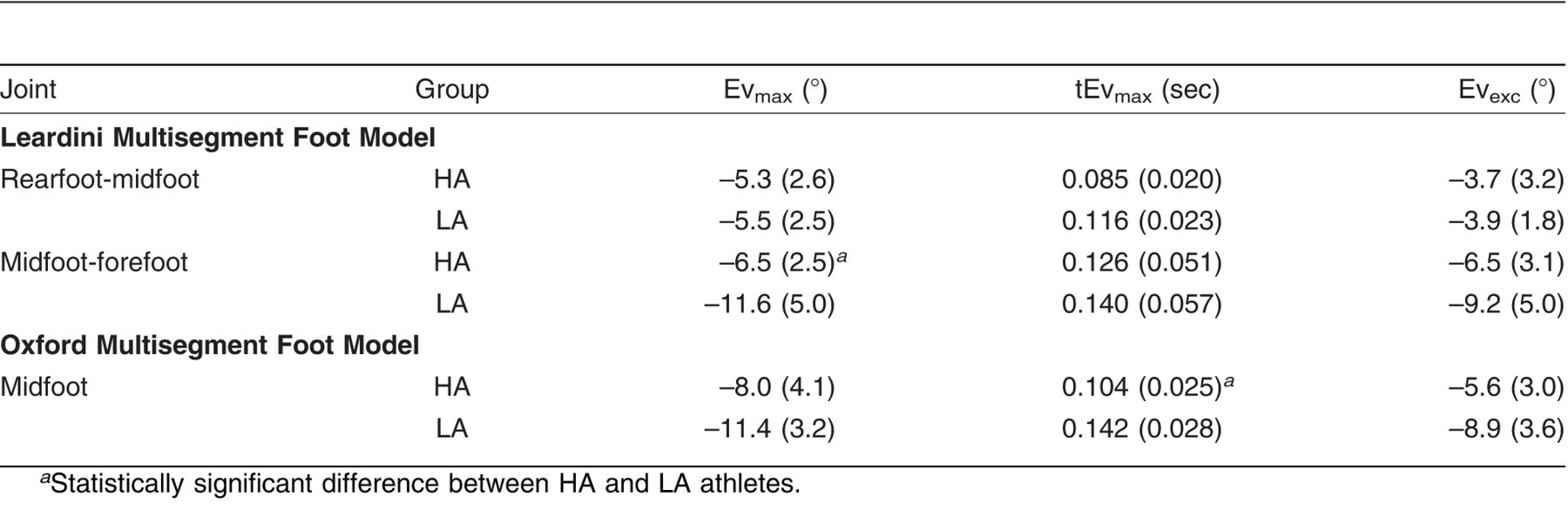
The high- and low-arched groups did not have significantly different mean ± SD walking velocities (high-arched: 1.36 ± 0.09 m/sec; low-arched: 1.36 ± 0.14 m/sec; P = .200) or running velocities (high-arched: 2.79 ± 0.28 m/sec; low-arched: 2.73 ± 0.29 m/sec; P = .356). The Leardini and Oxford multisegment foot models exhibited different findings in peak eversion angles in the feet of high- and low-arched athletes during walking (Table 1) and running (Table 2). The Leardini multisegment foot model demonstrated that high-arched athletes were less everted at the ankle (P =.012), rearfoot-midfoot (P = .031), and midfoot-forefoot (P = .013) joints than were low-arched athletes during walking. Furthermore, high-arched athletes exhibited less eversion than low-arched athletes at the ankle (P = .021) and midfoot-forefoot (P = .025) joints during running. The Oxford multisegment foot model also found significant differences between high- and low-arched athletes in peak ankle eversion angles in walking (P = .012) and running (P = .021). However, no differences were found between high- and low-arched athletes in peak midfoot eversion angles during walking (P = .073) or running (P = .072).
Table 1.
Peak Eversion (Evmax), Time to Peak Eversion (tEvmax), and Eversion Excursion (Evexc) Values in High- (HA) and Low-Arched (LA) Female Athletes During the Stance Phase of Walking
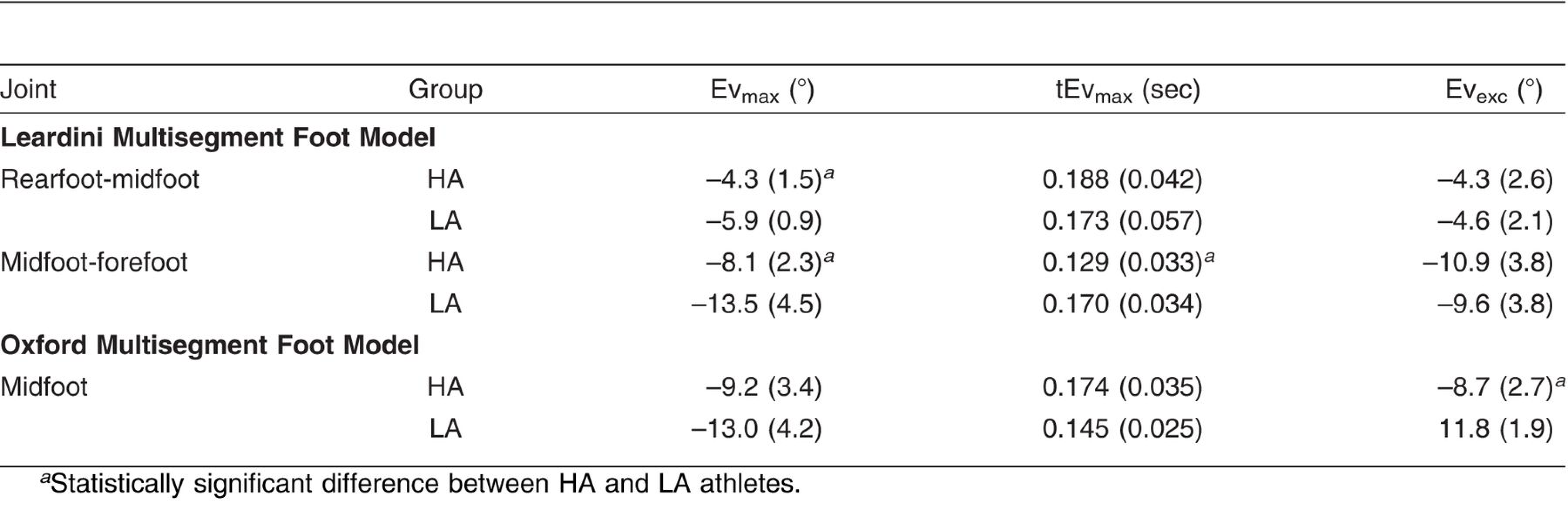
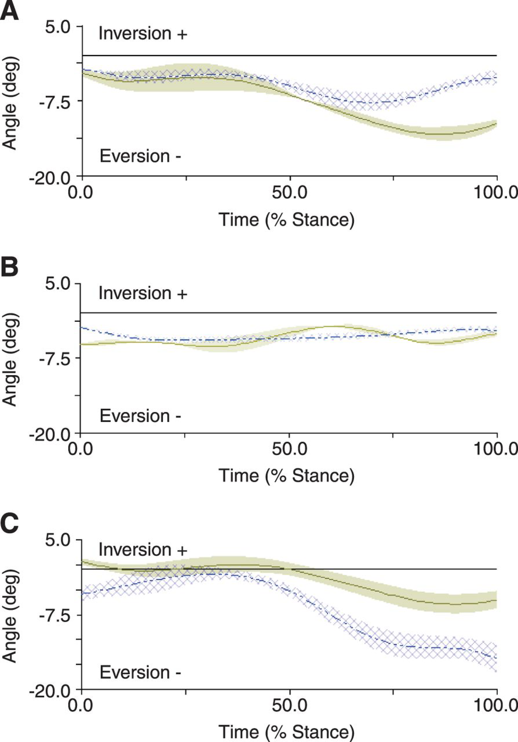
The significant differences between high- and low-arched athletes in times to peak eversion found using the Leardini multisegment foot model were different than those found using the Oxford multisegment foot model. The Leardini model demonstrated that high- and low-arched athletes exhibited similar times to peak eversion at the ankle and rearfoot-midfoot joints during walking (Table 1 and Fig. 1). High-arched athletes reached peak eversion in the midfoot-forefoot joint earlier than low-arched athletes during walking (P = .027). The Oxford model showed no differences in time to peak eversion at the ankle and midfoot joints. In running (Table 2 and Fig. 2), the Leardini multisegment foot model found no differences between high- and low-arched athletes at the ankle, rearfoot-midfoot, and midfoot-forefoot joints; however, the Oxford multisegment foot model revealed that high-arched athletes reached peak eversion in the midfoot joint earlier than low-arched athletes (P = .002).
Eversion excursion values were also different in the Leardini and Oxford multisegment foot models when comparing foot mechanics in high- and low-arched athletes during walking (Table 1 and Fig. 1) and running (Table 2 and Fig. 2). The Leardini model did not find differences in eversion excursion values between high- and low-arched athletes during walking (rearfoot-midfoot: P = .818; and midfoot-forefoot: P = .429). The Oxford midfoot joint exhibited a significant difference in eversion excursion between high- and low-arched athletes during walking (P = .018). Both models concluded that high- and low-arched athletes experience similar eversion excursions during running (Leardini rearfoot-midfoot: P = .123; Leardini midfoot-forefoot: P = .176; and Oxford midfoot: P = .087).
Table 2.
Peak Eversion (Evmax), Time to Peak Eversion (tEvmax), and Eversion Excursion (Evexc) Values in High- (HA) and Low-Arched (LA) Female Athletes During the Stance Phase of Running
Discussion
The Oxford and Leardini multisegment foot models track movement in the foot segment. The two models resulted in statistically significant differences in movement patterns in the foot between high-and low-arched athletes. The Leardini model[7,13] defines the shank, rearfoot, midfoot, and forefoot segments, and the Oxford model[11] defines the shank, rearfoot, and forefoot segments. The Oxford and Leardini multisegment foot models define the interaction between the shank and rearfoot similarly, resulting in similar kinematics at the ankle. The present study presents joint kinematic curves that are comparable with those presented in previous research studies using multisegment foot models.[6,11,13] Previous research has shown the midfoot joint of the Oxford model in healthy individuals to be inverted throughout the entirety of the stance phase of gait. However, in the present study, Oxford midfoot joint angles were everted in high- and low-arched athletes.[11] These differences in joint angles during stance may be due to structural differences of the feet used in the present study compared with previous research studies. The kinematics found using the Leardini model were similar to previous kinematic findings using a multisegment foot model and skin-mounted markers.[7] The similarities between these data and previous findings using these multisegment foot models support the validity of these data and the present findings.
Figure 1.
Ensemble curves of frontal plane angles of the Oxford midfoot (A), Leardini rearfoot-midfoot (B), and Leardini midfoot-forefoot (C) in high-arched (solid lines) and low-arched (dashed lines) female athletes during walking.
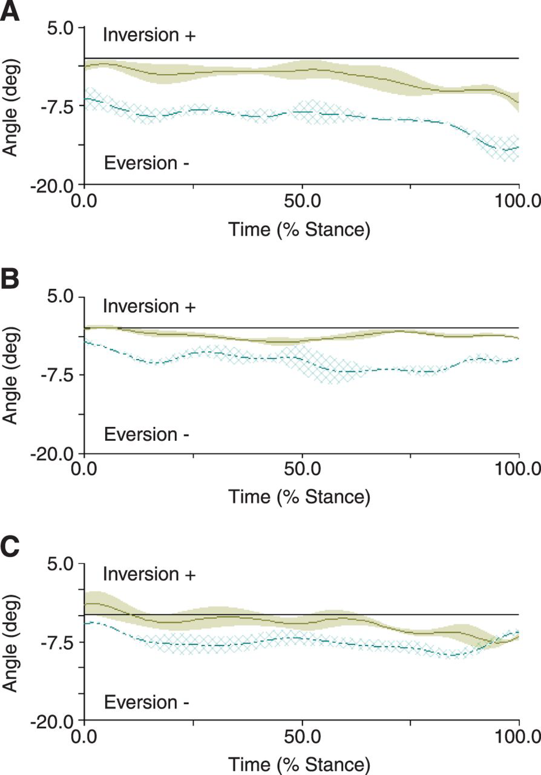
Figure 2.
Ensemble curves of frontal plane angles of the Oxford midfoot (A), Leardini rearfoot-midfoot (B), and Leardini midfoot-forefoot (C) in high-arched (solid lines) and low-arched (dashed lines) female athletes during running.
The purpose of the present study was to investigate the differences in frontal plane multisegment foot mechanics as measured by two multisegment foot models. The two models being examined defined ankle motion similarly, thus it will not be discussed. The multisegment foot kinematics reveal that the Leardini and Oxford models detected that high-arched athletes had significantly smaller peak ankle eversion angles than low-arched athletes during walking (Table 1) and running (Table 2). Similarly, neither the Oxford nor the Leardini model detected differences in eversion excursions between high- and low-arched athletes during the running movement. The two models, however, detected unique kinematic differences in the multisegment foot. During walking (Table 1 and Fig. 1) and running (Table 2 and Fig. 2), the Leardini model was more sensitive to differences in peak eversion angles compared with the Oxford multisegment foot model. The Leardini model has a defined midfoot segment that allows for tracking of the small movements in the midtarsal joint (rearfoot-midfoot joint). These data show that during walking there are statistically significant differences between high- and low-arched athletes in joint motion at the midtarsal joint, and, thus, midtarsal motion cannot be ignored when assessing foot function in these functionally different groups of athletes (Table 1). Conversely, in the running condition, no differences were observed between high- and low-arched athletes in rearfoot-midfoot motion (Table 2). The Oxford multisegment foot model was shown to be more sensitive to differences in ranges of motion measured by excursions during the walking task (Table 1). The Oxford multisegment foot model does not define a midfoot segment but defines the midfoot joint as the motion of the forefoot segment relative to the rearfoot segment. Therefore, the small motions in the midtarsal joint may have a cumulative or minimizing effect in the calculated joint excursions dependent on the rigidity and motions in the foot. The Oxford model may, therefore, result in greater ranges of motion and excursions in the flexible, low-arched foot.
The present study presents novel findings comparing multisegment foot kinematics in high- and low-arched athletes as measured by two different multisegment foot models. Previous research has presented conflicting results pertaining to the existence of differences in frontal plane motion between pes planus (low-arched) and healthy individuals.[4,19] However, another research study investigating multisegment foot motion of control individuals and individuals with adult acquired flatfoot, clinically known as tibialis posterior dysfunction, produced results similar to those found using the Leardini model. Specifically, individuals with tibialis posterior dysfunction had a trend toward exhibiting more everted mean frontal plane angles than control individuals.[23] Although these differences were not statistically significant, it is assumed that motion patterns in high-arched compared with low-arched feet would be more different than between low-arched feet and controls. At present, only a single research study has used the Leardini model[8] to directly investigate multisegment foot motion in high- and low-arched individuals during walking and running. Furthermore, no previous research, to our knowledge, has used the Oxford multisegment foot model to address the differences in high- and low-arched multisegment foot motion.
Although the findings of the present study are similar to those previously reported in invasive and noninvasive research studies, some limitations do exist. The largest limitation of the present study pertains to the use of skin-mounted markers to track the underlying skeletal structures. Although previous research has been conducted to validate the findings of these multisegment foot models, some skin-motion artifact is likely present and is a limitation of this study. Furthermore, an inherent limitation of using a multisegment foot model pertains to the calculation of kinetics in the foot and lower extremity. There are currently no validated protocols for calculating multisegment foot kinetics, which limits the application of multisegment foot studies. Finally, the sample size used in the present study was rather small and included only ten high-arched and ten low-arched participants. A larger sample size would have improved the statistical power within the study and may have revealed further differences between the two multisegment foot models.
In conclusion, the Leardini and Oxford multisegment foot models are capable of detecting differences in frontal plane motion between high- and low-arched athletes. However, the statistical differences found between the two functionally different groups were different between the Leardini and Oxford multisegment foot models. The Leardini model detected differences in peak joint angles in the frontal plane between high- and low-arched athletes, and the Oxford model found differences in frontal plane excursion values between high- and low-arched athletes. The present study also presents data that demonstrate that care should be taken in the interpretation and comparison of data collected using a multisegment foot model as different models may not provide similar findings in a given data set.
Financial Disclosure
None reported.
Conflict of Interest
None reported.
References
- Kaufman, KR, SKBrodine, RAShaffer, et al. :The effect of foot structure and range of motion on musculoskeletal overuse injuries. .Am J Sports Med27::585. ,1999. .
- WilliamsIII, DS, ISMcClay, and JHamill. :Arch structure and injury patterns in runners. .Clin Biomech16::341. ,2001. .
- WilliamsIII, DS, IMDavis, JPScholz, et al. :High-arched runners exhibit increased leg stiffness compared to low-arched runners. .Gait Posture19::263. ,2004. .
- Cobb, SC, LLTis, JTJohnson, et al. :The effect of low-mobile foot posture on multi-segment medial foot model gait kinematics. .Gait Posture30::334. ,2009. .
- Jenkyn, TR and ACNicol. :A multi-segment kinematic model of the foot with a novel definition of forefoot motion for use in clinical gait analysis during walking. .J Biomech40::3271. ,2007. .
- Leardini, A, MGBenedetti, LBerti, et al. :Rear-foot, mid-foot and fore-foot motion during the stance phase of gait. .Gait Posture25::453. ,2007. .
- Leardini, A, ZSawacha, GPaolini, et al. :A new anatomically based protocol for gait analysis in children. .Gait Posture26::560. ,2007. .
- Powell, DW, BLong, CEMilner, et al. :Frontal plane multi-segment foot kinematics in high- and low-arched females during dynamic loading tasks. .Hum Mov Sci30::105. ,2011. .
- Wolf, P, AStacoff, ALiu, et al. :Functional units of the human foot. .Gait Posture28::434. ,2008. .
- Kadaba, MP, HKRamakrishnan, MEWootten, et al. :Repeatability of kinematic, kinetic, and electromyographic data in normal adult gait. .J Orthop Res7::849. ,1989. .
- Carson, MC, MEHarrington, NThompson, et al. :Kinematic analysis of a multi-segment foot model for research and clinical applications: a repeatability analysis. .J Biomech34::1299. ,2001. .
- Hunt, AE, RMSmith, MTorode, et al. :Inter-segment foot motion and ground reaction forces over the stance phase of walking. .Clin Biomech16::592. ,2001. .
- Leardini, A, MGBenedetti, FCatani, et al. :An anatomically based protocol for the description of foot segment kinematics during gait. .Clin Biomech14::528. ,1999. .
- MacWilliams, BA, MCowley, and DENicholson. :Foot kinematics and kinetics during adolescent gait. .Gait Posture17::214. ,2003. .
- Myers, KA, MWang, RMMarks, et al. :Validation of a multisegment foot and ankle kinematic model for pediatric gait. .IEEE Trans Neural Syst Rehabil Eng12::122. ,2004. .
- Houck, JR, JMTome, and DANawoczenski. :Subtalar neutral position as an offset for a kinematic model of the foot during walking. .Gait Posture28::29. ,2008. .
- Stebbins, J, MHarrington, NThompson, et al. :Repeatability of a model for measuring multi-segment foot kinematics in children. .Gait Posture23::401. ,2006. .
- Woodburn, J, KMNelson, KLSiegel, et al. :Multisegment foot motion during gait: proof of concept in rheumatoid arthritis. .J Rheumatol31.:1918. ,2004. .
- Hunt, AE and RMSmith. :Mechanics and control of the flat versus normal foot during the stance phase of walking. .Clin Biomech19::391. ,2004. .
- Lundgren, P, CNester, ALiu, et al. :Invasive in vivo measurement of rear-, mid- and forefoot motion during walking. .Gait Posture28::93. ,2008. .
- WilliamsIII, DS and ISMcClay. :Measurements used to characterize the foot and the medial longitudinal arch: reliability and validity. .Phys Ther80::864. ,2000. .
- Zhang, S, DPowell, MKeefer, et al. :Foot and arch structure characteristics across age groups. . In:Proceedings of the 8th Footwear Biomechanics Symposium;June 27–29, 2007;Taipei, Taiwan.
- Rattanaprasert, U, RSmith, MSullivan, et al. :Three-dimensional kinematics of the forefoot, rearfoot, and leg without the function of tibialis posterior in comparison with normals during stance phase of walking. .Clin Biomech14::14. ,1999. .
© 2013 American Podiatric Medical Association