


Abstract
Xanthomas are described as deposits in the skin and subcutaneous tissues. Mostly known as pseudotumors, xanthomas consist of connective tissue containing mainly cholesterol, triglycerides, and numerous foamy macrophages. Bilateral Achilles tendon xanthomata is pathognomonic for cerebrotendinous xanthomatosis in the case of normal cholesterol levels but increased cholestanol levels in serum. In this article, we present findings regarding bilateral xanthomas of Achilles tendons in a patient with cerebrotendinous xanthomatosis.
Keywords:
n/a- Cerebrotendinous xanthomatosis (CTX) is a rare, late-onset, autosomal recessive lipid storage disease first described by van Bogaert et al[1] in 1937. It is caused by a mutation in the sterol 27-hydroxylase gene (CYP27) on chromosome 2q33-qter.[2,3,4,5] This mutation results in a defect in the activity of the hepatic mitochondrial enzyme sterol 27-hydroxylase. As a result, cholic acid synthesis is reduced, and no cholic acid or chenodeooxycholic acid is produced. Changes in bile acid biosynthesis result in accumulation of an excessive amount of cholestanol, which is a midsubstance in hepatic conversion of cholesterol to bile acids in the liver, in many different tissues. Biochemical diagnosis is made by determining excessive urinary excretion of bile alcohols and high serum cholestanol levels.[6,7] The onset of symptoms and signs in patients with CTX usually occurs in childhood or adolescence. Bilateral cataracts and diarrhea are the most frequently seen symptoms, followed by neurologic abnormalities and tendon xanthomas.[8,9] Neurologic symptoms include cerebellar and pyramidal dysfunction, cognitive impairment, dementia, epilepsy, and poly-neuropathy.[10,11] Other symptoms are premature atherosclerosis (mainly in coronary arteries) and osteoporosis.[12,13,14] Xanthomata affecting the Achilles tendon bilaterally with a normal serum cholesterol level are unusual and are usually associated with CTX. In this case, we describe a patient with CTX with bilateral Achilles tendon xanthomata.
Case Report
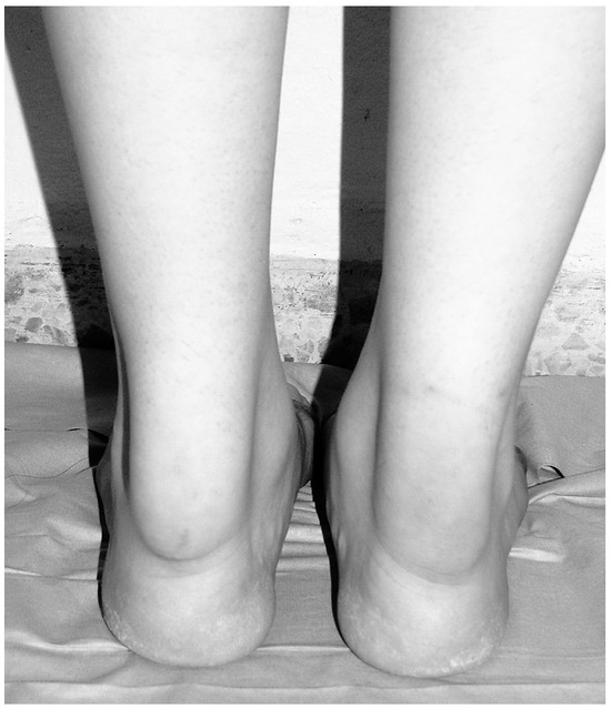
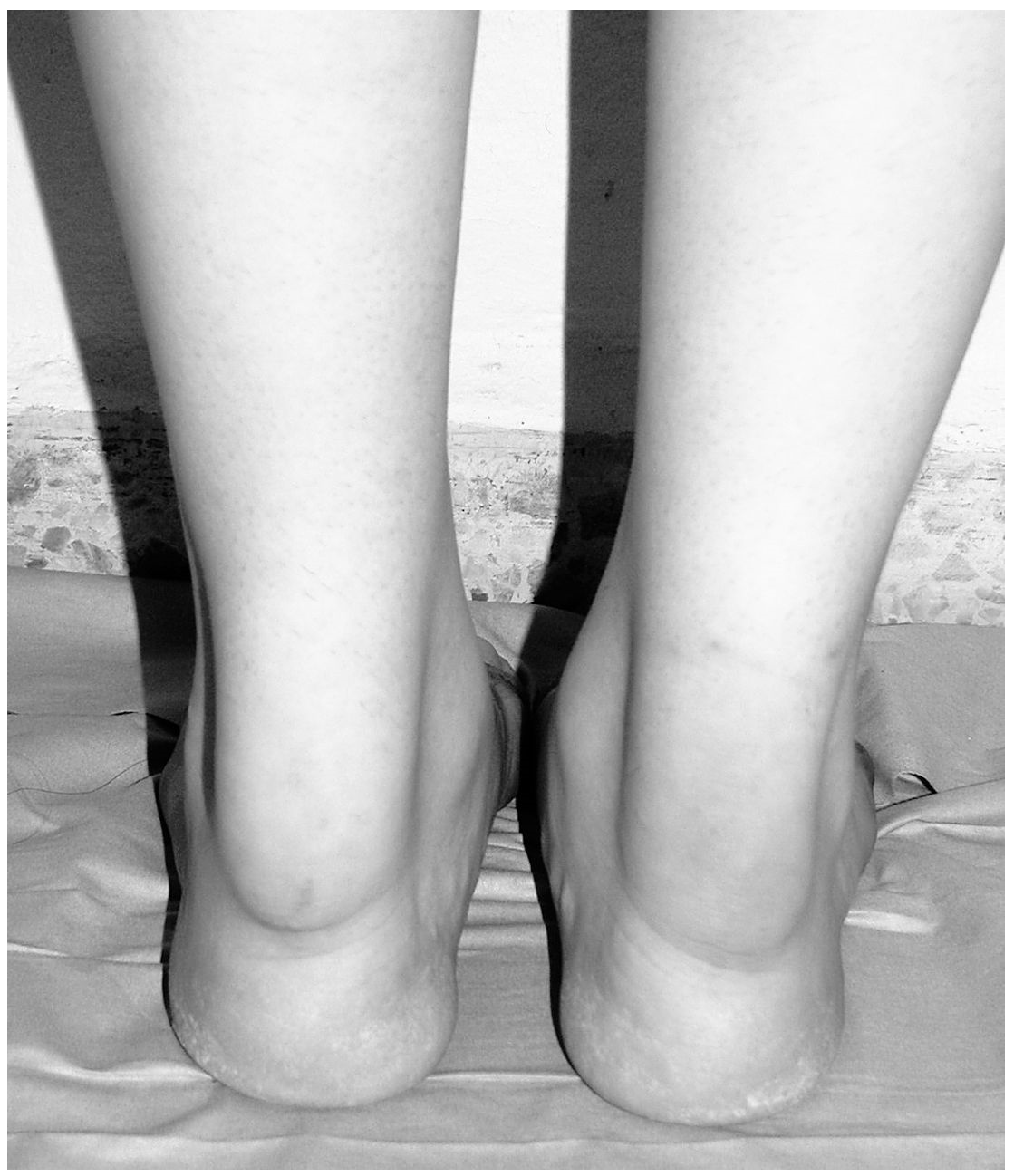
The patient in this case report was aware that data concerning the case would be submitted for publication. A 21-year-old female patient presented with a 6-year history of painless swellings in the posterior of both ankle joints. She had minimal concerns when walking, and walking distance was normal. The patient had undergone surgery in both eyes 6 years earlier owing to juvenile cataracts. At this time, she also noted some swellings in both Achilles tendons, and these progressed slowly up to now. On clinical examination, a nontender, painless, fusiform, hard, and symmetrical enlargement was seen in both Achilles tendons (
Figure 1
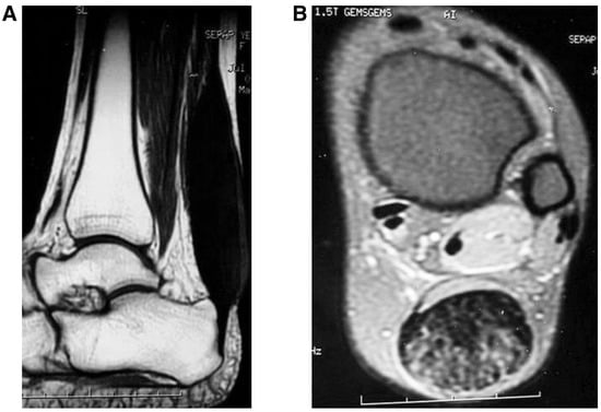
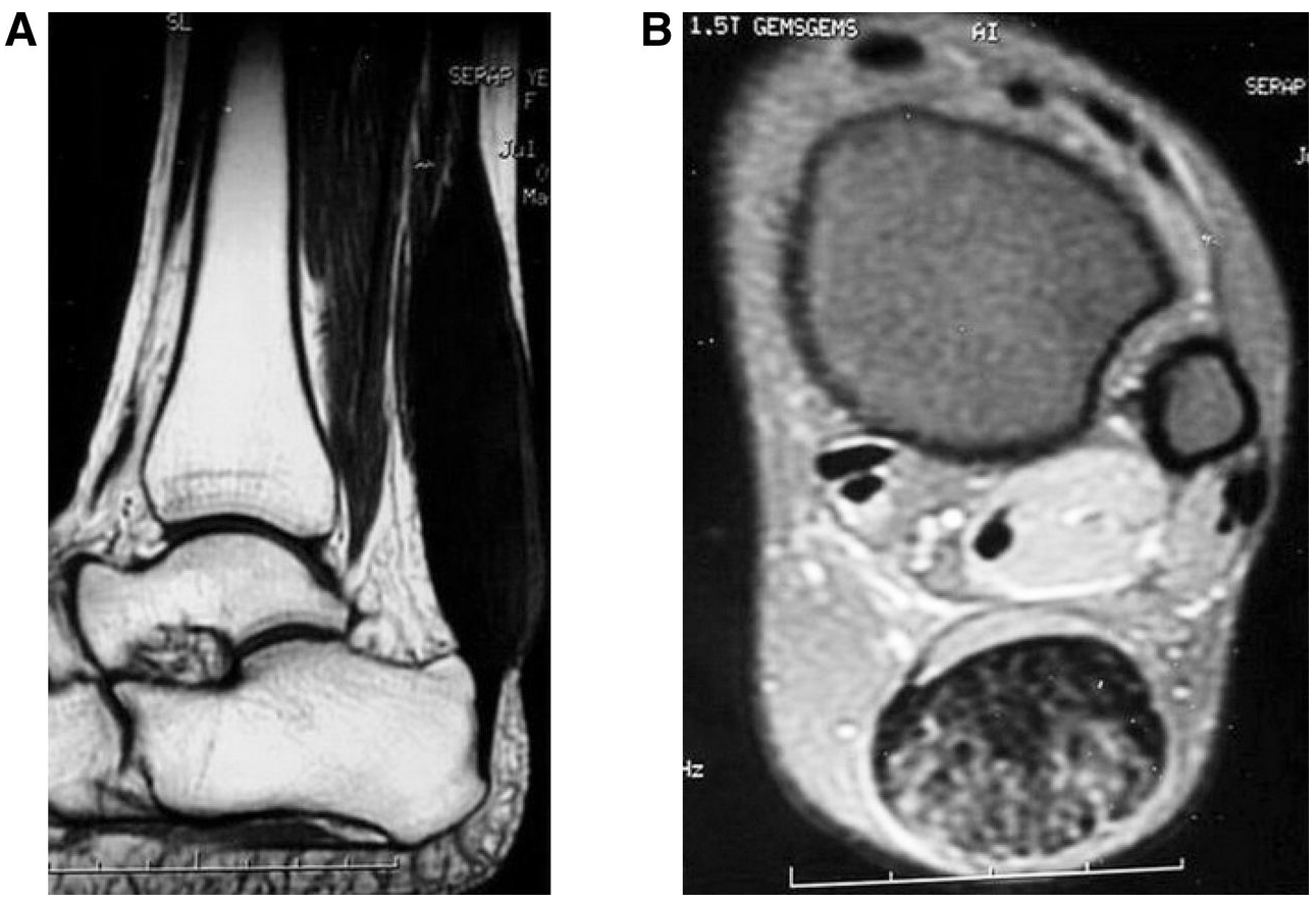
). The feet were plantigrade, and no deformity was seen. Dorsiflexion range of motion in both ankles was 158. The diameters of the masses were 60 × 35 mm (L × W) on the left side and 50 × 25 mm (L × W) on the right side. Magnetic resonance images of both ankles were acquired on a 1.5-T unit using the following sequences: sagittal T1- and T2-weighted spin echo, sagittal fast short tau inversion recovery, axial T1-weighted spin echo, and axial T2-weighted gradient echo. Conventional radiographs showed no bony abnormality. Magnetic resonance imaging demonstrated symmetrical fusiform enlargements involving both Achilles tendons bilaterally, extending along a segment just proximal to the insertion sites, measuring 110 × 23 mm on the left side and 110 × 25 mm on the right side. There were no signs of enthesopathy at the calcaneal insertions of the Achilles tendons. Tendon margins were well remained, and no abnormalities were seen involving the other ankle tendons and ankle joint. Bone and bone marrow signal changes were normal (
Figure 2
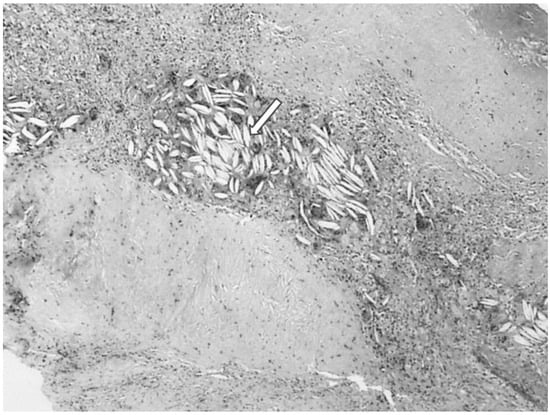
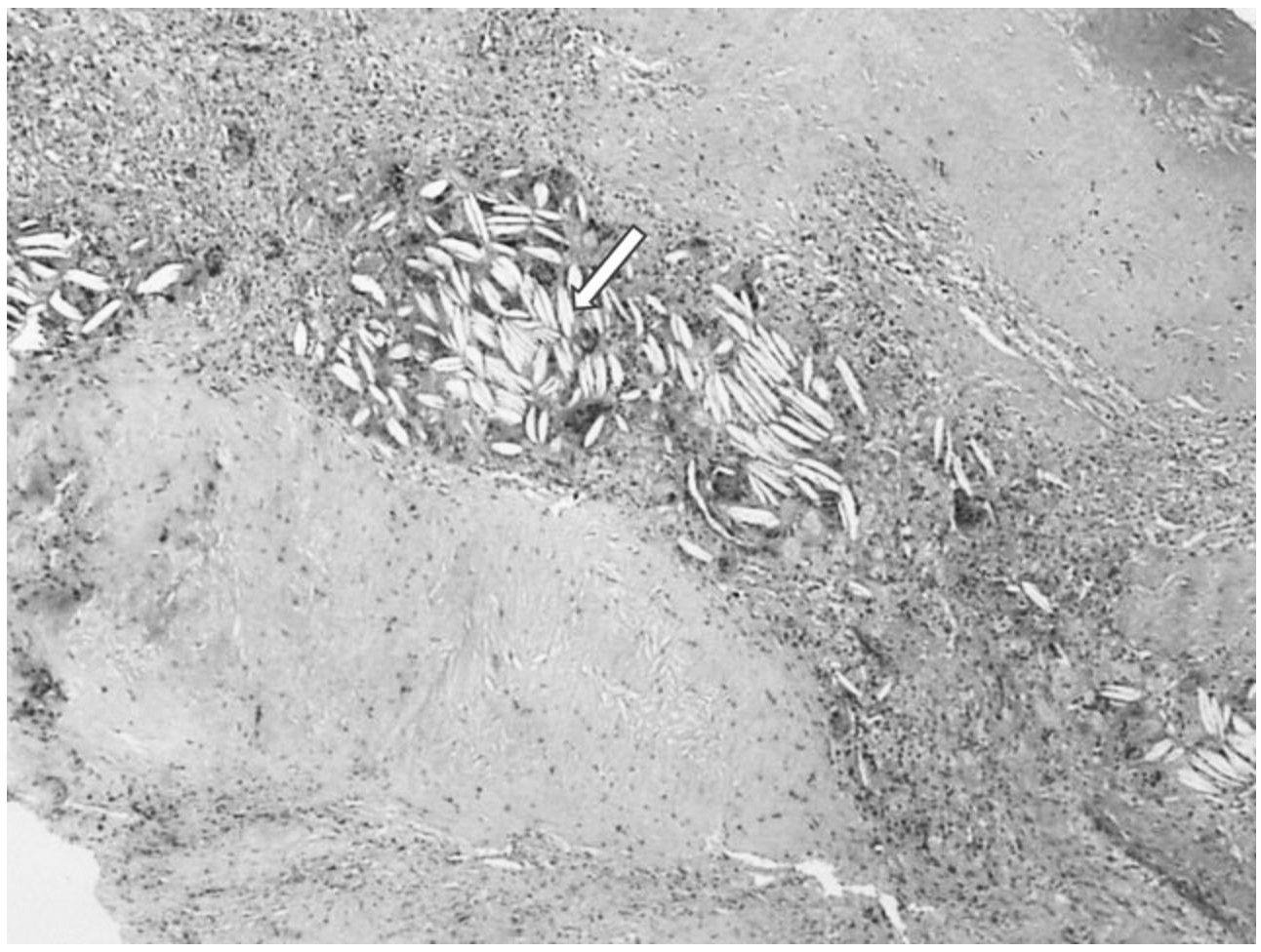
). An incisional biopsy of the right ankle was planned and performed. Macroscopically, a golden yellow tumor was seen infiltrating the tendon. Paraffin sections of biopsy specimens revealed a diagnosis of xanthoma. There were numerous foamy macrophages and cholesterol clefts surrounded by macrophages and giant cells in the connective tissue of the tendon (
Figure 3
). The patient was referred for biochemical assessment. Her serum cholesterol and other lipid levels were normal, but her serum cholestanol level was abnormal and increased. Laboratory parameters, including serum cholesterol, triglyceride, low-density lipoprotein, high-density lipoprotein, and serum cholestanol levels were 178, 66, 98, 65, and 2.8 mg/dL, respectively. The serum cholestanol level was 14 times the normal mean value of 0.2 mg/dL. These values confirmed the diagnosis of CTX. A magnetic resonance image of the brain showed no signs of pathologic central nervous system involvement. Findings from neurologic examination were normal except for some cognitive impairment. The patient was the oldest of three children. Her two brothers were healthy, and there was no family history of CTX or intermarriage. After the diagnosis of CTX, surgical excision and reconstruction of bilateral Achilles tendons were advised to the patient, but she refused the surgical treatment. Then, the patient consulted the endocrinology and metabolic diseases department and started bile acid replacement therapy with ursodeoxycholic acid and treatment with chenodeoxycholic acid. At the time of this writing, replacement therapy was continuing.
Figure 1.
Clinical photograph demonstrating bilateral fusiform swelling of the Achilles tendons.
Figure 2.
A, Sagittal T1-weighted magnetic resonance image demonstrating fusiform swelling of the right Achilles tendon just proximal to its insertion site. B, Axial T1-weighted image of the right Achilles tendon 2.5 cm proximal to the tibial plafond showing a heterogeneous signal in the tendon mass.
Figure 3.
Cholesterol clefts (white arrow) in the partly hyalinizated fibrous tissue surrounded by numerous foamy mononuclear and multinuclear giant cells (H&E, 340).
Discussion
Xanthomas are described as tumors or pseudotumors composed of connective tissue. This tissue contains foamy macrophages, giant cells, cholesterol, cholesterol esters, and triglycerides. When tendons are involved, the foam cells diffusely infiltrate the tendon tissue and spread between the collagen fibers. Xanthomas are usually seen with familial hyperlipidemias when plasma lipid levels are elevated. However, the Achilles tendon usually is not the primary target for this disease group. In CTX, the Achilles tendon is the primary site of xanthoma, and it is usually seen bilaterally. It is a rare genetic metabolic disorder of bile acid synthesis and metabolism. The disease is classically characterized by bilateral Achilles tendon xanthomata, bilateral cataract formation, and progressive neurologic deterioration (mainly pyramidal tract signs, cerebellar signs, and cognitive impairment).[15] The age at presentation is variable, but it is usually in the second or third decade of life. The first symptom of CTX is bilateral cataract formation, and this can be in association with neurologic dysfunction. Achilles tendon xanthomas develop later, usually in the fourth decade of life.[9] In the present patient, Achilles xanthomas were seen in the second decade of life, after bilateral cataract formation at 15 years old. This is different from the current literature and presented at an earlier age. We also observed no neurologic deterioration except some cognitive impairment. The diagnosis of CTX with routine biochemical markers of plasma lipid levels and urine analysis is difficult. This analysis often reveals normal findings, as seen in the present patient. Diagnosis depends on the finding of mainly elevated plasma cholestanol levels, and recently some new assessment techniques have been developed.[16] Medical treatment of CTX consists of bile acid replacement therapy with ursodeoxycholic acid and treatment with chenodeoxycholic acid. This treatment modality was first decribed by Berginer et al.[17] After this report, more studies were reported by others.[18,19] The therapeutic goal of treatment for CTX is reduction of the cholestanol level. Administration of chenodeoxycholic acid decreases the synthesis of bile alcohol and causes gradual falling of the cholestanol levels in serum and tissues. It has been demonstrated that long-term therapy with chenodeoxycholic acid can stop progression of the disease.[19] In the present patient, we have not seen any neurologic deterioration except some cognitive impairment. However, we also started therapy with chenodeoxycholic acid, but we do not know its effect on tendon xanthomas. In a recent report, there was no change in the size of the xanthoma despite more than 5 years of therapy with chenodeoxycholic acid.[20] The differential diagnosis for Achilles tendon xanthoma includes familial hypercholesterolemia, CTX, and sitosterolemia.[3] In familial hypercholes-terolemia, increased cholesterol levels are seen.
In conclusion, CTX may present with different signs and symptoms. The most commonly seen symptoms are bilateral Achilles tendon xanthomas and normal plasma lipid levels. These are commonly painful, but sometimes can be painless, as in the present patient. Early diagnosis is essential for patients with CTX because medical therapy can halt disease progression and prevent the devastating neurologic lesions.
Financial Disclosure
None reported.
Conflicts of Interest
None reported.
References
- VAN BOGAERT L, SCHERER HJ, Epstein E: Une Forme Cérébrale de la Cholestérinose Généralisée: (Type Particulier de Lipidose á Cholestérine), Mason et Cie, Paris, 1937.
- LEE MH, HAZARD S, CARPTEN JD, ET AL: Fine-mapping, mutation analyses, and structural mapping of cerebrotendinous xanthomatosis in U.S. pedigrees. J Lipid Res 2001, 42, 159. [CrossRef] [Scilit]
- BJORKEN I, BOBERG KM, LEITERSDOEF E: ‘‘Inborn Errors in Bile Acid Biosynthesis and Storage of Sterols Other than Cholesterol,’’. In The Metabolic and Molecular Bases of I˙nherited Disease; Scriver, C.R., Beaudet, A.L., Eds.; McGraw-Hill: New York, 2001; p. 2970. [Google Scholar]
- FEDERICO A, DOTTI MT: Cerebrotendinous xanthomatosis: clinical manifestations, diagnostic criteria, pathogenesis and therapy. J Child Neurol 2003, 18, 633. [CrossRef] [Scilit] [PubMed]
- GALLUS GN, DOTTI MT, FEDERICO A: Clinical and molecular diagnosis of cerebrotendinous xanthomatosis with a review of the mutations in the CYP27A1 gene. Neurol Sci 2006, 27, 143. [CrossRef] [Scilit] [PubMed]
- WOLTHERS BG, VOLMER M, VAN DER MOLEN JC, ET AL: Diagnosis of cerebrotendinous xanthomatosis (CTX) and effect of chenodeoxycholic acid therapy by analysis of urine, using capillary gas chromatography. Clin Chim Acta 1983, 131, 53. [CrossRef] [Scilit] [PubMed]
- WOLTHERS BG, WALRECHT HT, VAN DER MOLEN JC, ET AL: Use of determinations of 7-lathosterol (5 alpha-cholest-7-en3beta-ol) and other cholesterol precursors in serum in the study and treatment of disturbances of sterol metabolism, particularly cerebrotendinous xanthomatosis. J Lipid Res 1991, 32, 603.
- CRUYSBERG JR, WEVERS RA, TOLBOOM JJ: Juvenile cataract associated with chronic diarrhea in pediatric cerebrotendinous xanthomatosis. Am J Ophthalmol 1991, 112, 606. [CrossRef] [Scilit] [PubMed]
- CRUYSBERG JR, WEVERS RA, VAN ENGELEN BG, ET AL: Ocular and systemic manifestations of cerebrotendinous xanthomatosis. Am J Ophthalmol 1995, 120, 597. [CrossRef] [Scilit] [PubMed]
- KURITZKY A, BERGINER VM, KORCZYN AD: Peripheral neuropathy in cerebrotendinous xanthomatosis. Neurology 1979, 29, 880. [CrossRef] [Scilit] [PubMed]
- KATZ DA, SCHEINBERG L, HOROUPIAN DS, ET AL: Peripheral neuropathy in cerebrotendinous xanthomatosis. Arch Neurol 1985, 42, 1008. [CrossRef] [Scilit] [PubMed]
- BERGINER VM, SHANY S, ALKALAY D: Osteoporosis and increased bone fractures in cerebrotendinous xanthomatosis. Metabolism 1993, 42, 69. [CrossRef] [Scilit] [PubMed]
- CHANG WN, LUI CC: Failure in the treatment of longstanding osteoporosis in cerebrotendinous xanthomatosis. J Formos Med Assoc 1997, 96, 225.
- KURIYAMA M, FUJIYAMA J, KUBOTA R, ET AL: Osteoporosis and increased bone fractures in cerebrotendinous xanthomatosis. Metabolism 1993, 42, 1497.
- MOGHADASAIN MH: Cerebrotendinous xanthomatosis: clinical course, genotypes and metabolic backgrounds. Clin Invest Med 2004, 27, 42.
- HALPERIN G, ELISAF M, LEITERSDORF E, ET AL: A new method for determination of serum cholestanol by high-performance liquid chromatography with ultraviolet detection. J Chromatogr B 2000, 34, 345.
- BERGINER VM, SALEN G, SHEFER S: Long-term treatment of cerebrotendinous xanthomatosis with chenodeoxycholic acid. N Engl J Med 1984, 311, 1649. [CrossRef] [Scilit] [PubMed]
- MONDELLI M, ROSSI A, SCARPINI C, ET AL: Evoked potentials in cerebrotendinous xanthomatosis and effect induced by chenodeoxycholic acid. Arch Neurol 1992, 49, 469. [CrossRef] [Scilit] [PubMed]
- MONDELLI M, ROSSI A, SCARPINI C, ET AL: Cerebrotendinous xanthomatosis: 11 year treatment with chenodeoxycholic acid in five patients: an electrophysiological study. J Neurol Sci 2001, 190, 29. [CrossRef] [Scilit] [PubMed]
- BRODSKY JW, BEISCHER AD, ANAT D, ET AL: Cerebrotendi-nous xanthomatosis: a rare cause of bilateral Achilles tendon swelling and ataxia: a case report. J Bone Joint Surg Am 2006, 88, 1340.
© 2013 American Podiatric Medical Association