


Background:
Matrix metalloproteinases (MMPs) degrade extracellular matrix components. Increased MMP-9 content in diabetic skin contributes to skin vulnerability and refractory foot ulcers. To identify ways to decrease MMP-9 levels in skin, inhibition of MMP-9 expression in dermal fibroblasts using small interfering RNA was investigated in vitro.
Methods:
A full-thickness wound was created on the midback of streptozotocin-induced diabetic rats; skin biopsies were performed 3 days later. Skin MMP-9 expression was observed by immunohistochemical analysis. Dermal fibroblasts from 1-day-old normal Sprague Dawley rats cultured with high glucose and homocysteine concentrations were transfected with small interfering RNA complexes. Cells were collected 30, 48, and 72 hours after transfection, and reverse transcription–polymerase chain reaction, Western blot analysis, and gelatin zymography for MMP-9 were performed.
Results:
Expression of MMP-9 was increased in diabetic rat skin, especially around wounds. After 30-, 48-, and 72-hour transfection with each MMP-9–specific small interfering RNA, reverse transcription–polymerase chain reaction showed markedly decreased MMP-9 messenger RNA expression, protein abundance, and activity. Of four MMP-9 small interfering RNAs, one sequence had a stable high inhibition rate (>70% at 30 and 48 hours after transfection).
Conclusions:
Expression of MMP-9 was increased in diabetic rat skin, especially around wounds, and was markedly inhibited after MMP-9 small interfering RNA transfection in vitro (P < .05). These findings may provide new treatments for diabetic skin wounds. (J Am Podiatr Med Assoc 102(4): 299–308, 2012)
Diabetic foot ulcers cause considerable morbidity and mortality, and they pose a major health-care problem.[1] Because of the high incidence, great harm to the diabetic patient, and high treatment costs of diabetic foot ulcers, research on their prevention and treatment has become a hot spot in the field of diabetes. Several studies[2,3] have focused on characterizing the molecular mechanisms of chronic wound formation in diabetes, and compelling evidence has documented the role of matrix metalloproteinases (MMPs) in tissue repair, including in diabetic ulcers.[4]
The MMP family of proteases consists of zinc-dependent endopeptidases that can degrade essentially all extracellular matrix components. Of the growing family of MMPs, MMP-9 (gelatinase B, 92-kD type IV collagenase) is unique for its fibronectin-like collagen binding domains. The MMP-9 is thought to be responsible for detaching basal keratinocytes from the basement membrane, thus promoting their migration to cover exposed connective tissues. It can degrade many components of the skin matrix, such as collagen and elastin. Increased MMP-9 expression in diabetic foot ulcer tissues and wound effusions has been confirmed to contribute to the vulnerability of diabetic skin and the refractory nature of diabetic foot ulcers. In diabetic skin, MMP-9 expression is increased, and MMP-9 protein and activity are further increased in the wound after injury; in addition to degrading the extracellular matrix during diabetic foot ulcer healing, increased MMP-9 expression accelerates the degradation of the growth factor, integrin, and its receptors, which increases the inflammatory response in the wound, thus delaying healing.[5,6]
However, the location of MMPs in tissue remains controversial. In general, MMP-9 is produced by neutrophils, keratinocytes, and monocyte macrophages, whereas the role of fibroblasts is in doubt.[7,8]
As the main cells of the dermis, fibroblasts, whose quantity and function influence wound repair, participate in the whole process of ulcer healing.[9] As has been reported, there is little expression of MMP-9 in the fibroblasts of normal skin, whereas expression is increased in the fibroblasts of wounded skin.[10] During in vitro fibroblast culture models of wounding, simulating the diabetic condition with a high concentration of glucose, inflammatory factors, and homocysteine can increase MMP-9 production.[11–13] Lowering MMP-9 expression in diabetic skin may be a feasible way to accelerate diabetic foot wound healing, but no specific inhibitor of MMP-9 has yet been found.
Synthetic small interfering RNA (siRNA) as a means of mediating RNA interference has emerged as a powerful method to reduce gene-specific expression in vitro and in vivo,[14,15] and it has rapidly become a powerful tool in analyzing the function of genes in association with tumors, infectious diseases, and genetic abnormalities.[16] Until now, there have been few studies dealing with the inhibition of MMP-9 expression by RNA interference–mediated gene silencing in primary fibroblasts. To investigate a safe and effective way to decrease MMP-9 levels in diabetic skin, MMP-9 expression was inhibited in dermal fibroblasts of Sprague Dawley rats by RNA interference–mediated siRNA.
Methods
Animal Model
Male Sprague Dawley rats with an initial weight of 250 g were acclimatized for 1 week on arrival in the laboratory. Animals were randomized to the control or treatment group. Diabetes was induced by intra-abdominal injection of streptozotocin (40 mg/kg/d for 3 days) in 0.01 mol/L of citrate buffer (pH 4.5) after 12 hours of food deprivation. After 3 days, rats receiving streptozotocin were assessed for blood glucose levels and were included in the study if glucose concentrations were 16.7 mmol/L or greater in tail vein blood samples. Streptozotocin-treated rats were given daily injections of a small dose of protamine zinc insulin (1.0–3.0 U/d) as required to maintain blood glucose levels of 16.7 mmol/L or greater and to avoid acute crises.
All of the treated and control rats were maintained for 6 weeks with ad libitum access to water and a suitable rat diet. Rats were anesthetized, and the dorsal surface was shaved and washed with 75% alcohol. A sterilized template (1.3×1.3 cm) was placed on the midback, and a full-thickness excisional wound was created by removal of the skin and underlying panniculus carnosus. Wounds were allowed to form a scab. Skin biopsy specimens were obtained from the animals 3 days after injury. The entire wound, including a 5-mm margin of the surrounding normal skin, was excised to the level of the fascia for analysis.
There were three groups of skin samples for comparison: back skin of normal adult Sprague Dawley rats, back skin of streptozotocin-induced diabetic rats (6 weeks), and back skin around the wound (3 days after injury) of streptozotocin-induced diabetic rats. The distribution of MMP-9 was observed, and differences in MMP-9 expression were compared among the groups.
The care of animals and all of the procedures for animal experiments conformed to the guidelines of the Animal Care and Use Committee of the Guangzhou Biomedical and Health Institute, Chinese Academy of Sciences.
Immunohistochemical Analysis for MMP-9
Immunostaining was performed using streptavidin-peroxidase immunohistochemical analysis. Briefly, endogenous peroxidase was blocked by incubation with 3% hydrogen peroxide for 10 min at room temperature. After blocking nonspecific protein binding with 5% goat serum in phosphate-buffered saline, sections were incubated with monoclonal antibody against rat MMP-9 (Abcam, Cambridge, UK), followed by biotin-conjugated secondary antibody. Streptavidin-peroxidase was used to visualize the staining, with diaminobenzidine as the chromogenic substrate, and Harris hematoxylin was used as the counterstain. For negative controls, the primary antibody was replaced by phosphate-buffered saline.
Cell Culture
Fibroblasts were obtained from the dermis of 1-day-old normal Sprague Dawley rats and were grown in Dulbecco’s modified Eagle’s medium containing 10% fetal bovine serum in a CO2 incubator (37°C, 5% CO2). The third-generation cells were selected for phenotypic identification, which included cell shape evaluation with an electron microscope and Valentine presence using immunohistochemical analysis. Subcultures were digested with 0.25% trypsin when the cells were ready to harvest. After the third passage, cells were collected and stored in liquid nitrogen.
To mimic diabetic skin conditions, we established the model of high MMP-9 expression in the fibroblast cultures using high levels of glucose and hyperhomocysteine (Fluka, Milan, Italy). After 24 hours’ serum deprivation (0.5% fetal bovine serum), fibroblasts of 2 to 4 passages were cultured in Dulbecco’s modified Eagle’s medium containing 10% fetal bovine serum at two different glucose concentrations (5.5 and 22 mmol/L) and were treated with or without homocysteine (100 μmol/L) for 6 hours in a CO2 incubator (37°C, 5% CO2). Measurement of MMP-9 protein in the supernatant, as detected by gelatin zymography, was performed in cells cultured with low-serum (0.5% fetal bovine serum) Dulbecco’s modified Eagle’s medium.
MMP-9 Level Determination
MMP-9 messenger RNA levels were determined by reverse transcription–polymerase chain reaction. Total RNA was extracted by TRIzol (Invitrogen, Carlsbad, California) from cells to semiquantify MMP-9 messenger RNA levels by reverse transcription–polymerase chain reaction using glyceraldehyde-3-phosphate dehydrogenase (GAPDH) as the internal control. All of the specific primers were designed with Primer Premier 5.0 software (PREMIER Biosoft International, Palo Alto, California). Rat MMP-9 primers were as follows: sense, 5′-AGCCTGTGGTTGGTCAGAAG-3′; antisense, 5′-GTCCGGTTTCAGCATGTTTT-3′; product size, 452 bp. Rat GAPDH primers were as follows: sense, 5′-ACCACAGTCCATGCCATCAC-3′; antisense, 5′-TCCACCACCCTGTTGCTGT-3′; product size, 452 bp. Polymerase chain reaction was performed with 5 min of initial denaturation and then 31 cycles of 30 sec at 94°C (denaturation), 30 sec at 57°C (annealing), and 60 sec at 72°C (extension). The run was finished with one cycle at 72°C for 7 min. After amplification, products were visualized by gel electrophoresis and were photographed, followed by analysis with ImageQuant 5.2 software (Molecular Dynamics, Sunnyvale, California).
The MMP-9 protein was measured by Western blot analysis. Briefly, total protein was prepared and quantified with the MiniBCA method. Protein was extracted in radioimmunoprecipitation buffer (1× phosphate-buffered saline, 1% Nonidet P-40 [Caledon Laboratories Ltd, Georgetown, Ontario, Canada], 0.5% sodium deoxycholate, and 0.1% sodium dodecyl sulfate) containing freshly dissolved protease inhibitors. Denatured proteins (30 μg) were separated using sodium dodecyl sulfate–polyacrylamide gel electrophoresis on a 10% polyacrylamide gel and were electrically transferred onto polyvinylidene difluoride membranes (Qbiogene Inc, Irvine, California). After blocking the nonspecific background in skim milk, membranes were incubated with antibody for anti-rat monoclonal MMP-9 (1:1,000) for 8 hours at 4°C (Abcam), with goat anti-mouse horseradish peroxidase antibody (1:2,000) (Abcam) for 1 hour at room temperature before washing with Tris-buffered saline Tween-20 for 3 × 15 min. The GAPDH was used as the internal control. The samples were incubated with enhanced chemiluminescence reagent (Jingmei Biotech Co Ltd, Shenzhen, China) for 7 min at room temperature (3 min for the GAPDH internal control) and then was exposed to x-rays in a darkroom, followed by ImageQuant 5.2 image analysis. The MMP-9 protein content was expressed as a percentage of the internal control.
The MMP-9 activity in cell culture supernatants was assessed by gelatin zymography. Equal aliquots of conditioned culture media from an equal number of cells were fractionated with precast zymogram gels containing gelatin, according to the manufacturer’s protocol (Guangdong Chemical Reagent Inc, Guangzhou, China). After electrophoresis, gels were incubated in renaturing buffer (50 mmol/L of Tris-HCl [pH7.4], 2% [vol/vol] Triton X-100) for 30 min at room temperature and then were incubated in developing buffer (50 mmol/L of Tris-HCl [pH 8.0], 2.5 mmol/L of calcium chloride, 0.02% [wt/vol] Brij-35) for 72 hours at 37°C. Lytic bands corresponding to the latent form of MMP-9 (92 kDa) were analyzed as total activity and were visualized by staining with 0.5% (wt/vol) Coomassie Brilliant Blue solution. The gels were photographed, followed by analysis using ImageQuant 5.2 software.
Design and Construction of siRNA
The target sequence was chosen according to the rat MMP-9 gene in GenBank (GI: 13591992, locus: NM_031055), and four pairs of MMP-9 siRNAs were synthesized by Shanghai GenePharma Co Ltd (Shanghai, China), along with negative and positive controls. The sequences were as follows: siRNA1, 5′-CCUA UUGGAUCCAAAGCUA TT-3 ′ and 3 ′-GTGGAUAACCUAGGUUUCGAU-5′; siRNA2, 5′-GGGCUUAGAUCAUUCUUCATT-3′ and 3′-UGAAGAAUGAUCUAAGCCCAG-5′; siRNA3, 5′-CCACGUCUUUCACUACCAATT-3′ and 3′-GTGUUGCAGAAAGUGAUGGUU-5′; and siRNA4, 5′-GGCGUGUGAGUUUCCACAATT-3 ′ and 3 ′-GACCGCACACUCAAAGGUGUU-5′. All of the siRNA products were provided as annealed, freeze-dried powders that had undergone high-pressure liquid chromatography purification to remove the single chains of poor complementarity. These powders were dissolved in RNAse-free dilution buffer to a concentration of 50 μM as a stock solution.
Optimization of Transfection Conditions
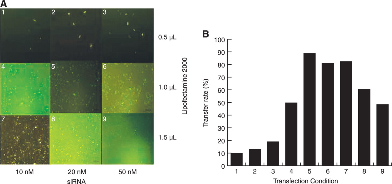
The optimal conditions for transfection were established by transfecting fluorescein isothiocyanate–labeled GAPDH into rat skin fibroblasts of 2 to 4 passages (normal glucose culture: 5.5 mmol/L of glucose) using Lipofectamine 2000 (Invitrogen). The transfer rate was detected by counting positive cells using inverted fluorescence microscopy, and cell viability was measured by MTT [3-(4,5-dimethylthiazol-2-yl)-2,5-diphenyltetrazolium bromide] under nine transfection conditions (Fig. 1A). The condition with a transfection rate of 75% and cell viability of 85% was selected for all subsequent transfections.
Transfection with MMP-9 siRNA
Fibroblasts of 2 to 4 passages were grown in Dulbecco’s modified Eagle’s medium containing 10% fetal bovine serum at 5.5 mmol/L of glucose. The cells were transfected with siRNA complex including the four pairs of MMP-9 siRNA, negative and positive controls, or transfection reagent only (mock transfection), under appropriate conditions. After 24 hours of serum deprivation (0.5% fetal bovine serum), the culture medium was replaced by Dulbecco’s modified Eagle’s medium containing 10% fetal bovine serum, 22 mmol/L of glucose, and 100 μmol/L of homocysteine and was cultured for 6, 24, and 48 hours.
Measurement of Inhibition Rate
Reverse transcription–polymerase chain reaction, Western blot analysis, and gelatin zymography for MMP-9 were performed to detect the percentages of inhibition of MMP-9 messenger RNA, protein expression, and activity, respectively, 30, 48, and 72 hours after transfection.
Figure 1.
A, Transfect fluorescein isothiocyanate–labeled glyceraldehyde-3-phosphate dehydrogenase small interfering RNA (siRNA) under nine transfection conditions to establish the optimal conditions for transfection. The final volume is 500 μL. The transfer rate was determined by positive cell count using inverted fluorescence microscopy after 24 hours. B, Histogram of the transfer rates of the nine trialed transfection conditions. Condition 5 (20 nM of siRNA and 1.0 μL of Lipofectamine 2000) had the highest transfer rate at 89.2% (P < .05).
Statistics
Data analysis was performed with Microsoft Excel 2003 (Microsoft Inc, Redmond, Washington) and SPSS 12.0 for Windows (SPSS Inc, Chicago, Illinois). Analysis of variance was used for repeated measurement data, in which the least significant difference method was used for the comparison between two groups. Statistical analyses were performed using the average results of three repeated experiments under identical conditions. Results are expressed as mean ± SD. Differences were considered significant at P < .05.
Results
Observation of MMP-9 Expression in Skin
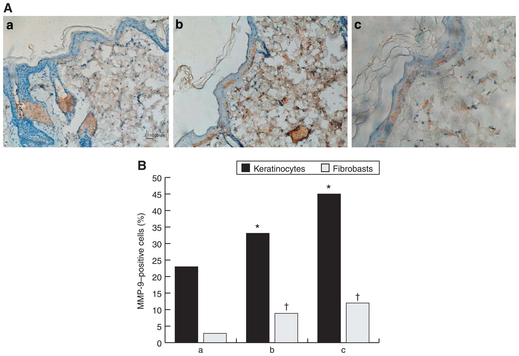
Immunohistochemical analysis showed MMP-9 to be present extensively in the epidermal layers of all rats. The mean ± SD percentage of MMP-9–positive cells was 22.89% ± 16.69% in the epidermis of normal skin, whereas in the epidermis of streptozotocin-induced diabetic rats, it was increased to 33.08% ± 18.16%, especially around the wound, where it was 44.92% ± 20.95%. There was no distribution of MMP-9 in the dermis of normal rats (mean ± SD percentage positive: 2.17% ± 2.02%), but MMP-9 in the dermis of streptozotocin-induced diabetic rats was observed (8.67% ± 2.76%), with an increase in the skin surrounding the wound (12.14% ± 4.44%) (P < .05) (Fig. 2).
Figure 2.
A, Immunohistochemical analysis for matrix metalloproteinase 9 (MMP-9) expression in skin from normal (a), diabetic (b), and wound periphery of diabetic (c) rats (positive results show as brown staining). B, The histogram indicates the percentages of MMP-9–positive cells in the three groups in part A. *P < .05 in epidermis; †P < .05 in dermis compared with the normal group.
Establishment of the Fibroblast Cell Model of High MMP-9 Expression
MMP-9 messenger RNA level, protein expression, and protease activity in the high glucose (22 mmol/L) group were 1.10-, 1.59-, and 1.34-fold, respectively, greater than those in the normal glucose group (P < .05). The effect was more pronounced when the cells were treated with hyperhomocysteine (100 μmol/L), with the corresponding levels being 1.25-, 2.63-, and 2.52-fold, respectively, greater than those in the normal group (P < .05) (Fig. 3).
Optimizing siRNA Transfection Conditions
By optimization, a transfection rate of up to 75%, with a cell viability of 85%, was obtained in cultured fibroblasts using Lipofectamine transfection reagent at 0.2% (vol/vol) and an siRNA concentration of 20 nM. Subsequent transfections with MMP-9 siRNA were performed under these optimal conditions (Fig. 1B).
Inhibition of MMP-9 Expression in Fibroblasts Using siRNA
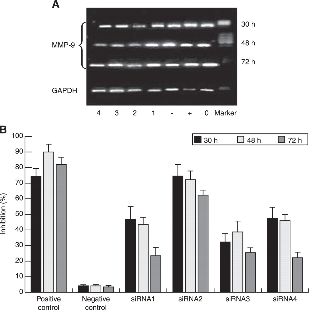
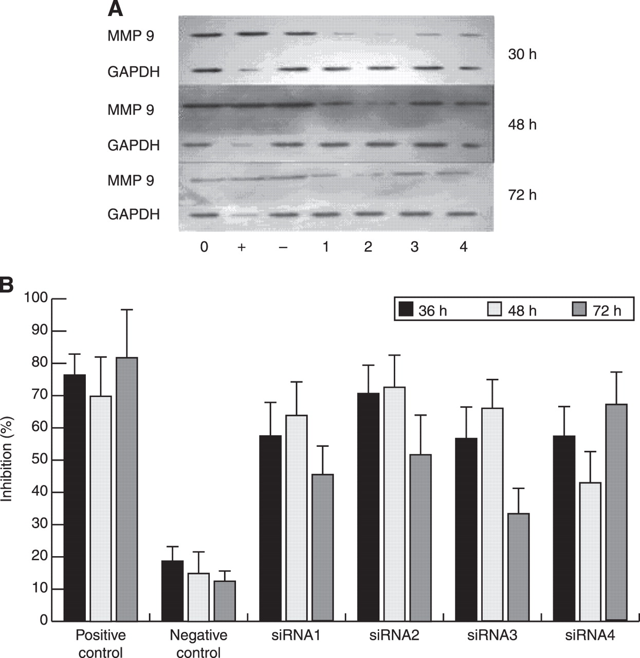
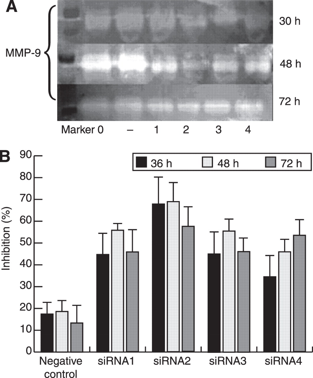
After 30-, 48-, and 72-hour transfection with 20 nmol/L of four MMP-9–specific siRNAs (siRNAs 1, 2, 3, and 4), fibroblasts were collected at different intervals, and reverse transcription–polymerase chain reaction, Western blot analysis, and gelatin zymography were performed. There was reduced MMP-9 messenger RNA expression in transfected cells compared with in cells undergoing mock transfection with Lipofectamine only. Mean ± SD inhibition rates of MMP-9 messenger RNA expression using the four siRNAs were 47.12% ± 7.60% with siRNA1, 74.23% ± 7.56% with siRNA2, 32.54% ± 5.02% with siRNA3, and 47.47% ± 6.80% with siRNA4 at 30 hours after transfection. At 48 hours after transfection, mean ± SD inhibition rates were 43.52% ± 4.30% with siRNA1, 71.85% ± 6.05% with siRNA2, 38.83% ± 6.87% with siRNA3, and 46.02% ± 3.08% with siRNA4; these values remained similar at 72 hours (Fig. 4). Furthermore, MMP-9 messenger RNA downregulation was paralleled by a reduction in MMP-9 protein abundance and activity. As shown in Figure 5A, mean ± SD inhibition rates of MMP-9 protein at 48 hours were 56.06% ± 10.05% with siRNA1, 73.17% ± 8.80% with siRNA2, 60.30% ± 10.22% with siRNA3, and 43.06% ± 9.01% with siRNA4; again, remaining almost constant at 72 hours (Fig. 5). Gelatin zymography showed the same trend; MMP-9 siRNA2 (5′-GGGCUUAGAUCAUUCUUCATT-3′ and 3 ′-UGAAGAAUGAUCUAAGCCCAG-5′) had the highest mean ± SD inhibition rates at 70.17% ± 4.83% at 30 hours and 69.40% ± 9.60% at 48 hours (Fig. 6).
Figure 3.
Effects of glucose and homocysteine (Hcy) on matrix metalloproteinase 9 (MMP-9) messenger RNA expression in cultured rat skin fibroblasts as measured by reverse transcription–polymerase chain reaction (A), Western blot analysis (B), and zymography analysis (C). GAPDH indicates glyceraldehyde-3-phosphate dehydrogenase. D, Histogram of the effects of glucose and Hcy on MMP-9. Data are given as mean ± SD. *P < .05 compared with the negative control group.
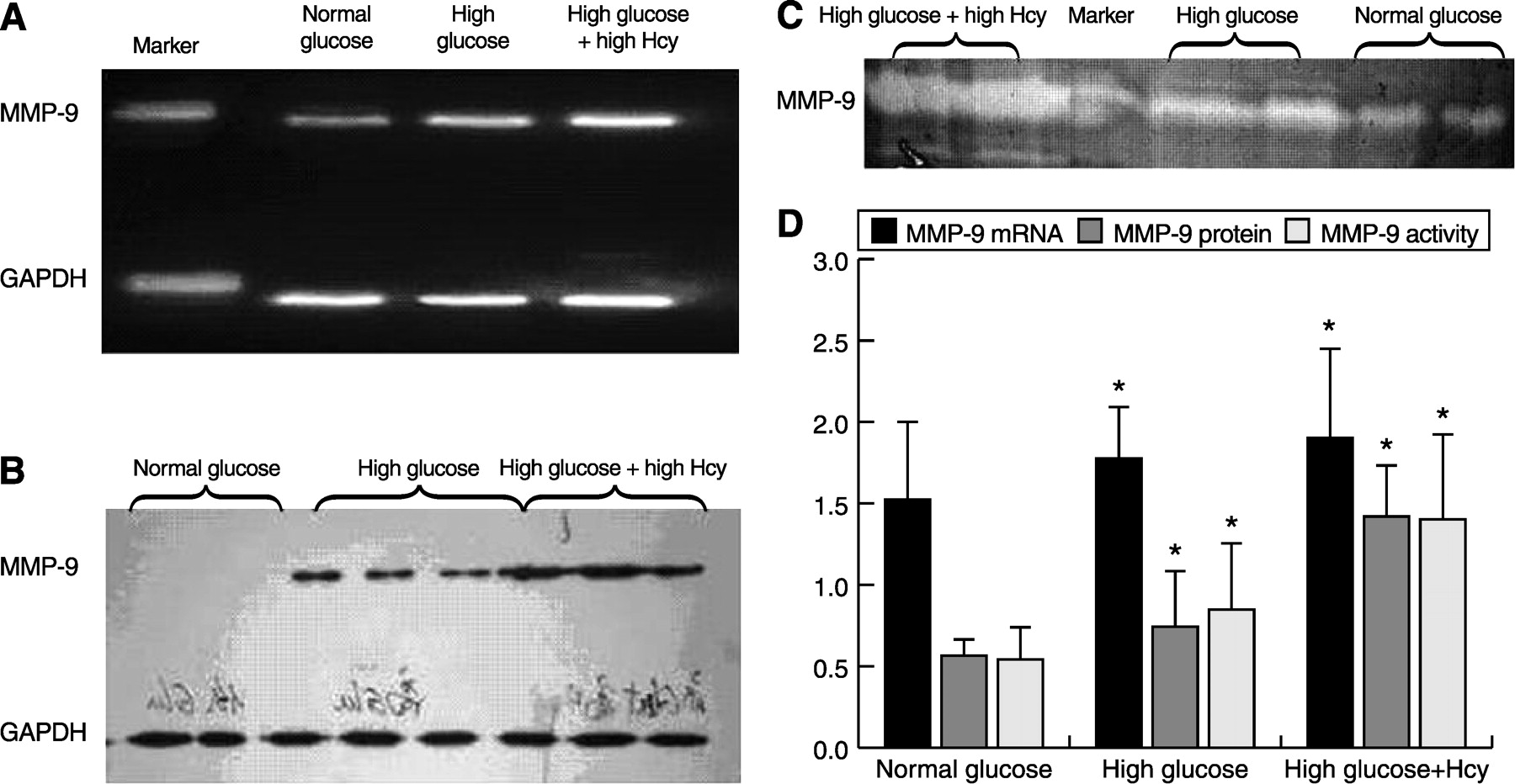
Figure 4.
A, Representative reverse transcription–polymerase chain reaction gel of expression of matrix metalloproteinase 9 (MMP-9) and glyceraldehyde-3-phosphate dehydrogenase (GAPDH) messenger RNA in cultured rat skin fibroblasts at different time points after transfection with small interfering RNA (siRNA). 0 indicates blank control; +, positive control; −, negative control; 1, siRNA1; 2, siRNA2; 3, siRNA3; and 4, siRNA4. B, Means of the densitometric analyses of all experiments for MMP-9 messenger RNA.
Discussion
The results of the present study demonstrated increased MMP-9 expression in the skin of diabetic rats, especially in the skin wound, and MMP-9 expression that could be markedly inhibited by treatment with MMP-9 siRNA (P < .05), especially at 48 hours after transfection. Furthermore, hyperhomocysteine increased MMP-9 production and enzymatic activity in fibroblasts, which were further increased by high glucose treatment (P < .05).
An elevated pro–MMP-9 level and, more significantly, an accumulation of active 82-kDa MMP-9 are associated with nonhealing of wounds. Based on its substrate preference for type IV collagen, which is present in the basement membrane matrix, the implications for dysregulation of this protease are clear.[8] Various cutaneous histologic and pathophysiologic alterations, composing the underlying disorder of diabetic skin, exist before injury. A set of local biochemical factors lead to the reduction of skin thickness and layers, denaturation of connective tissues, variations in cell morphological features, the decline of cell adhesion and proliferation, and abnormal collagen formation, among which, the imbalance of MMP-9 expression seems the most important.
The present study showed that during the healing of diabetic wounds, MMP-9 expression increased significantly compared with that of normal skin, which is consistent with the results of other studies. The abnormal increase in MMP-9 can degrade the growth factor, integrin, and its receptor, thus increasing the local inflammatory response and slowing wound healing. However, for the treatment of diabetic foot ulcers, it has been found that the simple application of growth factors does not accelerate ulcer healing; therefore, inhibition of MMP-9 expression in the local wound may be an approach to accelerate the healing of diabetic wounds.[17]
Figure 5.
A, Representative Western blot of expression of matrix metalloproteinase 9 (MMP-9) and glyceraldehyde-3-phosphate dehydrogenase (GAPDH) protein in cultured rat skin fibroblasts at different time points after transfection with small interfering RNA (siRNA). 0 indicates blank control; +, positive control; −, negative control; 1, siRNA1; 2, siRNA2; 3, siRNA3; and 4, siRNA4. B, Means of the densitometric analyses of all experiments for MMP-9 protein.
Furthermore, the research by Reiss et al[18] showed that MMP-9 could delay wound healing through interference with reepithelialization. They treated punch biopsy wounds on the backs of C57BL mice with exogenous MMP-9 and found that MMP-9 at a concentration paralleling that detected in human chronic wounds can delay wound healing in this animal model. This conclusion provided feasibility and possibility for improving wound healing by inhibiting the expression of MMP-9 in chronic wounds such as the diabetic foot.
Several methods of decreasing MMP-9 expression in diabetic skin have been tried, such as the use of all-trans retinoic acid, platelet-derived growth factor-BB, and nitric oxide donors.[10,13,19] However, these methods do not specifically inhibit MMP-9 expression. In the present study, RNA interference technology, which permits the simple, yet effective, knockdown of genes of interest,[14] was used to inhibit the abnormal increase of MMP-9 in diabetic skin. Until now, several techniques have been used to deliver siRNA to cell cultures in vitro[20,21]; of these, the liposome transfection method was chosen for its high tranfection efficiency and low cytotoxicity. Four sequence-specific siRNAs for rat MMP-9 were designed and synthesized in this study, and all were effective in downregulating the expression and activity of MMP-9 in rat dermal fibroblasts, with siRNA2 (5′-GGGCUUAGAUCAUUCUUCATT-3′ and 3′-UGAAGAAUGAUCUAAGCCCAG-5′) having the strongest inhibitory function. Whether downregulation of MMP-9 is effective in vivo in speeding up the wound process and what the appropriate inhibition level is need more affirmative study, which give us a valuable guide for further research.
The MMPs are regulated by a variety of inflammatory cytokines, hormones, and growth factors and by serum glucose and homocysteine, all of which are known to be operative in diabetic skin. Serum homocysteine is an important risk factor for diabetes with complications, and its detection is useful for prevention and treatment of the disease.[22,23] Meanwhile, research has shown that elevated homocysteine may impair wound healing by significantly decreasing wound nitric oxide and transforming growth factor-β bioactivity and, perhaps, also by modification of the provisional wound matrix formation.[24]
To avoid the complicating factors of the in vivo diabetic skin environment, high concentrations of glucose and homocysteine were used in the present study to mimic the local environment of diabetic skin in vitro. Enhanced MMP-9 production and activity were observed in fibroblast cells grown in the presence of high glucose. High glucose levels may increase MMP production and activity directly or indirectly (eg, via oxidative stress or advanced glycation products).[25] Homocysteine also enhanced MMP-9 production and activity in rat dermal fibroblasts. The results suggest that high serum homocysteine levels, in addition to high glucose levels, are involved in the regulation of MMP-9 expression in the diabetic condition. In addition to its effect on vessels and nerves, hyperhomocysteinemia may participate in the occurrence and development of diabetic foot ulcers via the regulation of MMP-9/TIMP1.
In the treatment of diabetic foot ulcers, there has been a wide range of dressings and different additives. In addition to antibacterial additives (eg, silver ions and iodine preparations), growth factors and cytokines have also been incorporated into dressings to promote wound healing. With the emergence and development of RNA interference technology, its use in the research of diabetic foot dressings may be a direction of great potential. The MMP-9 siRNA2, selected in the present study, is expected to be included in follow-up research dealing with diabetic foot dressings. In addition, fibroblasts, as the main repair cells, are significantly decreased in diabetic skin. Fibroblasts with MMP-9 inhibited by RNA interference could be reimplanted into the wound as a means of gene therapy in diabetic skin. This would not only increase the available cells but also change the local microenvironment of the diabetic foot ulcer, making it more conducive to tissue repair, potentially providing a feasible therapeutic method for diabetic foot ulcer treatment.[26]
Figure 6.
A, Representative gelatin zymograms of matrix metalloproteinase 9 (MMP-9) protease activity in cultured rat skin fibroblasts at different time points after transfection with small interfering RNA (siRNA). 0 indicates blank control; +, positive control; −, negative control; 1, siRNA1; 2, siRNA2; 3, siRNA3; and 4, siRNA4. B, Means of the densitometric analyses of all experiments for MMP-9 protease activity.
Financial Disclosure
The project was sponsored by the Guangdong Natural Science Fund.
Conflict of Interest
None reported.
References
- Singh, N, DGArmstrong, and BALipsky. :Preventing foot ulcers in patients with diabetes. .JAMA293::217. ,2005. .
- Jeffcoate, WJ. :Theories concerning the pathogenesis of the acute Charcot foot suggest future therapy. .Curr Diab Rep5::430. ,2005. .
- Hampton, S. :Caring for the diabetic patient with a foot ulcer. .Br J Nurs15::S22. ,2006. .
- Lobmann, R, AAmbrosch, GSchultz, et al. :Expression of matrix-metalloproteinases and their inhibitors in the wounds of diabetic and non-diabetic patients. .Diabetologia45::1011. ,2002. .
- Varani, J, RLWarner, MGharaee-Kermani, et al. :Vitamin A antagonizes decreased cell growth and elevated collagen-degrading matrix metalloproteinase and stimulates collagen accumulation in naturally aged human skin. .J Invest Dermatol114::480. ,2000. .
- Visse, R and HNagase. :Matrix metalloproteinases and tissue inhibitors of metalloproteinases: structure, function, and biochemistry. .Circ Res92::827. ,2003. .
- Iyer, V, KPumiglia, and CMDiPersio. :α3β1 integrin regulates MMP-9 mRNA stability in immortalized keratinocytes: a novel mechanism of integrin-mediated MMP gene expression. .J Cell Sci118 ( pt 6:):1185. ,2005. .
- Das, S, MMandal, TChakraborti, et al. :Structure and evolutionary aspects of matrix metallo-proteinases: a brief overview. .Mol Cell Biochem253::31. ,2003. .
- Andreea, SI, CMarietta, and DAnca. :AGEs and glucose levels modulate type I and III procollagen mRNA synthesis in dermal fibroblasts cells culture. .Exp Diabetes Res. [Published online ahead of print April 3, 2008; 10.1155/2008/473603].
- Lobmann, R, TPap, AAmbrosch, et al. :Differential effects of PDGF-BB on matrix metalloproteases and cytokine release in fibroblasts of Type 2 diabetic patients and normal controls in vitro. .J Diabetes Complications20::105. ,2006. .
- Lai, CF, VSeshadri, KHuang, et al. :An osteopontin–NADPH oxidase signaling cascade promotes pro–matrix metalloproteinase 9 activation in aortic mesenchymal cells. .Circ Res98::1479. ,2006. .
- Lee, CW, CCLin, SFLuo, et al. :TNF-α induced MMP-9 expression via activation of Src/EGFR, PDGFR/PI3K/Akt cascade and promotion of NF-κB/p300 binding in human tracheal smooth muscle cells. .Am J Physiol Lung Cell Mol Physiol292::L799. ,2007. .
- Solini, A, ESantini, MNannipieri, et al. :High glucose and homocysteine synergistically affect the metalloproteinases-tissue inhibitors of metalloproteinases pattern, but not TGFB expression, in human fibroblasts. .Diabetologia49::2499. ,2006. .
- Fire, A, SXu, MKMontgomery, et al. :Potent and specific genetic interference by double-stranded RNA in Caenorhabditis elegans. .Nature391::806. ,1998. .
- Elbashir, SM, JHarborth, WLendeckel, et al. :Duplexes of 21-nucleotide RNAs mediate RNA interference in cultured mammalian cells. .Nature411::494. ,2001. .
- Ren, JL, JSPan, TCheng, et al. :RNA interference inhibits hepatitis B virus gene expression and replication in HepG2-N10 cells. .Chin J Dig Dis7::230. ,2006. .
- Armstrong, DG and EBJude. :The role of matrix metalloproteinases in wound healing. .JAPMA92::12. ,2002. .
- Reiss, MJ, YPHan, EGarcia, et al. :Matrix metalloproteinase-9 delays wound healing in a murine wound model. .Surgery147::295. ,2010. .
- Burrow, JW, JAKoch, HChuang, et al. :Nitric oxide donors selectively reduce the expression of matrix metalloproteinases-8 and -9 by human diabetic skin fibroblasts. .J Surg Res140::90. ,2007. .
- Harborth, J, SMElbashir, KBechert, et al. :Identification of essential genes in cultured mammalian cells using small interfering RNAs. .J Cell Sci114::4557. ,2001. .
- Sui, G, CSoohoo, EBAffar, et al. :A DNA vector-based RNAi technology to suppress gene expression in mammalian cells. .Proc Natl Acad Sci U S A99::5515. ,2002. .
- Buysschaert, M, ASDramais, PEWallemacq, et al. :Hyperhomocysteinemia in type 2 diabetes: relation to macroangiopathy, nephropathy and insulin resistance. .Diabetes Care23.:1846. ,2000. .
- Fonseca, VA, AStone, MMunshi, et al. :Oxidative stress in diabetic macrovascular disease: does homocysteine play a role. .South Med J90::903. ,1997. .
- BoykinJr, JV and CBaylis. :Homocysteine: a stealth mediator of impaired wound healing: a preliminary study. .Wounds18::26. ,2006. .
- Death, AK, EJFisher, KCMcGrath, et al. :High glucose alters matrix metalloproteinase expression in two key vascular cells: potential impact on atherosclerosis in diabetes. .Atherosclerosis168::263. ,2003. .
- Branski, LK, CTPereira, DNHerndon, et al. :Gene therapy in wound healing: present status and future directions. .Gene Therapy14::1. ,2007. .