


Abstract
Turner syndrome is a genetic disorder that can present clinically with multiple concurrent comorbidities. This case report describes a 12-year-old girl with Turner syndrome who was referred for podiatric medical assessment and explores the application of optoelectronic stereophotogrammetry in the biomechanical assessment of the foot and lower limb. A four-segment kinematic foot model using 14-mm reflective markers was applied to the foot and lower limb of the patient to track motion at the tibia, rearfoot, forefoot, and hallux. Kinematic results presented in this case study illustrate evidence of excessive foot pronation throughout the stance phase of gait. Whether excessive pronation is a general characteristic of foot function in Turner syndrome remains to be confirmed, but the findings presented suggest that a comprehensive evaluation of foot biomechanics in patients with Turner syndrome may be warranted.
Turner syndrome is a genetic disorder associated with complete or partial absence of the second X chromosome in phenotypic females and is reported to occur in approximately 50 per 100,000 liveborn girls.[1] The syndrome can present with multiple concurrent comorbidities, which include congential malformation, lymphoedema, growth disturbance, ovarian failure, and musculoskeletal complications. The clinical management of girls with the syndrome requires specialist and integrated multidisciplinary care[1]; however, the role of the podiatric physician in the multidisciplinary team has been poorly acknowledged in the literature. In a descriptive study looking at foot problems in Turner syndrome, Findlay et al[2] characterized the foot as short, broad, and excessively pronated. The authors concluded that a variety of factors lead to foot abnormalities in these girls and, that podiatric medical examination should form part of their multidisciplinary care. The development of foot problems in Turner syndrome can be attributed to a combination of factors, including the aberrant mechanics and features specific to the syndrome (ie, lymphedema); however, this area requires further research. The study by Findlay et al[2] conducted static evaluation of the foot, and, consequently, further work is necessary to determine the characteristics of the dynamic foot and lower limb.
Clinical assessment of the pediatric foot and lower limb is often limited to subjective, static evaluation, but with advances in optoelectronic systems it is now possible to conduct comprehensive assessment of the foot during gait. Analysis of three-dimensional (3-D) gait data yields information that cannot be determined by clinical evaluation alone and can offer a variety of advantages when used in assessment as it allows for kinematic and kinetic data to be incorporated into clinical decision making.[3] Data derived from 3-D analysis can detail joint motion occurring in different planes and at different levels in the lower limb, thus providing a comprehensive understanding of the motion occurring during walking. Recently, a variety of foot models have emerged that allow for a greater understanding of foot function during gait.[3–5] It is beyond the remit of this report to discuss these models, and the reader is referred to the wider literature for further information.[6] Although 3-D modeling of the pediatric foot is still relatively novel, it yields data that can advance understanding of foot function and inform clinicial decision making; therefore, the aim of this case report was to present a 3-D biomechanical assessment of the foot in a girl with Turner syndrome.
Case Report and 3-D Analysis
A 12-year-old girl was referred for podiatric medical assessment after concerns were raised from the parents regarding the position of their daughter’s feet. On examination, characteristics of the foot in Turner syndrome were identified as hyperextension of the interphalangeal joints of both halluces and brachymetatarsia of the left foot with brachymetapody of the fourth and fifth toes. There was also evidence of lymphedema. In agreement with the previous research, both feet were broad but, in this case, large for skeletal height (UK size 8). Range and quality of motion at the ankle, subtalar, midtarsal, and first metatarsophalangeal joint were assessed, and an increase in range was noted at the ankle and midtarsal joints in both feet. The patient presented to the clinic in school attire, and the footwear was deemed suitable, although medial collapse of the uppers was noted along with excessive medial wear of the sole.
On static weightbearing assessment, there was flattening of the medial longitudinal arch and presentation of a planus foot type. The six-component Foot Posture Index was used to score the foot; a score of 10 was recorded for the left foot and 9 for the right. Clinical gait analysis revealed the feet to be pronated through the stance phase of gait. The gait pattern was tentative and apropulsive, with a reduced cadence and increased periods of double-limb support duration. At the time of assessment, there were no concerns regarding musculoskeletal pain or abnormalities. Clinical findings confirmed asymptomatic nondevelopmental flatfoot, and further analysis was conducted to evaluate the biomechanical function of the foot and lower limb.
Prior to data collection, reflective markers were applied to the foot and lower limb. For this, a Vicon plug-in gait marker set (Vicon Motion Systems Ltd, Oxford, England), similar to the Helen Hayes marker set,[7] was applied to the lower limbs, and the Oxford Foot Model[8] was applied to the dominant foot (right foot) by the same investigator (A.I.). A Ten camera Vicon 612 (Vicon Motion Systems Ltd, Oxford, UK) system was used to capture the coordinates of the reflective markers within the capture volume, sized appropriately to allow for one stride of the subject’s right foot to be tracked. Two force plates (Bertec, Model MIE Ltd, Leeds, UK) mounted within the laboratory floor recorded ground reaction forces (1000Hz) to determine gait events. The Oxford Foot Model defined the foot as four segments and measures relative motion of the tibia, hindfoot, forefoot, and hallux. Seventeen markers (14 mm) were placed at the following landmarks: lateral knee, lateral head of the fibula, tibial tuberosity, anterior aspect of the shin, lateral malleolus, medial malleolus, proximal posterior calcaneus, distal posterior calcaneus, a peg marker placed on the posterior calcaneus, lateral calcaneus, sustentaculum tali, first metatarsal–proximal dorsal, first metatarsal–distal medial, fifth metatarsal–proximal lateral, fifth metatarsal–distal lateral, midpoint of the distal heads of the second and third metatarsals, and the hallux at the proximal end of the first phalanx. A 3-D analysis was conducted using a ten-camera system (Vicon 612; Vicon Motion Systems Ltd) to track joint trajectories of the foot and lower limb.
After a static calibration capture, the patient was asked to walk at a comfortable, self-selected speed along the predetermined 8-m walkway, and ten successful trials were captured. A successful trial was defined as complete loading of the foot on the force platform. The data were processed in Vicon Polygon software (Vicon Motion Systems Ltd) to allow the normalization of joint motions over 100 time points from heel strike to heel strike, thus capturing stance and swing phase. Lower-limb joint and segmental kinematics were calculated based on the joint coordinate system[9] and were filtered using a Woltring filter with a mean square error value of 20.
Results of Kinematic Analysis
Table 1 summarizes the data captured for the case and presents mean segmental angles for the forefoot-hindfoot angle, forefoot-tibia angle, hind-foot-tibia angle, and hallux-forefoot angle at heel strike and toe off. At initial heel contact, the hindfoot (hindfoot-tibia angle) was plantarflexed and everted relative to the tibia and the forefoot (forefoot-hindfoot angle) pronated on the rearfoot. At toe-off, the hindfoot (hindfoot-tibia angle) maintained this everted and internally rotated position, similarly with the forefoot-tibia angle, which was prontated.
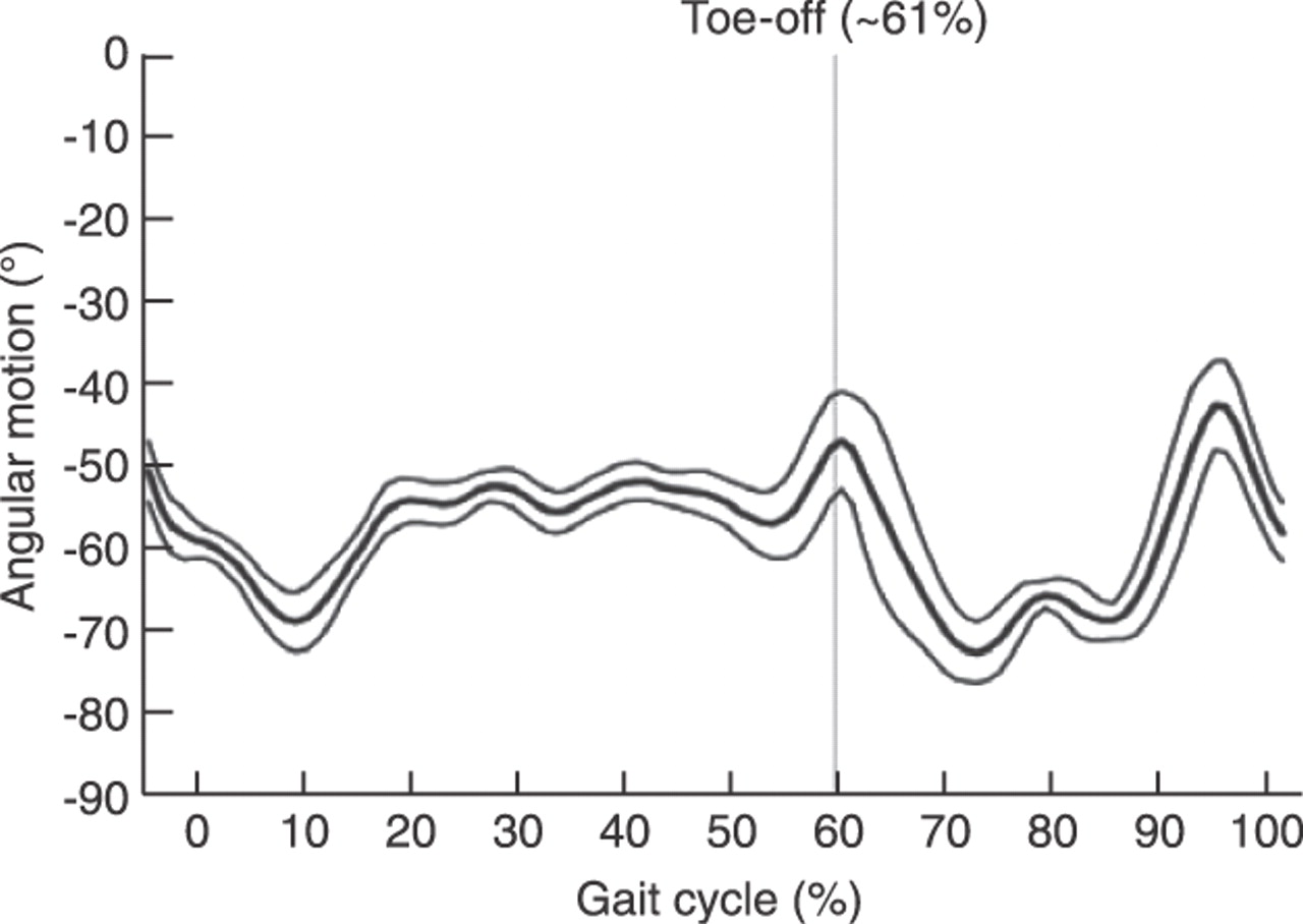
Figure 1 represents the movement of the forefoot relative to the tibia throughout the gait cycle. The pattern of motion of the forefoot relative to the tibia is similar to the data presented by Stebbins et al,[3] with one supination peak occurring around toe-off and one just before heel strike. The major variation between the studies is the angle offset, which can be attributable to anatomical differences between the patients (the patient in the present case study presented with pronated feet) or marker placement errors.
Table 1.
Intersegmental Angles Through the Stance Phase of Gait in the Sagittal (x), Frontal (y), and Transverse (z) Planes for the Right Foot
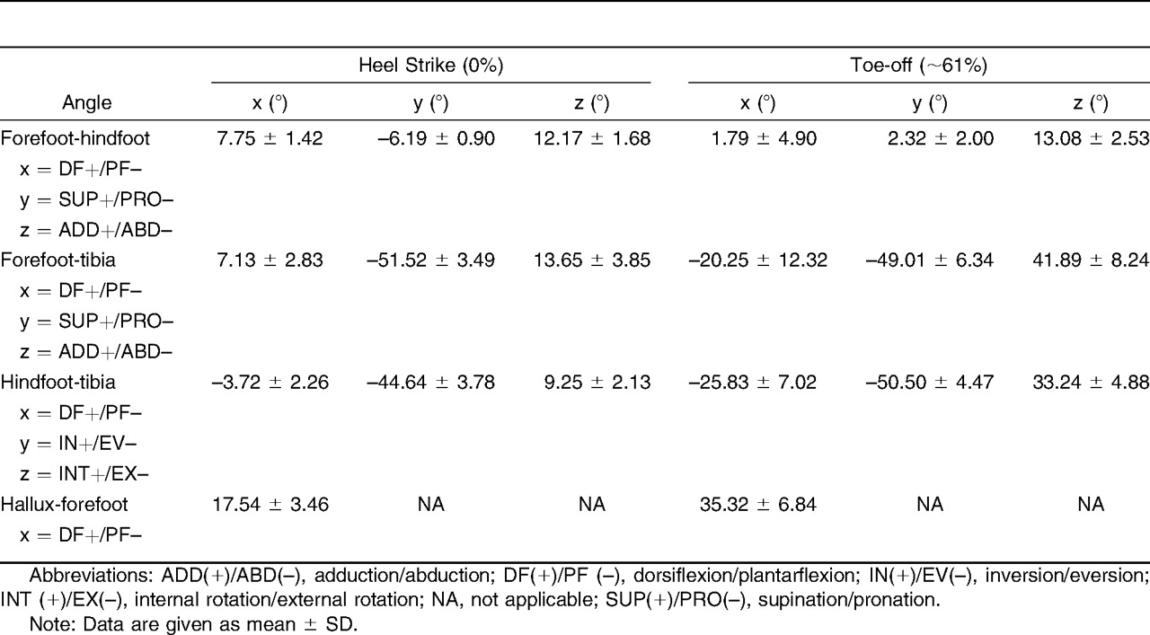
Figure 1.
Mean ± SD angular motion of the forefoot relative to the tibia throughout the gait cycle for the right foot.
Further analysis of the foot included range of motion for the gait events and maximum value for joint angles (Table 2). Range of motion of the forefoot relative to the hindfoot was greatest in the sagittal plane, with similar ranges in the frontal and transverse planes, corresponding to the findings of Stebbins et al.[3] When comparing the forefoot with the tibia, the greatest range of motion occurred in the sagittal plane, followed by the transverse and frontal planes. The forefoot was positioned in a high degree of pronation through the entire gait cycle. Variation between the trials was greatest for this segment, especially in the sagittal and transverse planes, possibly as a result of there being many joints in between the forefoot and the tibia where movement can occur. Motion of the hindfoot relative to the tibia was similar across all planes, with the hindfoot remaining everted through the gait cycle.
Discussion
This case report described a girl with Turner syndrome referred for podiatric medical evaluation after concerns about foot position and walking pattern. Clinical examination confirmed asymptomatic, nondevelopmental flatfoot, and 3-D gait analysis was incorporated into the patient assessment to determine the kinematic characteristics of the foot. After analysis, it was observed that mean segmental angles of the forefoot and hindfoot segments functioned in an everted/pronated alignment throughout the stance phase of gait (Table 1). At heel strike, the foot would be expected to be inverted and then move into an everted position throughout the loading response, but in this case, the rearfoot was everted at initial contact and maintained this position throughout the stance phase and at propulsion (Fig. 1). This is an important consideration as the foot would be expected to act as a rigid lever at propulsion and may, in this instance, reflect the apropulsive gait demonstrated during clinical examination.
Table 2.
Ranges of Motion and Maximum Values for the Segmental Angles in the Sagittal (x), Frontal (y), and Transverse (z) Planes
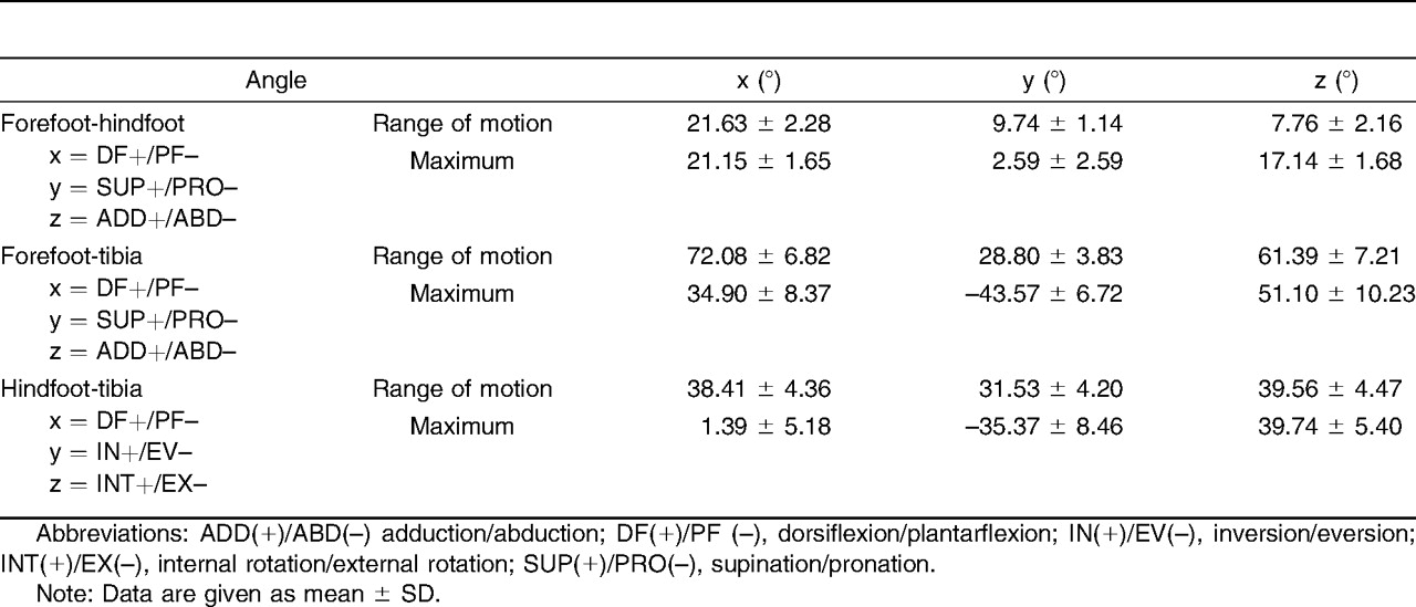
Although the findings of this case study are limited and cannot be extrapolated to the population of girls with Turner syndrome, comparison with other studies is warranted. Studies using the Oxford Foot Model in the assessment of the pediatric foot are limited, but some work has been conducted to determine the reliability of the model[3] and also to characterise the foot in cerebral palsy.[10] Compared with Stebbins et al,[3] in a study of 15 typically developing children (mean age, 9.5 years), this case study demonstrated considerably larger ranges of motion at the forefoot and rearfoot in all three planes; however, forefoot to rearfoot motion was comparable with these data. Clinical assessment indicated increased range of motion at the ankle joint, and this finding concurs with the 3-D measure of ankle range of motion, which showed twice as much ankle motion compared with the values published by Stebbins et al.[3] The maximal values for the present patient differed across all segment-to-segment comparisons with data from Stebbins et al,[3] which is indicative of an offset due to foot deformity in the static pose, especially true for forefoot pronation and rearfoot eversion. In line with the work presented by Stebbins et al,[3] each joint angle obtained throughout the dynamic trials was not referenced to the neutral position during the static trial. Although this may increase the variability in multiple-subject/session tests, it allows measurement of foot deformity, which may cause nonzero joint angles in the static trial. Further research is required to assess the kinematic differences between patients with Turner syndrome and healthy individuals using 3-D gait analysis methods to understand gait differences and systematic error due to marker placement.
Based on the clinical and biomechanical observations, a treatment plan was devised. The initial stage of the plan was foot care and footwear advice. In light of the syndrome, targeted foot care and footwear advice was given. Footwear advice was necessary as the footwear choices outside of school were not appropriate and failed to offer adequate support for the foot. Following discussion with the parents and daughter, it was agreed that prefabricated, medium-density orthotic devices be prescribed as a first-line treatment intervention with a view to casted orthoses being prescribed in the longer term. Initial progress with the prefabricated devices was positive and treatment and monitoring are currently ongoing.
The clinical presentation of rare conditions is often complex and requires management in a specialist, multidisciplinary team. This case report concurs with previous research regarding the foot in girls with Turner syndrome and supports the view that comprehensive evaluation of foot and lower-limb biomechanics in girls with Turner syndrome is warranted.
Funding
None reported.
Conflicts of Interest
None reported.
References
- Hjerrild, BE, KHMortensen, and CHGravholt. :Turner syndrome and clinical treatment. .Br Med Bull86::77. ,2008. .
- Findlay, CA, MDCDonaldson, and GWatt. :Foot problems in Turner’s syndrome. .J Pediatr138::775. ,2001. .
- Stebbins, J, MHarrington, NThompson, et al. :Repeatability of a model for measuring multi-segment foot kinematics in children. .Gait Posture23::401. ,2006. .
- MacWilliams, BA, MCowley, and DENicholson. :Foot kinematics and kinetics during adolescent gait. .Gait Posture17::214. ,2003. .
- Leardini, A, MGBenedetti, BDBettinelli, et al. :Rear-foot, mid-foot and fore-foot motion during the stance phase of gait. .Gait Posture25::453. ,2007. .
- Rankine, L, JLong, KCanseco, et al. :Multi-segmental foot modelling: a review. .Crit Rev Biomed Eng36::127. ,2008. .
- DavisIII, RB, SOunpuu, DTyburski, et al. :A gait data collection and reduction technique. .Hum Mov Sci10::575. ,1991. .
- Carson, MC, MEHarrington, NThompson, et al. :Kinematic analysis of a multi-segment foot model for research and clinical applications: a repeatability analysis. .J Biomech34::1299. ,2001. .
- Grood, E and WSuntay. :A joint coordinate system for the clinical description of three-dimensional motions: application to the knee. .J Biomech Eng105::136. ,1983. .
- Stebbins, J, MHarrington, NThompson, et al. :Gait compensations caused by foot deformity in cerebral palsy. .Gait Posture32::226. ,2010. .
© 2012 American Podiatric Medical Association