


Abstract
Background: We sought to develop new recombinant human epidermal growth factor (rhEGF)–containing hydrogels and to investigate their biological activity and therapeutic effects on wound healing in diabetic rats. Methods: Levels of rhEGF released from hydrogels were measured by enzyme-linked immunosorbent assay. The cellular proliferating activity of released rhEGF was evaluated by MTT assay. Fifty-six wounded diabetic rats were randomly divided into four groups with different topical treatment daily. The therapeutic effects were evaluated by wound area measurement, histologic analysis, immunohistochemical assessment of proliferating cell nuclear antigen and B-cell lymphoma/leukemia-2, and Western blotting of EGF receptor. Results: The rhEGF released from the hydrogel matrix kept its bioactivity on stimulating proliferation of the BALB/c3T3 cell line. Wound closure rates on postoperative day 14 were 75.8% in the negative control group, 82.83% in the group treated with hydrogel matrix, 85.87% in the group treated with rhEGF-containing hydrogel, and 81.18% in the group treated with rhEGF solution. Compared with hydrogel matrix, rhEGF-containing hydrogel had an additional effect on induction of EGF receptor expression (P < .05). Compared with negative controls, protein expression of B-cell lymphoma/leukemia-2 was higher in the rhEGF-containing groups (P < .05). Proliferating cell nuclear antigen was induced at its highest level on day 7 in the rhEGF-containing hydrogel–treated group (P < .05). Conclusions: These data from in vitro release and diabetic animal models highlight the efficacy of hydrogels as a controlled releasing system for topical application of EGFs. The rhEGF-containing hydrogel we developed holds the merits of prolonged and sustained releasing of bioactive rhEGF and therapeutic potential in enhancing diabetic wound healing.
Diabetes affects approximately 170 million people worldwide. Diabetic foot ulcer is a major complication of diabetes and a major morbidity associated with diabetes. In China, diabetic foot ulcer has become a leading cause of hospital admissions for diabetic patients. In addition, diabetic foot ulcers are expected to account for 84% of all diabetes-related lower-leg amputations because they are difficult to heal and are easily complicated with infection and gangrene.[1–3] Diabetic foot ulcers have been responsible for not only poor patient quality of life but also a heavy financial burden on society and the family. Currently, despite the existence of protocols to standardize care and the few Food and Drug Administration–approved therapies in the world, their limited efficacy and high expenditure prevent clinicians from implementing these modalities in routine clinical practice. As a result, diabetic foot ulcer is a big challenge for diabetic care staff.
Wound healing occurs as a series of dedicated cellular and molecular events responding to injury in which numerous growth factors are involved. One characteristic of poor-healing diabetic foot ulcers is decreased production or impaired action of growth factors. Epidermal growth factor (EGF) is a potent growth factor that plays an important role in promoting the process of wound healing through stimulating proliferation and migration of fibroblasts.[4,5] In vivo and in vitro pilot studies have demonstrated the potential efficacy of EGF in enhancing wound healing. Laato et al[6] confirmed that EGF is a potent mitogen for granuloma fibroblasts in a dose-dependent manner. In humans, Tsang et al[7] first demonstrated a therapeutic effect of human EGF on the healing of diabetic foot ulcers in a randomized, double-blind, controlled trial.[5] However, their therapeutic efficiency markedly depended on the implementing dosage and treatment duration. These may be partly attributable to the susceptible denaturation of the protein during storage and application or the rapid degradation by excessive matrix metalloproteinases in the wound. Advanced dressing combined with recombinant human EGF (rhEGF) treatment has been reported to possess an additional promising effect in promoting healing of chronic diabetic foot wounds.[8] At this point, the growth factor delivery system that can lead to sustained and stable release of growth factors must be promising in prompting wound healing. Hydrogels, with their high water absorbance and good breathability and biocompatibility, have been known as effective dressings to improve the healing of chronic wounds. Importantly, hydrogels have been extensively used in developing drug delivery systems in recent years.[9] Therefore, they are logically expected to be suitable carriers for growth factors.
In the present study, we postulated that rhEGF-containing hydrogel would be effective in enhancing the healing of diabetic foot ulcers. The biological activity of rhEGF-containing hydrogels and their therapeutic effects on wound healing in diabetic rats were investigated.
Materials and Methods
Preparation of rhEGF-Containing Hydrogels
Polyvinyl alcohol (5%) and sodium alginate (2%) were mixed (1:4, w/w) and stirred for 30 min at room temperature. Then, glycerol (2%, w/w), sodium borate (0.05%, w/w), and rhEGF (10 μg/g) (Cytolab Ltd, Rehovat, Israel) were added. Finally, in situ cross-linking was performed using cross-linking agents composed of 1% calcium chloride and 1% sodium benzoate. Each 5 g of hydrogel incorporated with rhEGF was sampled 24 hours, 7 days, and 14 days after hydrogel preparation for in vitro releasing tests. The previously mentioned chemical reagents were purchased from Guangzhou Chemical Plant in Guangzhou, China.
Estimation of In Vitro rhEGF Released from rhEGF-Containing Hydrogel
A small, conical flask containing 5 g of rhEGF-containing hydrogel (10 μg/g) was inverted into a beaker containing 100 mL of phosphate-buffered saline. After 5 min, 10 min, 20 min, 30 min, 1 hour, 1.5 hours, 2 hours, 4 hours, 6 hours, 8 hours, 10 hours, 12 hours, and 24 hours of incubation, 0.5 mL of the incubating solution was sampled at the same position. Levels of human EGF in these samples were measured by enzyme-linked immunosorbent assay (Shenxiong Science and Technology Co, Shanghai, China) according to the manufacturer’s instructions. The cumulative rhEGF release rate was calculated.
Evaluation of the Cellular Proliferating Activity of Released rhEGF from the Hydrogel
The released solutions were dissolved in Dulbecco’s modified Eagle’s medium (Jinuo Biomedical Technology Co, Hangzhou, China) containing 10% calf serum and co-cultured with the BALB/c3T3 cell line (Cell Bank, Chinese Academy of Sciences, Shanghai, China) in 96-well plates (200 μL for each well). After being cultured for 48 hours, 20 μL of MTT (5 g/ L; MB-chem, Jiangsu, China) was added to each well and incubated for another 4 hours. The reaction was stopped by adding 150 μL of dimethylsulfoxide (Sigma-Aldrich Corp, St. Louis, MO). Absorbance was measured at 492 nm, which could be used to evaluate the biological activity of released rhEGF from the hydrogel matrix.
Diabetic Animal Model and Generation of Full-thickness Wounds
Altogether, 56 male Sprague-Dawley rats obtained from Guangzhou University of Traditional Chinese Medicine with a start weight of 250 g were acclimated for 1 week on arrival in the laboratory. The Animal Care and Use Committee at Guangzhou University of Traditional Chinese Medicine approved all of the methods used in this study.
Diabetes was induced in rats via intra-abdominal injection of 35 mg/kg of streptozotocin (Sigma-Aldrich Corp) daily for 3 days. Serum glucose levels were measured from the tail vein daily using a glucometer (LifeScan Inc, a Johnson & Johnson company, Milpitas, California). Animals with blood glucose concentrations of 16.7 mmol/L or higher were considered diabetic. Diabetic animals received daily injections of protamine zinc insulin (1.0–2.0 U/d) as required to maintain blood glucose levels of 16.7 to 28.0 mmol/L and to avoid acute crisis for another 6 weeks before wounding.
Diabetic rats were anesthetized using pentobarbital sodium (30 mg/kg). The dorsal surface was shaved and disinfected with 70% ethanol. A sterilized template (1.3 × 1.3 cm) was placed on the midback, and the tissue was excised to the level of the panniculus carnosus using scissors and forceps. Skin biopsy specimens were obtained from the wound on the wounding day and 7 and 14 days after wounding. The entire wound, including a 5-mm margin of the surrounding normal skin, was excised to the level of fascia for analyses. Wounded animals were randomized into four groups (n = 7 each) with different topical treatments daily: no topical treatment, topical implementation of 0.5 g of hydrogel matrix, 0.5 g of rhEGF-containing hydrogel containing 5 μg of EGF, and 0.5 mL (5 μg) of rhEGF solution. A set of four groups was treated daily for 7 days, and another set was treated for 14 days.
In Vivo Evaluation of Wound Healing with the rhEGF-Containing Hydrogel
The therapeutic effects of rhEGF-containing hydrogel on wound healing were evaluated by wound area measurement and histologic analysis.
Wound size evaluation
Wound closure was documented with a digital camera (Canon A610; Canon, Tokyo, Japan) on days 0, 3, 5, 7, 10, and 14. Images were analyzed using ImageJ (1.36b) software. The percentage of wound closure was calculated as a percentage of the original wound size: [(initial area – final area)/initial area] ×100%.[10]
Histologic evaluation
Wound tissue was fixed in 4% polyformaldehyde. Tissue blocks were embedded in paraffin, sectioned at 4 μm, and stained by hematoxylin and eosin. Epidermal and dermal regeneration, thickness of granulation tissue, and angiogenesis were evaluated histologically. The criteria for evaluating histologic scores are described in the study by Galeano et al.[11]
Immunohistochemical Assessment of Epithelial Cell Proliferation and Apoptosis
Expression of proliferating cell nuclear antigen and B-cell lymphoma/leukemia-2 (BCL-2) on skin biopsy specimens from the margins of the wounds were evaluated by immunohistochemical analysis using the streptavidin-biotin complex technique. Briefly, endogenous peroxidase was blocked by incubation with 3% hydrogen peroxide. After antigen retrieval and blocking of nonspecific protein binding, sections were incubated with monoclonal mouse anti–proliferating cell nuclear antigen (1:100; Boster Biological Technology Ltd, Wuhan, China) at 4°C overnight, followed by biotin-conjugated secondary antibody goat anti-mouse IgG (1:200; Boster Biological Technology Ltd). Streptavidin-biotin complex was used to visualize the staining. Diaminobenzidine was used as the chromogenic substrate. To detect BCL-2 expression, we used monoclonal rabbit anti-BCL-2 (1:100; Boster Biological Technology Ltd) as the primary antibody and goat anti-rabbit IgG (1:200; Boster Biological Technology Ltd) as the secondary antibody. Data represent mean ± SD scores for which percentage positive is graded as follows: 0, none; 1, 25% or less; 2, 50% or less; 3, 75% or less; and 4, 100% or less. Intensity is rated as follows: 0, none; 1, weak; 2, moderate; and 3, intense. Final (composite) score = sum (percentage positive rating × intensity rating).[12]
Western Blotting of EGF Receptor
Western blotting was used to determine EGF receptor protein content. Skin specimens obtained 0, 7, and 14 days after wounding were homogenized in 1 mL of MT-CelLytics (Shenergy Biocolor BioScience & Technology Co, Shanghai) and 10 μL of MPIM (Shenergy Biocolor BioScience & Technology Co) and then were centrifuged at 15,000 ×g for 20 min at 4°. Protein concentrations were quantified by the MiniBCA (Bio-Rad, Hercules, CA) method. Equal amounts of the protein were separated on 10% sodium dodecyl sulfate–polyacrylamide gel and transferred to a polyvinylidene fluoride membrane. The membrane was blocked with a blocking buffer (5% bovine serum albumin in 1× Tris-buffered saline containing 0.1% Tween 20) for 2 hours at room temperature, incubated with rabbit anti–EGF receptor (1:1,000; Cell Signaling Technology Inc, Danvers, MA) at 4°C overnight, incubated with peroxidase-conjugated AffiniPure goat anti-rabbit IgG (1:2,000; Boster Biological Technology Ltd) for 1 hour at room temperature, and visualized using an enhanced chemiluminescent method kit (Boster Biological Technology Ltd). β-Actin was used as an internal control. Samples were exposed to radiography in a darkroom, followed by BandScan 4.3 software analysis (Glyko, Novato, California).
Statistical Analysis
Results are expressed as mean ± SD. Multivariate analysis of variance was used for repeated-measures data. Analysis of variance was used for non–repeated-measures data. The statistical software program SPSS version 13.0 (SPSS Inc, Chicago, Illinois) was used for calculations. Differences were considered significant at P < .05.
Results
In Vitro Release Model of rhEGF from rhEGF-Containing Hydrogel
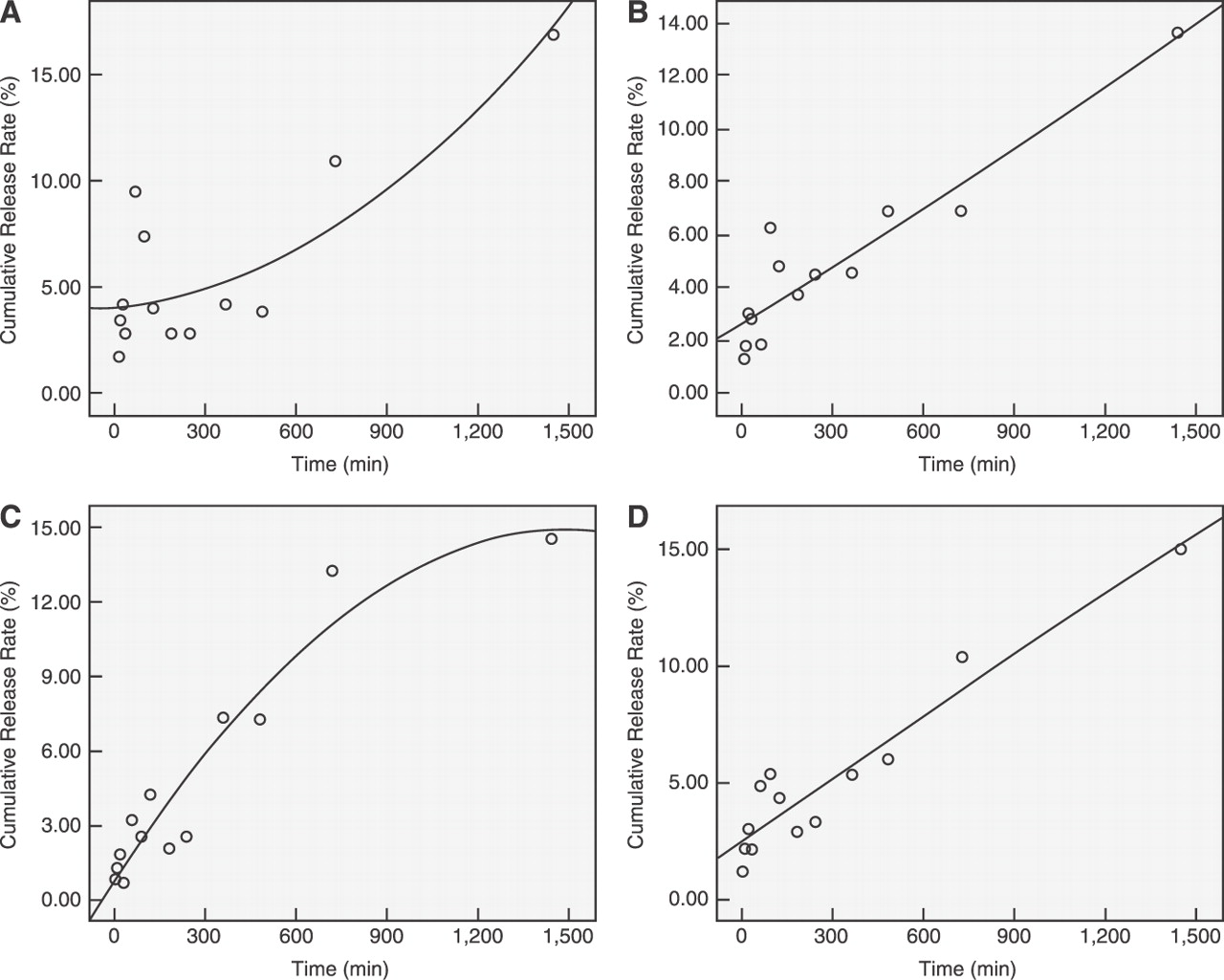
The 24-hour cumulative rhEGF release rates from the hydrogel matrix after the hydrogel was stored for 1, 7, and 14 days showed an initial burst release in the first 1 to 2 hours, followed by a sustained release rate of 16.9%, 13.67%, 14.57% in 24 hours, respectively, with a mean 24-hour release rate of 15.03% (Fig. 1). The quadratic for the average release model is Y = 2.504 + 0.009X − 2.3 × 10−7X2, according to which it needs 3, 7, and 27 days to attain release rates of 40%, 70%, and 100%, respectively.
In Vitro Biological Activity of Released rhEGF
The mean ± SD activity (absorbance) of the experimental groups (divided into groups 1d, 7d, and 14d based on the different storage times), the positive control group, and the negative control group were 0.90 ± 0.10, 0.86 ± 0.14, 0.80 ± 0.14, 0.87 ± 0.15, and 0.55 ± 0.10, respectively (Fig. 2). The higher ability to accelerate the proliferation of BALB/c3T3 in the experimental groups and the positive control group indicated that the released rhEGF possessed good biological activity.
In Vivo Effects of Wound Healing of Diabetic Rats
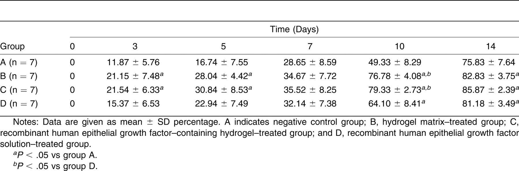
Processes of wound healing are documented in Table 1 and Figure 3. The rhEGF-containing hydrogel gained the best therapeutic effects on wound healing at different time points, followed by hydrogel matrix and rhEGF solution.
Further histologic analysis indicated that the angiogenesis composite scores for the rhEGF-treated groups were higher than those for groups without rhEGF treatment. Regarding reepithelialization, the hydrogel-treated groups had better scores than did the solution-treated groups. The group treated with rhEGF-containing hydrogel gained the highest composite scores for angiogenesis and reepithelialization. There were no statistically significant differences in enhancement of granulation between different topical agents (Table 2).
Immunohistochemical Assessment of Epithelial Proliferation and Apoptosis
Significant differences in proliferating cell nuclear antigen and BCL-2 expression were observed among groups during wound healing (Fig. 4). Day 7 after wounding, the proliferating cell nuclear antigen expression composite scores were higher in the rhEGF-containing hydrogel–treated group than in the negative control group, which indicates that rhEGF-containing hydrogel treatment significantly increased proliferation. Day 7 after wounding, the BCL-2 expression composite scores were higher in the rhEGF-containing hydrogel–treated group and the rhEGF solution–treated group than in the negative control group, which suggests that rhEGF up-regulated the expression of anti-apoptotic protein BCL-2 to restrain apoptosis to accelerate wound healing. No statistically significant differences were seen before wounding and on day 14 after wounding in the different groups.
Protein Abundance of EGF Receptor on Wounded Skin
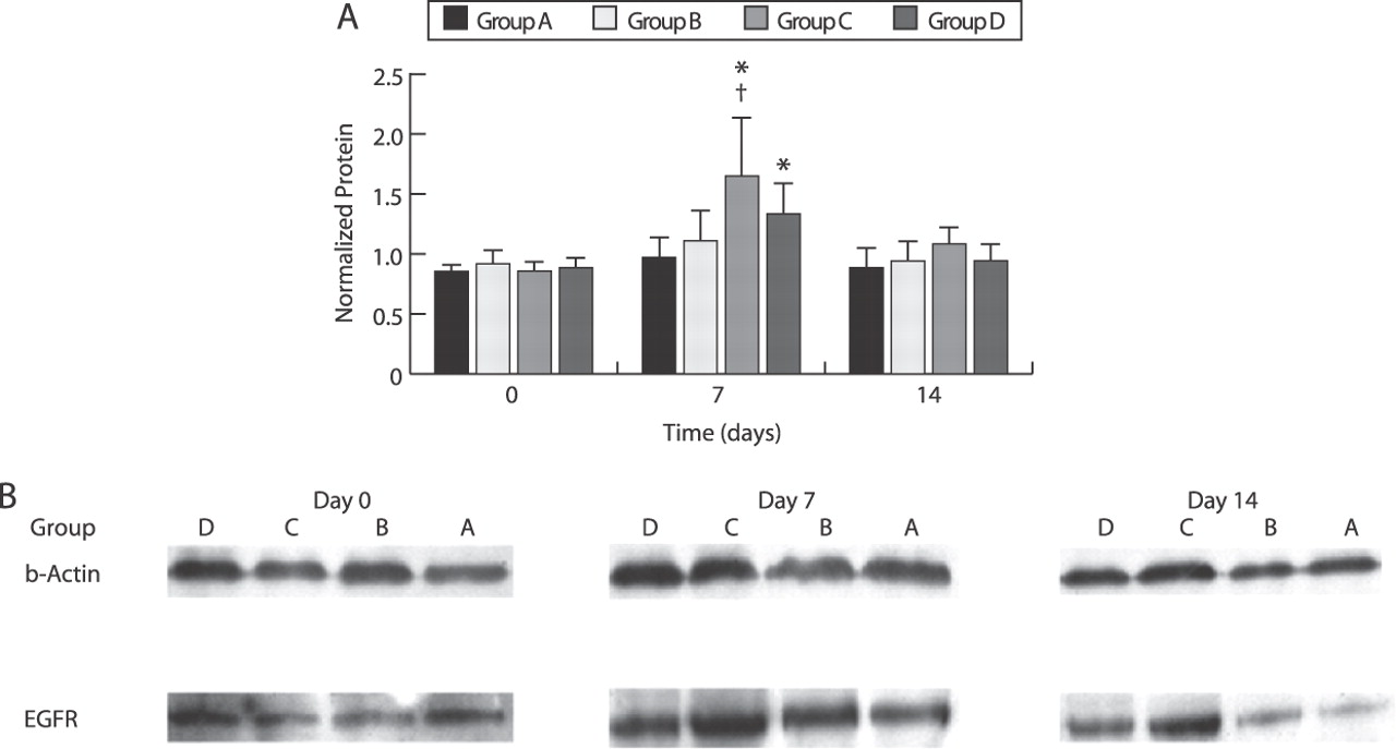
Day 7 after wounding, the relative values of EGF receptor expression in the rhEGF-containing hydrogel–treated group and the rhEGF solution–treated group were higher than those in the negative control and hydrogel matrix–treated groups, which indicates that rhEGF treatment significantly increased EGF receptor expression on wounds in the early days after operation (Fig. 5). The EGF receptor was induced at its highest level in the rhEGF-containing hydrogel–treated group. The time course of the induction of EGF receptor was similar to that of proliferating cell nuclear antigen and BCL-2.
Discussion
We developed rhEGF-containing hydrogels that can prolong the release of EGF with high bioactivity and accelerate wound healing in diabetic rats. In the in vitro releasing tests, there was an initial burst release in the first 1 to 2 hours, followed by a sustained release from the rhEGF-containing hydrogel. And the released rhEGF kept its bioactivity on stimulating in vitro proliferation of the BALB/c3T3 cell line even after 7 or 14 days’ storage of the hydrogel. Animal experiments showed that rhEGF-containing hydrogel had an additional effect on promoting wound healing.
Figure 1.
In vitro release model of recombinant human epidermal growth factor (rhEGF) from the rhEGF-containing hydrogel. Curve estimation (quadratic) was performed with time (X) as the independent variable and the cumulative release rate (Y) as the dependent variable. Shown are the rhEGF 24-hour cumulative release rates after treatment with rhEGF-containing hydrogel stored for 1 day (R = 0.84) (A), 7 days (R = 0.93) (B), and 14 days (R = 0.96) (C). D, The average release model, with Y = 2.504 + 0.009X −2.3 ×10−7X2 as the corresponding quadratic (R = 0.95).
Hydrogels had been one of the ideal materials for wound dressing. They could not only provide an optimal moisture environment for wound healing, which is believed to be the standard protocol, but also protect from the invasion of microorganisms and enhance macrophage function, epithelialization, and granulation tissue formation.[13–16] The present results coincide with these findings: rats treated with hydrogel matrix had a higher wound closure rate as early as 3 days after dressing with hydrogel and lasting 14 days compared with the negative control group. These results were at least partly attributable to better reepithelialization of the wound.
Compared with hydrogel matrix, rhEGF-containing hydrogel had an additional effect on inducing EGF receptor expression. Compared with the negative control group, BCL-2 expression was higher in the EGF-containing groups, including rhEGF-containing hydrogel and EGF solution. The epidermal proliferating marker proliferating cell nuclear antigen was induced at its highest level on day 7 in the group treated with rhEGF-containing hydrogel. These data indicate that rhEGF-containing hydrogel had additional potential in wound healing owing to the pleiotropic actions of EGF and the delivery system that lead to continuous release of the growth factor. Histologic analysis and measurements of proliferating cell nuclear antigen expression demonstrated that rhEGF could stimulate proliferation and migration and could accelerate reepithelialization and angiogenesis. The receptor tyrosine kinase EGF receptor had been implicated in this process because the data indicated that the time course of the induction of EGF receptor was similar to that of proliferating cell nuclear antigen and angiogenesis. Earlier studies[17] indicated that EGF receptor played an important role in wound healing through pleiotropic functions to enhance early healing after cutaneous incision. In addition, the present data also show that rhEGF upregulates expression of the anti-apoptotic protein BCL-2, which might restrain apoptosis and thereafter be good for wound healing. The EGF prevented the expected increase in apoptosis after small-bowel resection by shifting the ratio of bax to bcl-w in favor of cell survival.[18]
Figure 2.
In vitro study of biological activity on cell proliferation of released recombinant human epidermal growth factor (rhEGF) from rhEGF-containing hydrogel. The ability to accelerate BALB/c3T3 proliferation was higher in the experimental groups and the positive control group than in the negative control group.*P < .05 compared with the negative control group. Dulbecco’s modified Eagle’s medium containing 10% calf serum and 1 to 64 ng/L of rhEGF and Dulbecco’s modified Eagle’s medium containing 10% calf serum without rhEGF acted as positive and negative controls, respectively. Data are given as means; error bars represent SD.
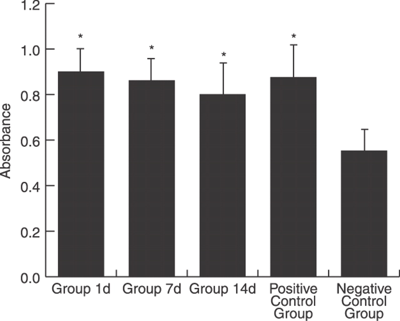
Table 1.
Wound Closure Rates at Different Time Points
If there is no suitable delivery system (which is the situation in the group treated with EGF solution), the closure rate of wounds is even worse than that of wounds treated with hydrogel matrix. Studies[19,20] have shown the importance of using adequate delivery systems when growth factors are used to enhance wound repair. The present data from an in vitro releasing model and a proliferating experiment indicate that hydrogels could prolong release of the growth factor and maintain identical biological activity on stimulating fibroblast proliferation. Because there are numerous physiologic impairments other than blunted secretion of EGF, such as other growth factor deficiencies, excessive matrix metalloproteinases, excessive inflammatory mediators, bioburden, and abnormal microcirculation, that can result in poor healing of diabetic wounds in vivo, it is rational that in the present experiments there were no higher wound closure rates observed in rats treated with rhEGF than in those treated with hydrogel matrix. Although contained in hydrogel, rhEGF showed additional effects on induction of EGF receptor expression and acceleration of angiogenesis, which were believed to be important for the success of wound healing. We can still see the advantage of rhEGF-containing hydrogels. Additional studies to optimize the composition of hydrogels and the concentration of EGF and to diminish the proteolytic activity at the site of application should be considered to improve efficacy. In addition, future studies need to assess the efficacy and safety of this rhEGF-containing hydrogel in humans.
Figure 3.
Recombinant human epidermal growth factor (rhEGF)–containing hydrogel accelerated the closure of full-thickness diabetic wounds. A, The kinetics of wound closure with different agents topically implemented. B, Representative pictures of the wounds topically treated with different agents at the indicated time points. Note that the group treated with rhEGF-containing hydrogel gained the highest rates of wound closure at different time points. Three and 5 days after wounding, wound closure rates were higher in the rhEGF-containing hydrogel group and the hydrogel matrix group than in negative controls (P < .05). Ten days after wounding, the wounds of rats treated with hydrogel healed better than those of rats treated without hydrogel (P < .05). Ten and 14 days after wounding, wound closure rates in the groups treated with hydrogel matrix, rhEGF-containing hydrogel, and rhEGF solution were higher than those in the negative control group (P < .05).
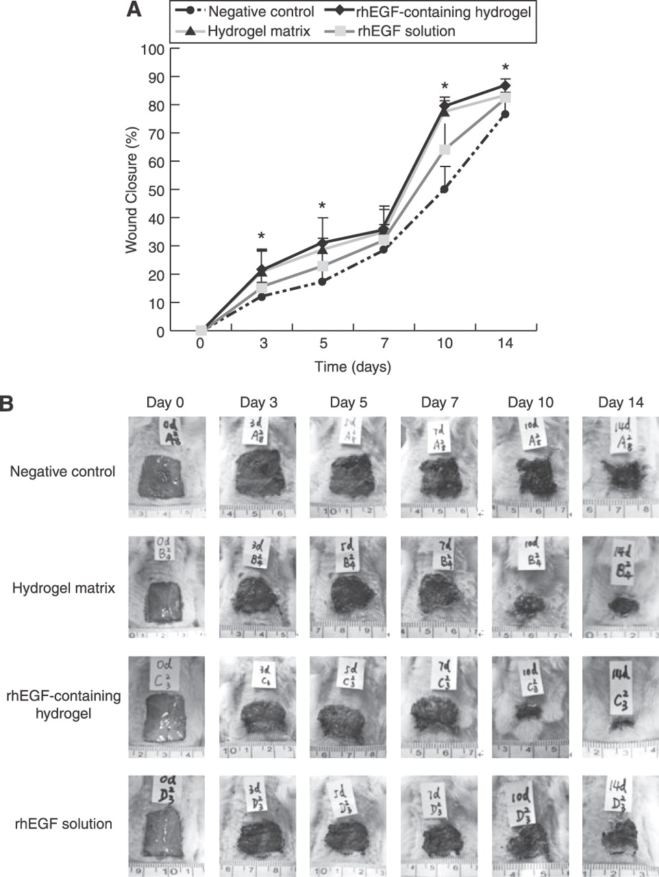
Table 2.
Comparison of Composite Scores of Histologic Evaluation in the Four Study Groups
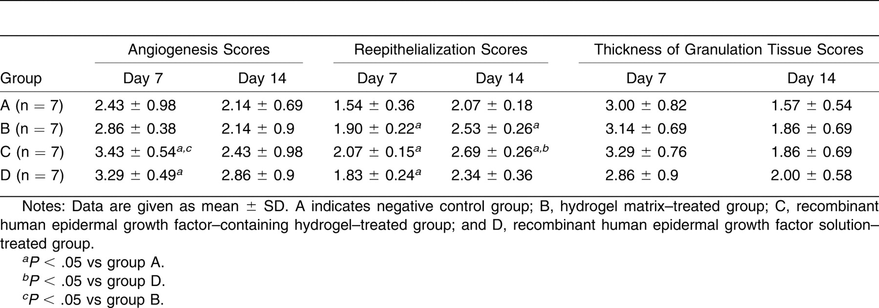
Figure 4.
The effect of treatments on proliferating cell nuclear antigen and B-cell lymphoma/leukemia-2 (BCL-2) immunoreactivity (IRS) in wounds. A, Recombinant human epidermal growth factor (rhEGF)–containing hydrogel treatment significantly increased the mean number of proliferating cell nuclear antigen–positive cells on postoperative day 7 (P < .05). B, The rhEGF-containing hydrogel and rhEGF solution treatments significantly increased the mean number of BCL-2–positive cells on postoperative day 7 (P < .05). Error bars represent SD. Group A indicates negative control group; group B, hydrogel matrix–treated group; group C, rhEGF-containing hydrogel–treated group; and group D, rhEGF solution–treated group.
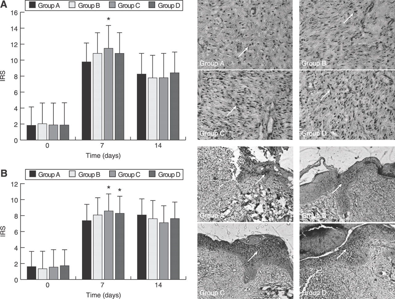
Figure 5.
The effect of treatments on epidermal growth factor receptor (EGFR) Western blotting in wounds on postoperative days 0, 7, and 14. A, Protein abundance of EGFR analysis data. B, Expression patterns of EGFR and β-actin. The EGFR expression level was significantly increased in the recombinant human EGF (rhEGF)–containing hydrogel–treated group on postoperative day 7. Group A indicates negative control group; group B, hydrogel matrix–treated group; group C, rhEGF-containing hydrogel–treated group; and group D, rhEGF solution–treated group. *P < .05 compared with the negative control group, †P < .05 compared with the hydrogel matrix–treated group. Error bars represent SD.
In conclusion, the present study highlights the efficacy of hydrogels as a controlled releasing system for topical application of growth factors. The rhEGF-containing hydrogel that we developed holds the merits of prolonged and sustained release of bioactive EGF and therapeutic potential in enhancing diabetic wound healing.
Funding
This work was supported by the Foundation for Building Ministry of Health Key Disciplines from the Ministry of Health of P.R. China and by the Guangdong Natural Science Fund Committee.
Conflicts of Interest
None reported.
References
- Chaturvedi, N, LKStevens, JHFuller, et al. :Risk factors, ethnic differences and mortality associated with lower-extremity gangrene and amputation in diabetes: the WHO Multinational Study of Vascular Disease in Diabetes. Diabetologia44 ( suppl 2): 65S, 2001.
- Jeffcoate, WJ and KGHarding. :Diabetic foot ulcers. Lancet361: 1545, 2003.
- Jeffcoate, WJ. :The incidence of amputation in diabetes. Acta Chir Belg105: 140, 2005.
- Tanaka, A, TNagate, and HMatsuda. :Acceleration of wound healing by gelatin film dressings with epidermal growth factor. J Vet Med Sci67: 909, 2005.
- Kwon, YB, HWKim, DHRoh, et al. :Topical application of epidermal growth factor accelerates wound healing by myofibroblast proliferation and collagen synthesis in rat. J Vet Sci7: 105, 2006.
- Laato, M, VMHahari, JNiinikoski, et al. :Epidermal growth factor increases collagen production in granulation tissue by stimulation of fibroblast proliferation and not by activation of procollagen genes. Biochem J247: 385, 1987.
- Tsang, MW, WKWong, CSHung, et al. :Human epidermal growth factor enhances healing of diabetic foot ulcers. Diabetes Care26.:1856. ,2003.
- Hong, JP, HDJung, and YWKim. :Recombinant human epidermal growth factor (EGF) to enhance healing for diabetic foot ulcers. Ann Plast Surg56: 394, 2006.
- Hoffman, AS. :Hydrogels for biomedical applications. Adv Drug Del Rev43: 3, 2002.
- Peppa, M, HBrem, PEhrlich, et al. :Adverse effects of dietary glycotoxins on wound healing in genetically diabetic mice. Diabetes52: 2805, 2003.
- Galeano, M, DAltavilla, DCucinotta, et al. :Recombinant human erythropoietin stimulates angiogenesis and wound healing in the genetically diabetic mouse. Diabetes53: 2509, 2004.
- Krajewska, M, SKrajewski, JIEpstein, et al. :Immunohistochemical analysis of bcl-2, bax, bcl-X, and mcl-1 expression in prostate cancers. Am J Pathol148: 1567, 1996.
- Winter, GD. :Formation of scab and the rate of epithelialization of superficial wounds in the skin of the young domestic pig. Nature193: 293, 1962.
- Hanna, JR and JAGiacopelli. :A review of wound healing and wound dressing products. J Foot Ankle Surg36: 2, 1997.
- Ovington, LG. :Advances in wound dressings. Clin Dermatol25: 33, 2007.
- Stashak, TS and EFarstvedt. :Update on wound dressings: indications and best use. Clin Tech Equine Pract3: 148, 2004.
- Repertinger, SK, ECampagnaro, JFuhrman, et al. :EGFR enhances early healing after cutaneous incisional wounding. J Invest Dermatol123: 982, 2004.
- Knott, AW, RJJuno, MDJarboe, et al. :EGF receptor signaling affects bcl-2 family gene expression and apoptosis after massive small bowel resection. J Pediatr Surg38: 875, 2003.
- Puolakkainen, PA, DRTwardzik, JERanchalis, et al. :The enhancement in wound healing by transforming growth factor-β 1 (TGF-β 1) depends on the topical delivery system. J Surg Res58: 321, 1995.
- Hori, K, CSotozono, and JHamuro. :Controlled-release of epidermal growth factor from cationized gelatin hydrogel enhances corneal epithelial wound healing. J Control Release118: 169, 2007.
© 2012 American Podiatric Medical Association