


Abstract
Molecular genetics is changing our understanding of the developmental translation of genotype to phenotype between and within different phylogenetic groups. Together with a growing understanding of our own evolutionary relationships to common ancestors, the epigenetic processes involved enforce a reexamination of what is regarded as a normal foot structure. A revised populationist approach is proposed and supported by paleoanthropologic evidence that reflects a picture of emerging suitability for bipedalism that is driven by natural genetic divergence. (J Am Podiatr Med Assoc 102(1): 64-70, 2012)
The propensity of the foot to deliver a series of normal functions during the gait cycle is frequently cited as being associated with morphology. Many models have been used to classify foot structure according to an ideal or optimal arrangement that defines normality and, if attained, is predicted to render an increased likelihood of normal function. Furthermore, individual intrinsic deviations from such an alignment are considered pathoetiologic due to positional and functional compensatory requirements. This approach to the description of foot morphology is a fundamentally essentialist one and stems from Platonic teachings. Essentialism, in which diversity in the biological world is accounted for by the interaction of natural form with interfering forces, postulates the concept that in biology there is but one true value. Variability in nature is, thus, accounted for as a deviation from what is natural by interfering forces that frustrate a natural tendency. The corollary to essentialist thinking is that deviations from this one true value represent “abnormal” values.
A converse approach is found in populationist thinking that stresses the uniqueness of all variable forms and features that can be collectively described in statistical terms. Individual values from any given population can be gathered to establish an arithmetic mean that serves only as a statistical abstraction, and only the individual values of which the population is composed can be said to exist. This compares with essentialism in which only the idealized form exists.
This philosophical difference is not simply a tautological argument; in podiatric medicine and other disciplines, foot typing has principally used an essentialist approach to assess foot morphology (subtalar neutral positioning, arch height, etc) to assess a causal link with function. Regarding foot morphology in this way is a fundamental and questionable assumption and may underpin the reported poor predictive ability of correlating foot structural features to function.[1] A more unified approach to describing foot morphology integrated with more functional-based approaches to the evaluation and treatment of foot abnormalities has been proposed.[2] Although this theoretical integration of ideological paradigms seems eminently sensible and noteworthy, the foot morphology theory aspect of this paper is supported by reference to Rootian principles only, which encapsulate a classic essentialist approach to defining normality and the reliability and validity of which has been questioned.[3]
Intuitively, we might sense that several questions present themselves if a more populationist approach to morphologic variance is adopted: Might anatomical variance viewed in this way provide insights into functional patterns? Do such functional patterns influence symptoms? Are such explanations validated by empirical findings? Are such perspectives commensurate with other related anatomical and physiologic control mechanisms?
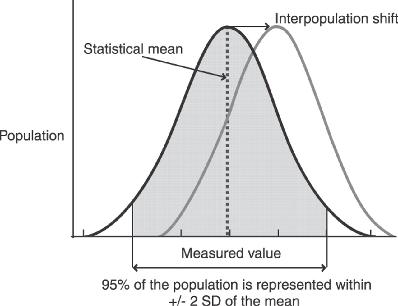
In his Natural Inheritance of 1889, Frances Galton completed his investigations of the correlation coefficient and introduced the term normal law as a nascent term to describe the normal distribution of values found in nature. Now an established medical statistical method of setting value ranges in a population, natural biological features can be assessed on interval scales to reveal the normal or Gaussian (bell-shaped) distribution curve of values that present a mean value for the population in question, 95% of which will fall within 2 SD of this theoretical point. The fundamental issue here is that positions in this range do not represent mistakes made by natural or observer fallibility but are statistical expressions of heterogeneic variability. Essentialists and populationists agree that averages exist, both granting variation phenomena. For essentialists, however, variability represents an aberration from normal, whereas the populationist viewpoint is that any aberration (within 2 SD) from the purely hypothetical position of the mean is normal (Fig. 1).
Figure 1.
All biological values demonstrate a continuum for a given population.
Although essentialist arguments are as much entitled to appeal to the principle of tenacity as any other scientific hypothesis, Galton’s cousin, Charles Darwin, having completed On the Origin of Species by Means of Natural Selection, posited the concept that natural biological variation was the fuel of evolutionary change, thus seriously weakening the grip of essentialist thinking on biological thought. The evolutionary change of pedal structure has been the subject of much attention in the scientific literature and has involved the consideration of adaptation that reflects the acquisition of bipedalism in modern humans. However, paleoanthropological evidence is rarely cited in recent podiatric medical literature, which is, perhaps, due to the lack of a contemporary framework that could integrate clinical and nonclinical disciplines and still avoid a somewhat esoteric outcome. This might prove possible if such important contributions are viewed in a different light. Darwin summarized evolution as “descent with modification.” For the purposes of this paper and for reasons that should become apparent, this description is perhaps more specifically and better understood as “evolution is genetics plus time.”
Perspectives on Human Foot Evolution
Interest in the development of apomorphic features in the human foot is long-standing, and readers are encouraged to consider the references for a more detailed account of specific findings. What follows is a brief outline of approaches to the subject of anatomy and paleoanthropology regarding the foot that present a remarkably cohesive picture of the acquisition of key functional attributes linked to morphology.
In 1931, George Williams[4] concluded that the human foot was the result of a gradual evolutionary transition that retained anatomical similarities with other groups when he reported findings of what he described as an atavistic human foot. The outstanding features of his morphologic example included arrested tarsal development; a divergent, highly independent hallux; and the retention of intertarsal and tarsometatarsal mobility. Such findings, he concluded, supported Keith’s earlier contention[5] that the hallux was a key contribution to the formation of the essentially human medial longitudinal arch. Keith’s insight was followed by Morton’s treatise of the acquired functional features of the longitudinal arch through evolution,[6] with further published conclusions regarding foot dysfunction associated with first-ray shortness or hypermobility.[7] Having proposed a functional association between the first ray and rearfoot pronation and the establishment of an altered leverage arm between the first and second metatarsals, Morton espoused a link between anatomically connected pedal structures to achieve a shared combination of roles, ie, pronation and line of progression in propulsion.
In an early detailed study of the plantar aponeurosis in relation to the arch of the foot, Hicks[8] first described a fundamentally passive use of metatar-sophalangeal extension to produce a rise in the medial longitudinal arch, inversion (supination) of the rearfoot, and lateral leg rotation with tightness of the plantar aponeurosis itself. With extension of the forefoot joints, the first ray demonstrates greatest (plantar) flexion, and the analogy of a windlass mechanism was introduced. In a revision of this subject, Bojsen-Møller and Flagstad[9,10] considered the anatomical and functional apomorphic features of human feet compared with the feet of other extant apes. Here, true anatomical variances of the subjects were noted, particularly in relation to the shape of the calcaneocuboid joint, which renders the midtarsus stable before heel-lift having undergone a reduction in congruity during initial contact loading of the rearfoot. The functional significance of metatarsal lengths was also presented, and a “high gear” (medial) propulsive optimum was proposed to maximize the windlass effect. Bojsen-Møller refers to the evolutionary polysegmentation remodeling of the forefoot (adduction of the two medial metatarsals and hallux with pronation of the lateral three) that serves as an echo of Dudley Morton’s reminder[6] that nature is a remodeler rather than a creator. In a recent critique of Morton’s proposal, Glasoe and Coughlin[11] gathered contemporary clinical and research findings to question the validity of some of Morton’s conclusions: first-ray shortness not being necessarily pathoetiological, although a functional transfer of weightbearing to the lateral forefoot was supported in cases of hypermobility and hallux valgus deformity.
The phylogenetic similarities of human feet to those of other related genera have been considered by a variety of authors, including Gebo[12] who, in 1992, compared and contrasted anatomical foot structures of humans with those of extant African apes, specifically focusing on bone size, orientation, and function in relation to general anatomical requirements for their respective environments. The larger evolved modifications of the human foot received detailed consideration from Kidd,[13] who reiterated the changes in size and proportion of osseous elements. Specifically, from a human perspective, these modifications include a larger tarsus with smaller digits; the absence of a divergent, opposable first ray; and the presence of longitudinal and transverse arches. This work, drawing on the earlier work of researchers[14–16] examining the evolutionary significance of human foot joints compared with those of extinct hominoids and extant apes, also notes the intense modifications to the midtarsal joints and offers some unique podiatric medical perspectives: First, the anatomical changes gradually effected in the rearfoot included an increase in calcaneal inclination and formed the proximal end of the medial longitudinal arch. Consequential changes to talar orientation conferred a torque conversion ratio into the subtalar joint due to a concomitant change in joint axis alignments. Such modifications offer insights into the evolution of a stable arch configuration that can confer a degree of stability that is spatially and temporally variable in relation to osseous positions. Second, the gradual reduction in first-ray divergence and position composed the distal element of the arch, in essence defining the arch structure as a complex of traits that gradually evolved through transitional stages that are observable in the fossils of human ancestors. In further similar work, Kidd[17] proposed that formation of the lateral arch with greater calcaneocuboid stability was already present in early hominoid bipeds, with subsequent equivalent changes occurring in the medial side of the foot during a series of selectively advantageous changes. Moreover, the implication is that contemporary biomechanical foot dysfunction could be viewed as a morphologically and functionally atavistic state.
The comparative paleoanthropological and taxologic evidence that exists is subject to several interpretive constraints. The elucidation of discovered fossils (which seems likely to continue) suggests that hominoid ancestors do not represent a straightforward lineage from Australopithecus afarensis through incremental “improvements” (via Homo habilis and Homo ergaster) to “arrive” via a variety of intermediates at Homo sapiens.[18] The use of taxonomic terms is an artificial construct that hides the temporally continuous morphologic and functional variances of extinct species, and, furthermore, the brachiation of ancestral predecessors into different extant species did not arrest further evolutionary change. The continuing analyses of fossil structure,[19–22] the comparative dynamic functional attributes of early bipedal ancestors,[23–30] and the paleopathologic study of foot deformity[31] offer significant insights into the evolving suitability of the human foot to upright propulsive gait despite a propensity for injurious biomechanical failure. Although future studies of phylogeography may assist in our further understanding of the evolution of modern humans, the replacement of regional populations by a small population of a recent common ancestor from East Africa approximately 150,000 years ago (“Out-of-Africa 2”) is gaining favor.[32] During such a period, what captures the attention are the broad similarities of retained morphologic features, the differences between which are explained by sequential heterochronic[33] and heterotopic changes. That is to say, the sequencing and timing of developmental growth can be used to explain features that cannot be assessed by the metrics of size and shape alone. It is suggested that interspecies and intraspecies differences in hominoid foot morphology are better explained in terms of spatiotemporal alterations in the genetic expression of common features. Chimpanzees and bonobos share the closest genetic similarities with modern humans, sharing more than 98% of identical DNA; yet, the evolutionary divergence in developmental, morphologic, and functional pathways is likely to have been brought about by small (yet highly significant) shifts in regulatory genes.[34]
The Genetic Influence on Morphologic Variation
After their morphogenetic study of ossification patterns in rabbits in 1948, Crary and Sawin[35] suggested potential forthcoming clues as to the process of the differentiation of the mammalian skeleton, the contemporaneous discussion of which largely remained the remit of biological philosophy. In the past 40 years, there has been an accelerating role for molecular biologists in the empirical testing of gene theory in the regulation of developmental systems. Evolutionary developmental biology posits an integration of disciplines that conflicts with previous assumptions that “genes” program species-specific characters in a particulate manner (Mendelian inheritance). That is to say, it does not necessarily follow that phenotype follows genotype. Physical traits have been identified to be under the control of hierarchically organized (regulatory) routines of gene expression[36] that may, under certain intrinsic (minor nonfatal mutations) and extrinsic (environmentally selectively advantageous) conditions, combine to become genetically diverse between isolated populations. Such cumulative changes will ultimately lead to a “speciation event.” In accepting this paradigm, each new generation must possess a new gene pool that must be viable for the next, and retained characteristics are likely to show quantitative variation within a continuum.
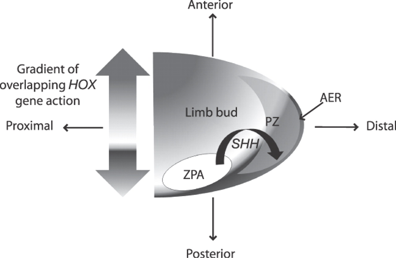
For the past decade, the specificity of empirical research has improved our understanding of the ontogenic development of the human limb. Anteroposterior and proximal-distal limb patterning at the zone of polarizing activity and the apical ectodermal ridge have been found to be under the influence of several genes, including sonic hedgehog (SHH), the early target of which is bone morphogenesis. The profound nature of these gene expressions is that their restriction results in significant limb dysmorphologies (Fig. 2).[37,38]
Figure 2.
Regulatory gene activity and interactions govern temporospatial limb development. AER indicates apical ectodermal ridge; PZ, polarizing zone; SHH, sonic hedgehog; and ZPA, zone of polarizing activity.
The homeobox (HOX) gene family consists of highly conserved transcription factors that are retained in clusters from nematodes to all vertebrates. They are responsible for the anteroposterior patterning of the body axis and limb development, each one displaying a pattern of expression that depends on its relative position in the cluster. There are four such clusters in the human genome, each having a separate location: HOXA (chromosome 7), HOXB (chromosome 17), HOXC (chromosome 12), and HOXD (chromosome 2).[39] Morphologic identities of different genera via control of the expression of a body plan, determination of cell fate, and other developmental and evolutionary processes through HOX gene action are well reported.[40–42] After analytical studies of their interaction, it is clear that the axes of development cannot be considered in isolation, as are Cartesian coordinate approaches. It remains probable that proximal-distal extension and limb patterning of the distal elements are largely influenced by a second phase of HOX function, “downstream” of SHH,[43] including expression in the mesenchyme adjacent to the growing cartilage.[44] In further research, the nutritional influence of folate (required for chondrocyte growth and differentiation) and retinoic acid (limb growth and patterning) was demonstrated on HOX gene–controlled phenotypes, demonstrating not only the beneficial effect of diet on skeletogenesis but implying a nutritional effect on skeletal variation through regulatory gene expression.[45,46] In a detailed study of HOXC13 and HOXD13 function, the osteogenic differentiation of bone marrow–derived mesenchyme stem cells has been suggested.[47]
Thus, HOX genes act at several levels in the genetic pathway that control limb development, including the foot,[48] and are believed to regulate overlapping sets of target genes by binding to specific DNA sequences in cis-regulatory elements; furthermore, mutations in HOXD13 have been found to cause human foot malformations, such as synpolydactyly. At later stages, they directly regulate the expression of genes involved in the formation of joints.[49] In the context of evolutionary development, there is clear potential to investigate how molecular biology shapes genetic variation of patterning and growth genes in closely related taxa. Moreover, it may prove to be a powerful tool in understanding developmental variation within human populations.
Figure 3.
Gene regulatory proteins can block the action of RNA polymerase from attaching to specific protein-building genes.
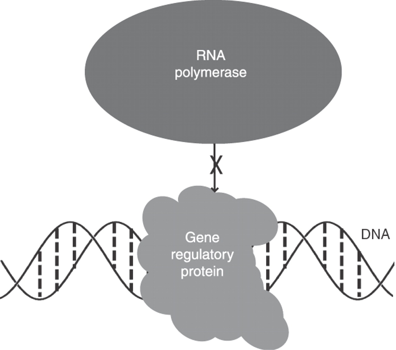
The interactive sequence involving SHH, HOX genes, and other target genes has been found to be influential in determining several key features in a dose-dependent manner (Fig. 3), including the digit identity and number and the characteristic patterns of growth rate and segmentation of digital rays in primates.[50] Researchers concluded that the patterning of digit formation was primarily responsible for such adaptations in primates and that such change was the result of alterations in a network of regulatory and patterning genes. The variation within and between taxa of shared morphologic traits has been the subject of much recent investigation[51–57]; results suggest either direct mutagenesis of protein synthesis or alterations in the regulation of genetic expression through quantitative trait loci: these loci are not necessarily genes but are stretches of DNA that are closely linked to the genes that underlie the trait in question. They demonstrate the capacity to promote correlations within structures and inversely correlate between them through epistasis (a genetically interactive effect). The acquisition of key morphologic and functional traits is a highly complex and, as yet, poorly understood process.
The predominance of work in this area has focused on larger taxonomic differences of gene expression. To understand natural variations within our own populations, we require a clearer picture of differences within our species that do not invoke the macromutations of larger limb dysmorphologies. One such picture has emerged of how small differences in HOX expression result in more modest morphologic variation.[58] The attractive implication is that subtle differences in HOX regulation and morphology within a species group are potentially representative examples from within a continuum of normal and that threshold values exist for significant deformity. The evolution of complex human traits has been one of incremental changes that are more than 50% polygenic,[59] and, although many mutagenic changes are neutral, some produce change; this has the potential to alter anatomical features that confer altered function or a preadaptive change that may be exploited at a later stage.[60,61] For modern humans, the adoption of an upright stance has been assigned primacy in the scientific literature concerning evolutionary anthropomorphic change. The retention and development of physical traits that help define our species are no longer exposed to the selective pressures that previously governed their inherent success. However, the variable phenotypic expressions of such traits form the structures on which the environment continues to act and produce the symptoms with which we are confronted.
Conclusions
The complex relationship between genes and their regulatory expression that has influenced our evolution and continues to influence population variance will be explored in part 2 of this paper, to be published in a subsequent issue. In considering the structure-function complex of the foot, it remains implicit that our genetic and epigenetic makeup is likely to prove to be a significant factor in determining any statistical likelihood of ensuing functional entropy. Therefore, to develop a coherent statistical correlation between anatomical alignment and the resulting forces from movement, an assimilation of contributory elements must first be determined. Furthermore, moving toward an understanding of the consequential outcomes of such a relationship and the potential for pathoetiology will require an inclusive approach to models, perhaps initiated through the work of other disciplines. The fact that these disciplines may not always be clinically based should not prevent us from incorporating important scientific evidence into a more integrated evidence-based framework.
Financial Disclosure
None reported.
Conflicts of Interest
None reported.
References
- Razeghi, M and ME Batt: Foot type classification: a critical review of current methods. Gait Posture 15: 282, 2002.
- Harradine, P and L Bevan: A review of the theoretical unified approach to podiatric biomechanics in relation to foot orthoses therapy. JAPMA 99: 317, 2009.
- Picciano, AM, MS Rowlands, and TW Worrell: Reliability of open and closed kinetic chain subtalar joint neutral positions and navicular drop test. J Orthop Sports Phys Ther 18: 553, 1993.
- Williams, GA: Atavistic human foot: its developmental significance. Am J Phys Anthropol XVI: 1, 1931.
- Keith, A: Hunterian lectures on man’s posture: its evolution and disorders. Br Med J 1: 545, 1923.
- Morton, DJ: Evolution of the longitudinal arch of the human foot. J Bone Joint Surg Am 6: 56, 1924.
- Morton, DJ: Hypermobility of the first metatarsal bone: the interlinking factor between metatarsalgia and longitudinal arch strains. J Bone Joint Surg Am 10: 187, 1928.
- Hicks, JH: The mechanics of the foot. J Anat 88: 25, 1954.
- Bojsen-Møller, F and KE Flagstad: Plantar aponeurosis and internal architecture of the ball of the foot. J Anat 121: 599, 1976.
- Bojsen-Møller, F: Calcaneocuboid joint and stability of the longitudinal arch of the foot at high and low gear push off. J Anat 129: 165, 1979.
- Glasoe, WM and MJ Coughlin: A critical analysis of Dudley Morton’s concept of disordered foot function. J Foot Ankle Surg 45: 147, 2006.
- Gebo, DL: Plantigrady and foot adaption in African apes: implications for hominid origins. Am J Phys Anthropol 89: 29, 1992.
- Kidd, R: Evolution of the rearfoot: a model of adaptation with evidence from the fossil record. JAPMA 89: 2, 1999.
- Elftman, H and J Manter: The evolution of the human foot, with especial reference to the joints. J Anat 70: 56, 1935.
- Lewis, OJ: The joints of the evolving foot: part III. The fossil evidence. J Anat 131: 275, 1980.
- Kidd, RS, P O’Higgins, and CE Oxnard: The OH8 foot: a reappraisal of the functional morphology of the hindfoot utilizing a multivariate analysis. J Hum Evol 31: 269, 1996.
- Kidd, R: The past is the key to the present: thoughts on the origins of human foot structure, function and dysfunction as seen from the fossil record. The Foot 8: 75, 1998.
- Harcourt-Smith, WEH and LC Aiello: Fossils, feet and the evolution of human bipedal locomotion. J Anat 204: 403, 2004.
- Susman, RL and DJ de Ruiter: New hominin first metatarsal (SK 1813) from Swartkrans. J Hum Evol 47: 171, 2004.
- Kidd, R and C Oxnard: Little foot and big thoughts: a reevaluation of the Stw573 foot from Sterkfontein, South Africa. Homo 55: 189, 2005.
- McHenry, HM and AL Jones: Hallucial convergence in early hominids. J Hum Evol 50: 534, 2006.
- Zipfel, B, JM DeSilva, and RS Kidd: Earliest complete hominin fifth metatarsal: implications for the evolution of the lateral column of the foot. Am J Phys Anthropol 140: 532, 2009.
- Meldrum, DJ: Kinematics of the cercopithecine foot on arboreal and terrestrial substrates with implications for the interpretation of hominid terrestrial adaptations. Am J Phys Anthropol 84: 273, 1991.
- Schmitt, D and SG Larson: Heel contact as a function of substrate type and speed in primates. Am J Phys Anthropol 96: 39, 1995.
- Wang, WJ, RH Crompton, Y Li, et al: Energy transformation during erect and bent-hip, bent-knee walking by humans with implications for the evolution of bipedalism. J Hum Evol 44: 563, 2003.
- Wang, W, RH Crompton, TS Carey, et al: Comparison of inverse-dynamics musculo-skeletal models of AL 288-1 Australopithecus afarensis and KNM-WT 15000 Homo ergaster to modern humans, with implications for the evolution of bipedalism. J Hum Evol 44: 453, 2004.
- Wang, WJ and RH Crompton: Analysis of the human and ape foot during bipedal standing with implications for the evolution of the foot. J Biomech 37: 1831, 2004.
- Preuschoft, H: Mechanisms for the acquisition of habitual bipedality: are there biomechanical reasons for the acquisition of upright bipedal posture? J Anat 204: 363, 2004.
- Bennett, MR, JWK Harris, BG Richmond, et al: Early hominin foot morphology based on 1.5 million-year-old footprints from Ileret, Kenya. Science 323: 1197, 2009.
- Lovejoy, CO, B Latimer, G Suwa, et al: Combining prehension and propulsion: the foot of Ardipithecus ramidus: Science 326: 72e1, 2009.
- Mays, SA: Paleopathological study of hallux valgus. Am J Phys Anthropol 126: 139, 2005.
- Finlayson, C: Biogeography and evolution of the genus Homo. Trends Ecol Evol 20: 457, 2005.
- Blomquist, GE: Brief communication: methods of sequence heterochrony for describing modular developmental changes in human evolution. Am J Phys Anthropol 138: 231, 2009.
- Zollikofer, CP and MS de Leon Ponce: The evolution of hominin ontogenies. Semin Cell Dev Biol 21: 441, 2010.
- Crary, DD and PB Sawin: Morphogenetic studies in the rabbit: VI. Genetics factors influencing the ossification pattern of the limbs. Genetics 34: 508, 1949.
- Newman, SA: Developmental mechanisms: putting genes in their place. J Biosci 27: 97, 2002.
- Roberts, DJ and C Tabin: The genetics of human limb development. Am J Hum Genet 55: 1, 1994.
- Hill, RE, SJ Heaney, and LA Lettice: Sonic hedgehog: restricted expression and limb dysmorphologies. J Anat 202: 13, 2003.
- Lappin, TR, DG Grier, A Thompson, et al: HOX genes: seductive science, mysterious mechanisms. Ulster Med J 75: 23, 2006.
- Zhang, J and N Nei: Evolution of antennapedia-class homeo-box genes. Genetics 142: 295, 1996.
- Iimura, T and O Pourquie: Hox genes in time and space during vertebrate body formation. Dev Growth Differ 49: 265, 2007.
- Leomons, D and W McGinnis: Genomic evolution of Hox gene clusters. Science 313: 1918, 2006.
- Zakany, J and D Duboule: The role of Hox genes during vertebrate limb development. Curr Opin Genet Dev 17: 359, 2007.
- Suzuki, M and A Kuroiwa: Transition of Hox expression during limb cartilage development. Mech Dev 118: 241, 2002.
- Kappen, C, MA Mello, and RH Finnell: Folate modulates Hox gene-controlled skeletal phenotypes. Genesis 39: 155, 2004.
- Tamura, K, S Yonei-Tamura, T Yano, et al: The autopod: its formation during limb development. Dev Growth Differ 50: 177, 2008.
- Chae, SW, BK Jee, JY Lee, et al: Hox gene analysis in the osteogenic differentiation of human mesenchymal stem cells. Genet Mol Biol 31: 815, 2008.
- Barham, G and NM Clarke: Genetic regulation of embryological limb development with relation to congenital limb deformity in humans. J Child Orthop 2: 1, 2008.
- Salsi, V, MA Vigano, F Cocchiarella, et al: Hoxd 13 binds in vivo and regulates the expression of genes acting in key pathways for early limb and skeletal patterning. Dev Biol 317: 497, 2008.
- Chiu, C and MW Hamrick: Evolution and development of the primate limb skeleton. Evol Anthropol 11: 94, 2002.
- Carroll, SB: Homeotic genes and the evolution of arthropods and chordates. Nature 376: 479, 1995.
- Shapiro, MD, ME Marks, CL Peichel, et al: Genetic and developmental basis of evolutionary pelvic reduction in threespine sticklebacks. Nature 428: 717, 2004.
- Shubin, N, C Tabin, and S Carroll: Fossils, genes and the evolution of animal limbs. Nature 388: 639, 1997.
- Nadeau, JH and WN Frankel: The roads from phenotypic variation to gene discovery: mutagenesis versus QTLs. Nat Genet 25: 381, 2000.
- Chase, K, DR Carrier, FR Adler, et al: Genetic basis for systems of skeletal quantitative traits: principal component analysis of the canid skeleton. Proc Natl Acad Sci U S A 99: 9930, 2002.
- Ellegren, H and BC Sheldon: Genetic basis of fitness differences in natural populations. Nature 452: 169, 2008.
- Reno, PL, MA McCollum, MJ Cohn, et al: Patterns of correlation and covariation of anthropoid distal fore-limb segments correspond to Hoxd expression territories. J Exp Zool B Mol Dev Evol 310: 240, 2008.
- Carroll, SB: Endless forms: the evolution of gene regulation and morphological diversity. Cell 101: 577, 2000.
- Carroll, SB: Genetics and the making of Homo sapiens. Nature 422: 849, 2003.
- Averof, M: Arthropod Hox genes: insights on the evolutionary forces that shape gene functions. Curr Opin Genet Dev 12: 386, 2002.
- Bridgham, JT, SM Carroll, and JW Thornton: Evolution of hormone-receptor complexity by molecular exploitation. Science 312: 97, 2006.
© 2012 American Podiatric Medical Association