


Abstract
Navicular stress injuries in athletes can be devastating. Clinical findings are frequently nonspecific until significant progression of the abnormality has occurred. The use of diagnostic imaging techniques early in the discovery period increases the likelihood of establishing an immediate diagnosis and avoids frank fracture of the navicular bone. Delayed diagnosis of navicular stress injuries in athletes can cause dire consequences. The physician must be aware of the injury in establishing a high index of clinical suspicion. The timing and sequencing of diagnostic imaging studies is essential in establishing a diagnosis to manage the patient and minimize time away from competition. This case study examines the history and management of an elite high school track athlete who sustained a navicular stress injury. The timing and use of diagnostic imaging studies is reviewed.
Stress fractures of the lower extremity are injuries that commonly occur in runners. It is accepted that stress injury to the bone results from a disturbance in the balance of bone resorption and remodeling due to repetitive weightbearing coupled with an increase in the intensity of training.[1–3] The intense repetition of mechanical loading of the foot leaves competitive athletes more vulnerable to this type of injury. Studies[1, 4–6] conducted on the distribution of stress fractures in track and field runners conclude that the navicular bone is the second most common site of injury in this population.
Navicular stress injuries remain a diagnostic challenge due to a lack of symptom specificity combined with the ambiguity seen with individual diagnostic imaging modalities. As a consequence, these injuries are often misdiagnosed or delayed.[7] Failure to make a timely diagnosis of navicular stress injury can lead to progression of the condition, resulting in unnecessary disability and downtime due to complications such as delayed union or nonunion, avascular necrosis, and juxtaarticular arthritis. These complications are magnified as the timeframe from injury to treatment increases, and they may be career ending if through-and-through fracture occurs.[8] To prevent these complications, navicular stress injuries should be pursued aggressively in individuals with a high index of clinical suspicion. Through a good history, a physical examination, and an appropriate combination of diagnostic studies, a delayed diagnosis of navicular stress injuries can be avoided, minimizing downtime and returning the athlete to competition as soon as possible.
Case Study
One month into the spring track season, an 18-year-old male track athlete presented to the primary author’s (R.M.Y.) office with concerns about pain in the area of the sinus tarsi on the left foot of 2 weeks’ duration. The athlete competed in sprinting events. He related increased pain with activity until reaching a level that affected his workouts and performance. Clinically, there was mild swelling in the area of the sinus tarsus on the affected foot and a bilateral gastrocnemius and soleus equinus. Radiographs of the left foot were negative for fracture but identified an accessory ossicle in the sinus tarsi (Fig. 1). The initial diagnosis was left foot sinus tarsitis. Treatment included decreasing workouts, anti-inflammatory medication, heel cord stretching exercises, and physical therapy. No improvement was seen as the patient continued participating in track meets.
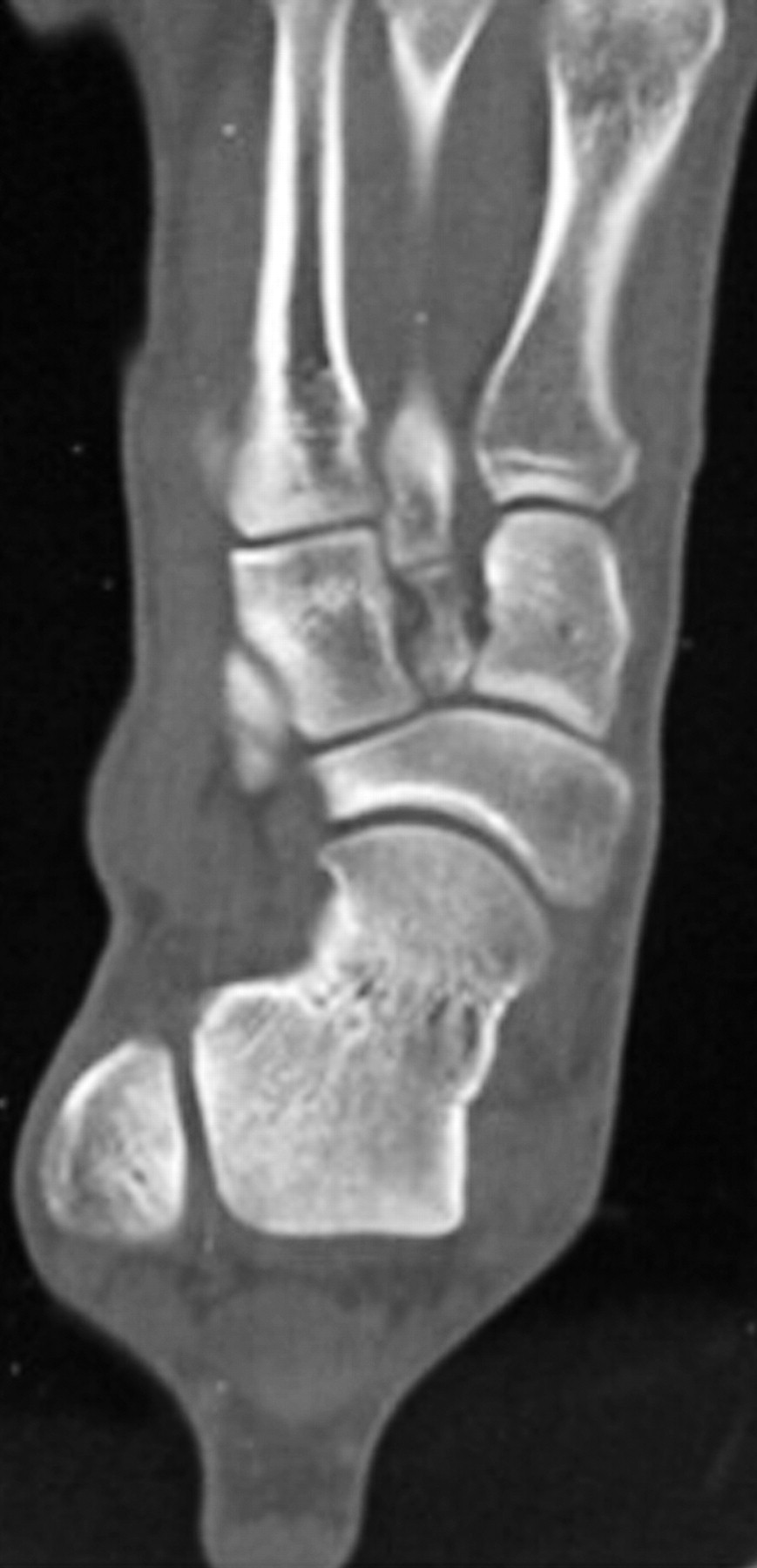
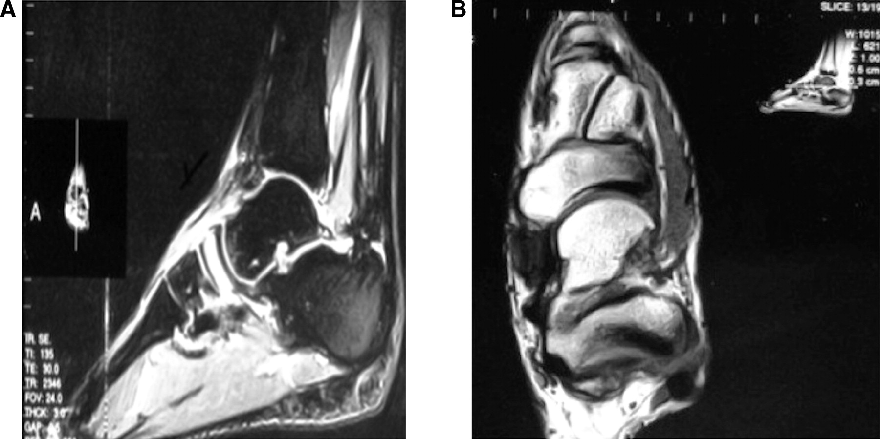
The 2-week follow-up appointment revealed pain on compression of the navicular bone, a positive “N” sign. Only mild swelling was noted on clinical examination. A computed tomographic (CT) scan showed no evidence of fracture or gross alteration in bone density. The lateral aspect of the navicular bone was noted to be thinner, and the presence of an accessory ossicle was seen in the area of the sinus tarsi (Fig. 2). Magnetic resonance images (MRIs) demonstrated a homogenous increase in signal intensity of the entire left navicular bone without evidence of fracture (Fig. 3). The patient was diagnosed as having a severe stress reaction of the left navicular bone. He was placed in serial nonweightbearing below-the-knee casts for 6 weeks, followed by a nonweightbearing posterior splint for 2 weeks and an additional 2 weeks in a below-the-knee walking boot. Muscle strengthening and range-of-motion exercises were prescribed during use of the posterior splint and walking boot. The patient was able to begin fall baseball practice as a scholarship athlete at a Division I baseball program without restrictions.
Figure 1.
Medial oblique (A) and anteroposterior (B) radiographs displaying an accessory ossicle and no clear navicular abnormalities.
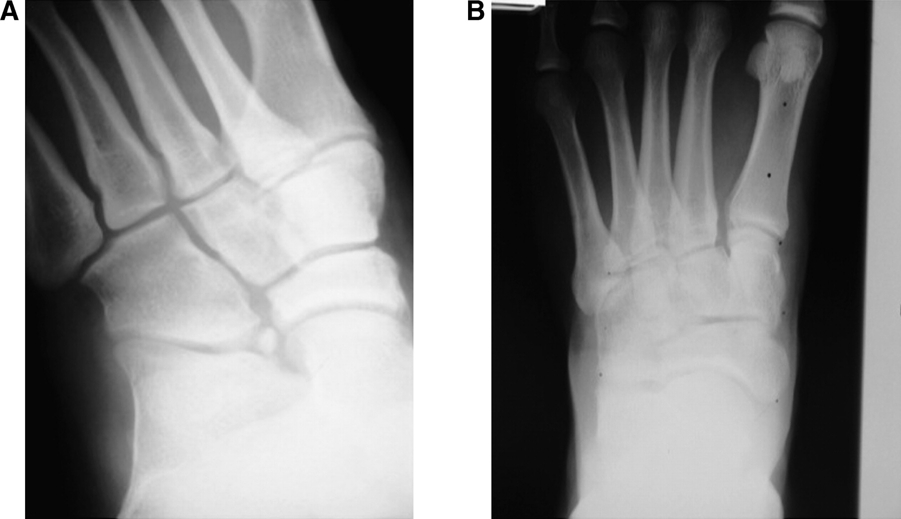
Figure 2.
Computed tomographic scan revealing no evidence of navicular fracture or alterations in bone density but narrowing of the lateral aspect of the navicular bone.
Discussion
Navicular stress fractures of the “fatigue” type are characterized by the mechanical failure of normal bone subjected to repetitive stresses over time.[8–14] The foot mechanics and mechanisms of injury are well documented.[8–14] Anatomical variations, including a cavus foot type, metatarsus adductus, a short first metatarsal bone, talar beaking, limited subtalar motion, limited ankle dorsiflexion, and medial narrowing of the talonavicular joint, have been suggested as risk factors for navicular injury.[7–12] Competitive runners are especially susceptible to stress fracture of the navicular bone due to the high impact of the sport coupled with repetitive overuse during training.
Figure 3.
Sagittal (A) and transverse (B) short tau inversion recovery magnetic resonance images depict a homogenous increase in signal intensity through the navicular bone indicative of inflammation and edema.
Clinical assessment is challenging. The symptoms of a navicular stress fracture are insidious and are often initially misdiagnosed.[15, 16] Anterior tibial tendonitis, midfoot sprain, rheumatologic disease, idiopathic ischemic necrosis of the navicular bone (Kohler disease), and plantar fascial injury all display similar symptoms.[2, 8] Pain is present during running, jumping, and other weightbearing activities, subsiding with rest and ice.[16] Pain may also radiate laterally along the cuboid or sinus tarsi or distally along the first and second rays, although swelling and discoloration are often not present.[15] Examination of a patient presenting with a navicular stress fracture will yield tenderness over the proximal dorsal aspect of the navicular bone, the N sign.[17, 18] The N sign is located in the central area of the bone between the extensor hallucis longus and the anterior tibial tendon.[2] A positive N sign presents a high degree of suspicion and warrants further testing with multiple imaging modalities.
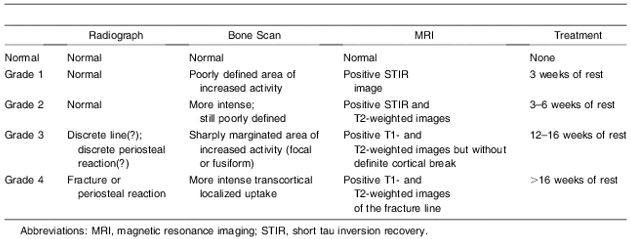
The clinical difficulty of assessing navicular stress fractures is magnified by the variety of imaging modalities available for assessment compounded with its failure to show up on radiographs. It has been recommended that a positive three-phase technetium-99 bone scan followed by a CT scan provides sufficient diagnostic information when injury to the bone has advanced to the point where a fracture line is present.[8, 14] In a study by Burne et al,[19] CT was much more effective than was MRI in the detection of chronic stress fracture lines during follow-up. In cases in which an acute stress reaction injury to the navicular bone is suspected, the use of MRI should not be overlooked[19]; MRI not only is more specific and sensitive than other imaging modalities but also shows marrow changes in the bone, good anatomical resolution of the fracture pattern, and bony edema on T2-weighted images.[20, 21] Note that MRI does not differentiate edema caused by avascular necrosis from edema related to navicular stress injuries[8]; furthermore, the thick proximal cortex of the navicular bone at the talonavicular joint can prevent visualization of fracture lines.[19] Therefore, CT along with MRI are justifiable when diagnosing a navicular stress reaction or fracture. Arendt and Griffiths[7] described a radiologic and management algorithm of stress phenomena and stress fractures for overuse bone injuries (Table 1).
The patient in this case study was a highly competitive track athlete with an athletic scholarship to a Division I baseball program. He was diagnosed as having a severe left navicular bone stress reaction or grade 2/3 injury. Owing to a high index of clinical suspicion based on the patient’s history and physical examination findings, the timely use of diagnostic imaging studies (Figs. 1–3) permitted the clinician to establish a diagnosis and implement conservative treatment before development of a true fracture. In this instance, although a technetium-99 bone scan could have been useful, the clinical findings suggested more specific diagnostic studies.
Table 1.
Radiologic and Management Algorithm of Stress Phenomena and Stress Fractures for Overuse Bone Injuries[7]
Conclusions
This case study demonstrates the importance of having an awareness of navicular stress injuries in competitive athletes. When the history of the concern and the physical findings, subtle as they might be, are present, the clinician is obligated to aggressively investigate the possibility of navicular injury with diagnostic imaging techniques. Radiographs are typically negative for osseous abnormalities. A technetium-99 bone scan is a good initial study to determine whether the pathologic finding is osseous or soft tissue in nature. A positive bone scan finding should be followed immediately by an MRI, including T1-weighted, T2-weighted, and short tau inversion recovery images. If the physical findings suggest a navicular injury and the radiographic findings are negative, going directly to the MRI is an acceptable approach in providing detailed evaluation of osseous and soft tissues. Also, CT may be used to evaluate the navicular bone for frank fracture and changes in bone density or to monitor healing; however, MRI remains the study of choice for evaluating anatomical and structural integrity. The goal in navicular stress injuries is to establish a diagnosis in the earliest stage to implement appropriate treatment, minimizing time away from competition to avoid long-term sequelae associated with the presence of a fracture line.
Financial Disclosure: None reported. | |
Conflict of Interest: None reported. |
References
- Arendt, E, JAgel, CHeikes, et al. :Stress injuries to bone in college athletes: a retrospective review of experience at a single institution. Am J Sports Med 31:959, 2003.
- Ostlie, DK and SMSimons. :Tarsal navicular stress fracture in young athlete: case report with clinical, radiologic, and pathophysiologic correlations. J Am Board Fam Pract 14:381, 2001.
- Goergen, TG, EAVenn-Watson, DJRossman, et al. :Tarsal navicular stress fractures in runners. AJR Am J Roentgenol 136:201, 1981.
- Bennell, KL, SAMalcolm, SAThomas, et al. :The incidence and distribution of stress fractures in competitive track and field athletes. Am J Sports Med 24:211, 1996.
- Bennell, KL and PDBrukner. :Epidemiology and site specificity of stress fractures. Clin Sports Med 16:179, 1997.
- Khan, KM, PDBrukner, CKearney, et al. :Tarsal navicular stress fracture in athletes. Sports Med 17:65, 1994.
- Arendt, EA and HJGriffiths. :The use of MR imaging in the assessment and clinical management of stress reactions of bone in high-performance athletes. Clin Sports Med 16:291, 1997.
- Lee, S and RBAnderson. :Stress fractures of the tarsal navicular. Foot Ankle Clin 9:85, 2004.
- Anderson, MW and AGreenspan. :Stress fractures. Radiology 199:1, 1996.
- Matheson, GO, DBClement, DCMckenzie, et al. :Stress fractures in athletes: a study of 320 cases. Am J Sports Med 15:46, 1987.
- Digiovanni, CW. :Fractures of the navicular. Foot Ankle Clin 9:25, 2004.
- Van Langelaan, EJ. :A kinematical analysis of the tarsal joints: an x-ray photogrammetric study. Acta Orthop Scand Suppl 204:1, 1983.
- Orava, S, JKarpakka, AHulkko, et al. :Stress avulsion fracture of the tarsal navicular: an uncommon sports-related overuse injury. Am J Sports Med 19:392, 1991.
- Haverstock, BD. :Stress fractures of the foot and ankle. Clin Podiatr Med Surg 18:273, 2001.
- Torg, JS, HPavlov, LHCooley, et al. :Stress fractures of the tarsal navicular: a retrospective review of twenty-one cases. J Bone Joint Surg Am 64:700, 1982.
- Fitch, KD, JBBlackwell, and WNGilmour. :Operation for non-union of stress fracture of the tarsal navicular. J Bone Joint Surg Br 71:105, 1989.
- Potter, NJ, PDBrukner, MMakdissi, et al. :Navicular stress fractures: outcomes of surgical and conservative management. Br J Sports Med 40:692, 2006.
- Khan, KM, PJFuller, PDBrukner, et al. :Outcome of conservative and surgical management of navicular stress fracture in athletes: 86 cases proven with computerized tomography. Am J Sports Med 20:657, 1992.
- Burne, SG, CHMahoney, BBForster, et al. :Tarsal navicular stress injury: long-term outcome and clinico-radiological correlation using both computed tomography and magnetic resonance imaging. Am J Sports Med 33.:1875, 2005.
- Sizensky, JA and RMMarks. :Imaging of the navicular. Foot Ankle Clin 9:181, 2004.
- Ariyoshi, M, KNagata, MKubo, et al. :MRI monitoring of tarsal navicular stress fracture healing: a case report. Kurume Med J 45:223, 1998.
© 2011 American Podiatric Medical Association