


Abstract
Cutaneous squamous cell carcinoma is the second most common form of skin cancer and accounts for 20% of cutaneous malignancies. We report the case of a patient who presented with a complaint of nonhealing wounds following radiation therapy for the treatment of noninvasive squamous cell carcinoma of both lower extremities. Initial biopsies of the wounds were benign. However, a second biopsy performed approximately 2 months later was found to be positive for invasive squamous cell carcinoma. This case uniquely exemplifies that all nonhealing wounds should be viewed with a critical eye for possible malignancy even in the presence of previous negative biopsy. This is especially true for radiation wounds that may be prone to malignant transformation or recurrence.
Cutaneous squamous cell carcinoma is the second most common form of skin cancer and accounts for 20% of cutaneous malignancies.[1] General risk factors associated with the development of squamous cell carcinoma include: age older than 50, fair skin, history of prior nonmelanoma skin cancer, exposure to UV light, exposure to chemical carcinogens, exposure to ionizing radiation, and chronic immunosuppression.[1–3] It occurs most frequently on the head and neck; however, upper and lower extremities are the second most commonly affected area.[3]
If squamous cell carcinoma is suspected, it is imperative that a definitive biopsy is obtained in a timely fashion. Proper technique must be used to ensure reliable results. Occasionally it becomes necessary to repeat a biopsy despite previous negative findings. Important factors to take into account when considering a second biopsy include time between biopsies, previous biopsy technique, change in appearance of lesion and perhaps most important, clinical intuition.
Once the diagnosis of squamous cell carcinoma is made, there are a variety of treatment modalities available. Radiation therapy is often used when the size or location of a tumor prevents surgical intervention or the patient is medically at risk for surgery. In these cases, a focused beam of x-rays is used to target and destroy cancer cells, while limiting damage to healthy cells. However, damage to noncancer cells may occur. On a cellular level, radiation can cause the release of free radicals or direct damage to cellular DNA. Possible sequela of radiation therapy includes: fibrosis, pigmentation change, small vessel damage, and nonhealing wound. Risks also include recurrence or new onset of malignancy. The true incidence of radiation injury to normal tissue and the long-term complication rate is unknown.[4]
The following reports the case of a patient who presented with a complaint of nonhealing wounds following radiation therapy for the treatment of noninvasive squamous cell carcinoma of both lower extremities. Initial biopsies of the wounds were benign. However, a second biopsy performed approximately 2 months later was found to be positive for invasive squamous cell carcinoma.
Case Report
An 87-year-old fair skinned, white female presented to the Saint Barnabas Medical Center Wound Care Center (Livingston, New Jersey) with the complaint of two nonhealing, radiation wounds of the left leg. The patient reported that the more proximal wound had been growing in size and becoming painful. The patient had completed a 6-week course of fractional radiation (6000cGy total) for the treatment of noninvasive squamous cell carcinoma of both lower extremities 8 months prior. Since that time, she had developed nonhealing wounds of the medial ankle and more proximal left leg. The patient had a history of rheumatoid arthritis and had been treated with methotrexate but was currently taking no medications. She had no other significant medical history. The patient, who was a retired pediatrician, denied any history of significant sun exposure or other risk factors for developing cutaneous carcinoma.
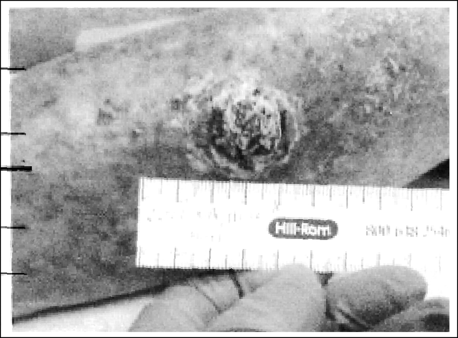
Physical examination of the areas of chief complaint revealed a wound at the medial aspect of the left ankle. The full thickness wound measured 2.3 cm x 1.3 cm. There was granulation tissue at the base. The margins were attached without any rolling or other irregularities. The second wound at the anterior aspect of the leg, however, was quite suspicious in appearance. The wound measured approximately 2.5 cm in diameter and was hyperpigmented. There was a raised, scaling roof with eschar and active bleeding (Fig. 1). The patient was noted to have mild left-sided femoral lymph adenopathy. Physical exam was otherwise unremarkable.
Figure 1.
Initial presentation of anterior leg wound.
The patient had previously sought treatment at another wound-care center where the medicine, vascular, and dermatology departments evaluated her. Local wound care was initiated and biopsies of both lesions were performed 2 months prior to her presentation described here. Biopsy of the distal ulcer revealed inflammatory cells and all findings were benign. However, the biopsy of the proximal lesion was more equivocal in nature. The findings were benign, but the pathology report noted that there was not enough full thickness specimen available to make a definitive diagnosis.
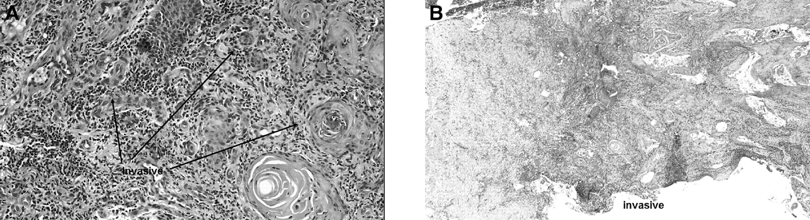
Given the patient’s history and clinical findings, it was deemed necessary to perform a new biopsy of the proximal lesion. However, taking into account the patient’s age, location and size of tumor, and the fact that the site had been previously irradiated, excisional biopsy would not be practical. Therefore, multiple incisional biopsies were performed. Firstly, the entire roof of the mass was excised and sent for pathological review. Once the roof of the lesion was removed, three full-thickness 5mm-punch biopsies were obtained. The first specimen, which was the roof of the lesion, came back from pathology with a diagnosis of keratin and eschar (benign findings) similar to the results of the previous biopsy. However, the full-thickness punch biopsies were positive for invasive squamous cell carcinoma (Fig. 2).
The patient was subsequently referred to the oncology department. The decision was made to treat the patient nonsurgically with a relatively conservative course of chemotherapy.
Figure 2.
A, High-magnification (x 360) and B, lower magnification (x 40) photomicrograph showing invasive squamous cell carcinoma (cancer cells breaching the basement membrane of the epidermal layer and entering the dermis thus gaining the capacity to metastasize).
Discussion
There are likely several different pathways that may lead to recurrence of cutaneous carcinoma following primary irradiation. Analogous to surgery, radiation therapy may leave cancer cells untreated. Failure to obtain “clean margins,” so to speak, would allow recurrence of cancer cells with the same genetic make up as the original cancer. Radiation therapy may also have the undesirable effect of causing malignant transformation of previously healthy cells. The same radiation that was used to destroy cancer cells can cause DNA damage to healthy cells and lead to a new unrelated malignancy. This new cancer would be genetically unrelated when compared to the primary tumor. Finally, radiation therapy may not affect all tumor cells equally and, therefore, may inadvertently select for more aggressive tumor cells. Heterogenicity in cancer cells subpopulations might allow different responses to radiation. Thus, it is likely that irradiation might select for more aggressive and radioresistant tumors. The persistence/recurrent cancer caused by radiotherapy selection, therefore, might be genetically related, but different from the corresponding irradiated primary tumor.[5] The case discussed here, may be an example of radiotherapy selecting for more aggressive tumor cells. However, it is impossible to determine this definitively without genetic testing of initial and recurrent cancer cells.
It is especially important to consider possible recurrence of cutaneous malignancy when treating radiation wounds. Although biopsy of these wounds can be performed without significant increased risk,[6] great care must be taken due to the significant injury that has already been done to surrounding cellular DNA. Proper technique must be used to prevent false negatives requiring further potentially harmful repeated biopsy. An excisional biopsy is usually the technique of choice. However, when this is not practical, appropriate full-thickness incisional biopsies are needed. Incision biopsy may be performed with surgical scalpel or circular blade (“punch”). When using a precalibrated punch, the hub of the instrument must be allowed to rest on the skin thus allowing the edge of the instrument to pass full thickness through the dermis. In this case, previous biopsy may have yielded false negative because the thick roof of the lesion did not allow the punch biopsy blade to pass. If the punch is not allowed to pass full thickness, results will be similar to the scrape technique and will be unreliable. By deroofing the lesion prior to performing the punch biopsy a more accurate specimen was obtained.
Conclusion
All nonhealing wounds should be viewed with a critical eye for possible malignancy, even in the presence of previous negative biopsy. This is especially true for radiation wounds that may be prone to malignant transformation or recurrence.
Funding
None reported.
Acknowledgment
The authors would like to thank Rajiv Pulinthanathu, MD, Department of Pathology, Saint Barnabas Medical Center.
Conflicts of Interest
None reported.
References
- Johnson, TM, DERowe, BRNelson, et al. :Squamous cell carcinoma of the skin (excluding lip and oral mucosa). .J Am Acad Dermatol26::467. ,1992. .
- Sams, WM and PLynch. :Principles and Practice of Dermatology. ,Churchhill Livingstone. ,New York, NY. ,1990. .
- Dormand, EL, HRidha, and MVesely. :Long-term outcome of squamous cell carcinoma of the upper and lower limbs. .J Plast Reconstr Aesthet Surg63::1705. ,2010. .
- Mathews, M and HWilliams. :Radiation Ulcers .Available at: http://emedicine.medscape.com/article/1298239-overview. .Accessed May 18, 2011.
- Deganello, A, FAlessandro, SIacopo, et al. :Genetic alterations between primary head and neck squamous cell carcinoma and recurrence after radiotherapy. .Cancer116::1291. ,2010. .
- Brown, A, DWendler, KCamphausen, et al. :Performing nondiagnostic research biopsies in irradiated tissue: a review of scientific, clinical, and ethical considerations. .J Clin Oncol26::3987. ,2008. .
© 2011 American Podiatric Medical Association