


Abstract
Background: Sever’s disease, also known as calcaneal apophysitis, is thought to be an inflammation of the apophysis of the heel, which is open in childhood. This condition has been commented on and looked at in a retrospective manner but has not been examined systematically. We assembled the most commonly cited theoretical causative models identified from the literature and tested them to determine whether any were risk factors. Methods: Children with Sever’s disease were compared with a similarly aged nonsymptomatic population to determine whether identifiable risk factors exist for the onset of Sever’s disease. Areas raised in the literature and, hence, compared were biomechanical foot malalignment, as measured by Root et al–type foot measurements and the Foot Posture Index; ankle joint dorsiflexion, measured with a modified apparatus; body mass index; and total activity and types of sport played. Results: Statistically significant but small odds ratios were found in forefoot to rearfoot determination and left ankle joint dorsiflexion. Conclusions: This study suggests that there is no evidence to support that weight and activity levels are risk factors for Sever’s disease. The statistically significant but clinically negligible odds ratio (0.93) on the left side for decreased ankle joint dorsiflexion and statistically significant and clinically stronger odds ratio bilaterally for forefoot to rearfoot malalignment suggest that biomechanical malalignment is an area for further investigation.
Sever’s disease, also known as calcaneal apophysitis, is an inflammation of the apophysis of the heel, which is open in childhood. [1,2] It is a condition that has been commented on and looked at in a retrospective manner but that has not been examined in a systematic manner to date. Sever’s disease was first described by Haglund [3] in 1907, although Sever [4] in 1912 received more credit for describing the condition. Sever’s disease is a condition that has been described in various ways and attributed to a variety of causative factors. It affects children while bony growth is occurring at the calcaneal epiphysis, generally between ages 7 and 15 years in boys and 5 and 13 years in girls. Sever’s disease is reported to compose 2% to 16% of musculoskeletal injuries in children. [5]
The literature suggests that the clinical picture of a patient with Sever’s disease is an active sport-playing child aged 10 to 12 years, generally male, often presenting at the beginning of a sport season, in the midst of a growth spurt, and experiencing pain over the apophyseal area of one or both heels. [6-10] The child will generally be found to have limited ankle joint dorsiflexion, an underlying biomechanical deformity of variable nature, and worse pain with increased activity. [4,6,7,9-15] The pain is often severe enough that at the end of a sporting activity the child will actively limp to take weight off the affected heel(s). [4,6,7,9,10]
However, all of these traits arise from anecdotal and observational surmisings of the authors [4,10-12] or from data that have involved multiple practitioners’ assessments and retrospective analysis of clinical notes. These retrospective data may have been collected up to 10 years before writing the publication. [6-9,14,15] No authors, to our knowledge, have reported measurement of the time of onset of the discomfort in sporting activities. There is no mention in any of these articles of the methods used to measure the biomechanical variables mentioned. Reliability or validity of methods used to obtain the ankle joint dorsiflexion or biomechanical malalignment data are not commented on, thus reducing the strength of the data.
The aims of this study, therefore, were to assemble the most commonly cited theoretical causative models identified in the literature and to test these parameters on a group of children with Sever’s disease and on a group of similarly aged controls without Sever’s disease. Our null hypothesis was that there are no identifiable risk factors in these parameters, as identified by odds ratios, between the two groups.
Methods
Design and Sample
This study used a prospective case-control comparison design to establish whether there were identifiable risk factors in the sporting, biomechanical, and physical characteristics of children with and without Sever’s disease. A simple comparison of data was also performed to compare the two groups on a variety of measures. Subjects were not excluded from the non–Sever’s disease group if they had other musculoskeletal pain. Greater numbers of individuals without Sever’s disease were recruited to increase the power of the odds ratios calculated, which suggested that at least a 3:1 ratio was necessary to increase the power of the calculations above 0.8.
Ethics Approval
Ethics approval was received from the Human Research Ethics Committee of the University of South Australia before recruitment of participants. Ethical approval was also granted by the South Australian Department of Education and Children Services for access to children in the government school sector and from the South Australian Catholic Education office for children in the Catholic school sector. Other nongovernment schools granted approval for the study on an individual basis.
Inclusion and Exclusion Criteria
Patients with Sever’s disease were considered eligible for participation if they met the following criteria: male or female children aged 8 to 14 years experiencing unilateral or bilateral heel pain corresponding to a Sever’s disease diagnosis, ie, pain over the apophyseal area on the squeeze test. [7] Individuals without Sever’s disease were considered eligible for participation if they met the following criteria: male or female children aged 8 to 14 years without reported heel pain consistent with a potential diagnosis of Sever’s disease.
Patients were excluded from both groups if they met any of the following criteria: 1) medically diagnosed conditions that may confound diagnosis, including, but not restricted to, spondyloarthropathies, rheumatoid arthritis, complex regional pain syndrome, and fractures of the calcaneus; 2) recent laceration or puncture to the heel that was reported to be symptomatic; 3) nonambulatory; and 4) unable to communicate in English.
Sever’s Disease Sample
Patients with Sever’s disease were recruited from the University of South Australia Podiatry Clinic, the private practice of one of us (R.W.S.), the primary schools selected for recruitment of participants without Sever’s disease, metropolitan Adelaide private podiatrists, and Australian Rules football and soccer clubs in metropolitan Adelaide. If there was concurrent pain in other areas, such as the Achilles tendon or the arch of the foot, the participant was still deemed suitable for inclusion in the study because the criteria for a Sever’s disease diagnosis were still met.
Non–Sever’s Disease Sample
Participants without Sever’s disease were recruited from selected schools in the Adelaide metropolitan area. An examination of the proportionate distribution of government to nongovernment school student numbers for the year levels of interest (years 5–7 to match the highest incidence ages in the literature [7,8]) was calculated from the Australian Bureau of Statistics Schools Document 4221.0, 2001. From this, it was calculated that the ratio of children in nongovernment to government schools was 1:2.25. Schools were then randomly and blindly selected from a list of all metropolitan primary schools obtained from the Department of Education and Child Services in Adelaide and were placed into one of five categories representing suburbs north, south, east, west, and central (within 5 km of the General Post Office) until each had four schools, and seven of the total number of schools were nongovernment to reflect this ratio.
Schools were approached by letter for inclusion in the study, followed by a telephone call to the principal. However, many schools did not wish to take part in the study, and numbers of students willing to participate varied considerably from school to school. This occurred to the extent that the process of school selection became recruitment from the schools that agreed to participate. In addition, recruitment of schools not on the original list was undertaken to increase numbers of participants; so, although a random sample was the aim, this was not achieved.
Subject Examination Locations and Procedures
Participants without Sever’s disease were examined at their respective schools. Patients with Sever’s disease were seen at one of the following venues at their convenience: the University of South Australia Podiatry Clinic, the private podiatric medicine practice of one of us (R.W.S.), the participant’s home, or the participant’s school.
Participants provided signed parental consent forms, and parents and participants were given the opportunity to ask questions about the study. All contact with the study participants was provided by one of us (R.W.S.). Participants were questioned specifically regarding the presence of Sever’s disease symptoms and other inclusion and exclusion criteria and were grouped accordingly. If Sever’s disease was present, it was noted whether it was unilateral or bilateral. Participants were then interviewed using an activity questionnaire. [16,17] A physical examination of the participant then took place.
The following information was collected from each participant: sporting activities and amount thereof (activity questionnaire), resting calcaneal stance position, neutral calcaneal stance position, forefoot to rearfoot relationship, eight-item Foot Posture Index score, ankle joint dorsiflexion (performed following the method of Scharfbillig and Scutter [18]), height (in meters), and weight (in kilograms). The first four parameters were assessed in the same manner as described by Evans et al. [19] Height was determined with the participant standing without shoes against a wall or door frame. A book was placed on the participant’s head, and a mark was made on the vertical surface. Using a 150-cm plastic measuring tape, the distance from the mark to the supporting surface was measured. Weight was measured with the participant standing without shoes on a Soehnle (Nassau, Germany) digital measuring scale that displayed the weight to one decimal point on an LCD readout. Body mass index was calculated as weight in kilograms divided by the square of the height in meters.
Reliability and Validity of Measures
Activity Questionnaire.
A reliable and validated questionnaire [16,17] was used as a template for questions selected to gather these data. It was used in an interview format rather than in the self-report format of the original questionnaire owing to the wide age range of participants and to ensure that the participants understood the questions.
Ankle Joint Dorsiflexion.
This was measured with the modified Lidcombe template, which showed excellent reliability and good face validity, the discussion of which appeared in an earlier article. [18]
Traditional Foot Measures.
The reliability testing of the examiner was conducted in an earlier study, [19] and the results are included in the “Results” section herein. The validity of these measures is acknowledged as questionable at best owing to the difficulties of identifying subtalar neutral and the 2:1 ratio of movement, as identified by Menz, [20] but because these are still the most common measures used in the podiatric medical community, by our assessment, they were deemed important to be included.
Foot Posture Index.
The eight-item Foot Posture Index was originally investigated as an alternative foot measure to the traditional Root et al–type [21] measures because of their well-documented issues with reliability and validity, as commented on previously herein. The inability to validate parts of the eight-item Foot Posture Index in an earlier experiment [22] places caveats on its use as a tool. Redmond et al [23] recently reported validity testing for an amended six-criteria Foot Posture Index using computer modeling, which, similar to our experiment, looks at gross states of resting, pronated and supinated, and has found acceptable validity of movement between these states. There has been no advancement in showing the validity of either of the Foot Posture Index versions as a measure of small movements; coupling this with the cumulative problems noted by Evans et al, [19] the Foot Posture Index must still be used with caution, in our opinion. There have been no further attempts at criterion-based validation of individual criteria of the Foot Posture Index; however, Menz and Munteanu [24] compared the eight-item Foot Posture Index with three arch parameters and found a moderate correlation between them. The relative infancy of the Foot Posture Index means that further testing is needed before a full assessment can be made of this measurement tool. Because this experiment was conducted before the introduction of the six-item Foot Posture Index and because reliability and validity testing was performed on the eight-item Foot Posture Index, the eight-item version was used.
Reliability testing [19] found mixed reliability for the measures used in this experiment, which is acknowledged as a limitation of this study when drawing extrapolations. The reliability results are presented in Table 1.

Table 1.
Reliability of the Tester for Foot Measures

Analysis
Data were analyzed with SPSS version 12.0 (SPSS Inc, Chicago, Illinois). Data from the physical measures of height, weight, and traditional foot measures (resting and neutral calcaneal stance positions, forefoot to rearfoot relationship, and ankle joint dorsiflexion) were interval in nature and were analyzed accordingly. Data from the Foot Posture Index, while ordinal in nature, were treated as interval data for the sake of analysis, as done in the earlier reliability study [19] and in keeping with some current statistical literature. [25-27] Data were considered for either the left or right side in keeping with Menz’s recommendations. [28]
Data from the activity questionnaire were mixed in their classification. Total activity hours was interval data, but the type of activity undertaken was nominal data. Each type was analyzed appropriate to its form. Age, although interval in nature, was analyzed as ordinal data initially to show separation and then was used as interval data in the regression statistics.
The foot measures, body mass index, and activity levels were examined with simple descriptive statistics. Data from the forefoot position measurement and the difference between the resting and neutral calcaneal stance positions were analyzed as absolute values because the varus and valgus orientations were not deemed important compared with the numerical measure and difference, respectively. Activity levels for the activities for each participant were combined to produce the number of activity hours used in the analysis.
The Sever’s disease and non–Sever’s disease groups were then compared via binary logistic regression with grouped covariates. This was to ensure that significance was not explained by another related variable and to determine whether any of the factors studied were predictive of risk of Sever’s disease. [29] Alpha level was set at 0.05. The Foot Posture Index, although being a summed scale without determined interval qualities in its individual criteria, was considered as interval data for the sake of this part of the analysis also for the aforementioned reasons. Each criterion of the Foot Posture Index was also investigated with descriptive statistics and binary logistic regression.
To assess the homogeneity of the groups, the age and sex distributions of the participants in the two groups were compared with χ2 analysis. A significant difference was found for both factors on analysis; thus, sex was included as a covariate in all of the regression analyses, and age was included as a covariate in its standardized (age minus mean of all ages in sample) and standardized squared ([age minus mean of all ages in sample] squared) forms. These calculations were used to reduce the risk of the standard error being increased and to reduce the risk of collinearity effects on the results.
The Hosmer and Lemeshow [29] test was calculated in each of the binary logistic regressions as an indication of the “goodness of fit” of the analysis of the variables in a linear model. Where this test returned a significant result, indicating a lack of goodness of fit, attempts were made to find a better model, eg, loglinear or curvilinear, to match the data, but this was unsuccessful, and, as such, the linear model was used as the best model available.
The sports played by all of the participants were analyzed via simple frequencies, with χ2 analysis used to determine whether a difference existed between the groups studied. Sports that commonly presented, such as Australian Rules football and basketball, were individually grouped, whereas those that did not have many participants, such as hockey and lacrosse, were grouped into a category of “other sports.” Schoolyard games, such as “chasey” (tag) and handball, were grouped together, and activities, such as walking and riding a bike to school, were collected under the heading of “other.” Any physical education lesson was classed as PE, regardless of the sports played. The stage at which patients with Sever’s disease first experienced pain during sports participation was analyzed via frequency data.
Results
Reliability
The investigator’s reliability of the foot measures undertaken is presented in Table 1.
Sample
There were 236 participants without Sever’s disease. Sixty-seven patients with Sever’s disease were recruited from the investigator’s private practice (n = 8), the University of South Australia Podiatry Clinic (n = 2), South Australian podiatric medical practices (n = 26), schools involved in the provision of study participants without Sever’s disease (n = 26), and Australian Rules football and soccer clubs in metropolitan Adelaide (n = 5).
Age
Figure 1 shows the percentage distribution of ages in the two groups and demonstrates the dissimilar distribution between them. The Sever’s disease group is more evenly spread across the age groups compared with the non–Sever’s disease group, which is more bell shaped in its distribution. In contrast, the mean (SD) ages of the two groups show little difference (Sever’s disease group: 10.87 [1.526] years; non–Sever’s disease group: 10.86 [0.968] years; and both groups: 10.86 [1.113] years). However, on χ2 analysis (P < .01), there was a significant difference between the two groups, which highlights the difference seen in Figure 1. Consequently, it was necessary to consider age as a covariate in further analysis, as mentioned in the “Analysis” subsection.
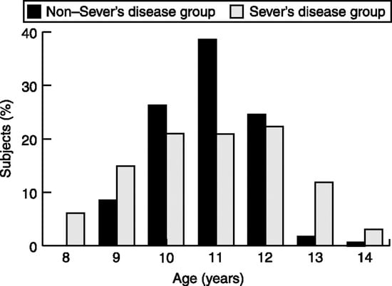
Figure 1.
The age distributions of the Sever’s disease and non–Sever’s disease groups.
Sex
Figure 2 shows the percentage sex distribution in the two groups. A marked difference is seen in the percentages of boys to girls, with the Sever’s disease group having a 71.6% male composition and the non–Sever’s disease group having a 53.4% male composition. On χ2 analysis, this showed a significant difference (P < .01); hence, sex was also included as a covariate in all binary logistic analyses (as discussed in the “Analysis” subsection).
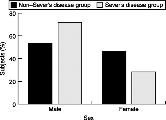
Figure 2.
The sex distributions of the Sever’s disease and non–Sever’s disease groups.
Heel Distribution and Forefoot to Rearfoot Alignment
In the Sever’s disease group, 30 patients (44.8%) had unilateral heel involvement and 37 (55.2%) had bilateral involvement. Malalignments in the Sever’s disease group were measured in 34 participants with forefoot varus (one subject had a rearfoot and forefoot malalignment) (mean [SD], 6.33 [2.31]°), three with rearfoot varus (mean [SD], 6.00 [1.73]°); and 29 with forefoot valgus (mean [SD], 4.67 [0.58]°); two participants had no malalignment recorded.
Foot Measurements, Body Mass Index, and Activity Data
Table 2 provides descriptive statistics for the foot measurements, body mass index, and activity data for the two study groups. Examination of the raw summary data shows that many of the variables were similar in their means and ranges.
Binary logistic regression was then undertaken with all appropriate variables included as covariates. This enabled determination of the variables that did not have their significance explained by the inclusion of other variables as covariates. The left and right sides were considered separately and included standardized age, standardized age squared, and sex as covariates (Table 3). This test revealed appropriate fit to the linear model as indicated by the Hosmer and Lemeshow test.
The variables of forefoot position and ankle joint dorsiflexion on the left side produced statistically significant risk factors. Left ankle dorsiflexion showed a small odds ratio, suggesting that the significance, while present statistically, is clinically small. The forefoot position bilaterally was 1.5 times more likely to be greater in magnitude in the symptomatic group, suggesting greater biomechanical malalignment in the Sever’s disease group.
Body mass index was also examined for each age level in the Sever’s disease and non–Sever’s disease groups for comparison with normal values for these age groups (Table 4). [30] Although there was no statistically significant difference between body mass index values when examined in whole groups (Table 3), comparison of body mass index data against overweight thresholds for age [30] suggest that in children aged 8 to 11 years in the Sever’s disease group were overweight or very close to it, although this trend is not continued in the older age group.
Each individual criterion of the Foot Posture Index was also examined, with the aforementioned covariates for each side (Table 5). The medial longitudinal arch criterion was a significant risk factor, as was the talar head palpation criterion on both sides. The medial longitudinal arch result suggests that patients with Sever’s disease were approximately half as likely to have a higher reading on this criterion as their non–Sever’s disease counterparts. The talar head palpation criterion suggested that the Sever’s disease group was twice as likely to have a higher reading on this criterion as their non–Sever’s disease counterparts. This negated their effect when looked at as a summed score in the whole Foot Posture Index, as seen in Table 3.
Table 2.
Descriptive Statistics for Foot Measurements, BMI, and Activity Levelsa
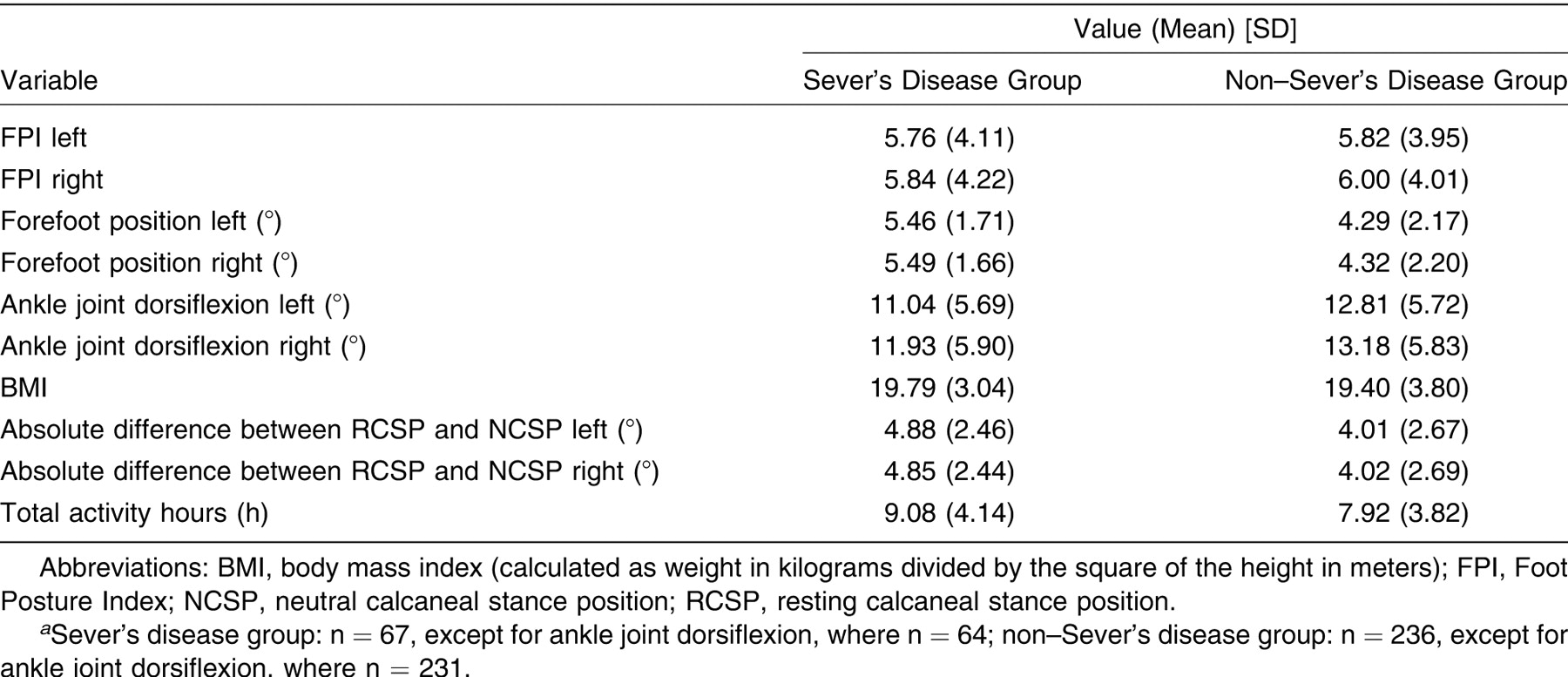
Table 3.
Binary Logistic Regression of All Left- and Right-Sided Foot Measurements, BMI, and Total Activity
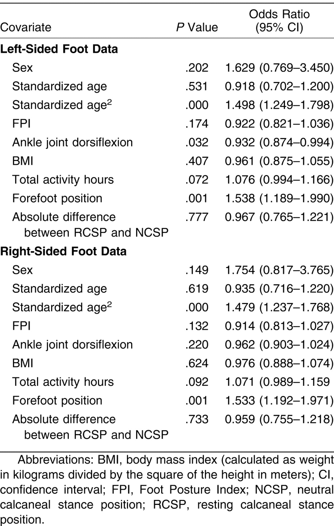
Table 4.
Descriptive Statistics of BMI Data for Separate Age Groups and Comparison with Age-Adjusted Levels of Overweight and Obesity Thresholds
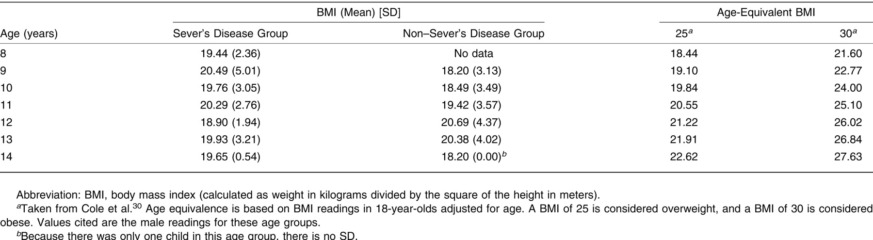
Table 5.
Binary Logistic Regression of All Left and Right Side FPI Criteria
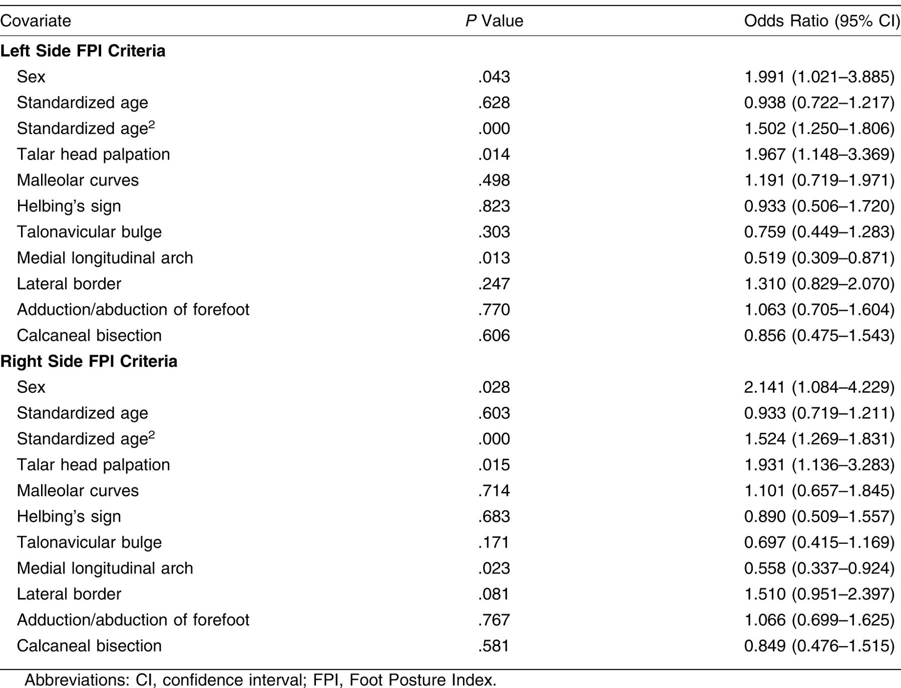
Type of Sports Played
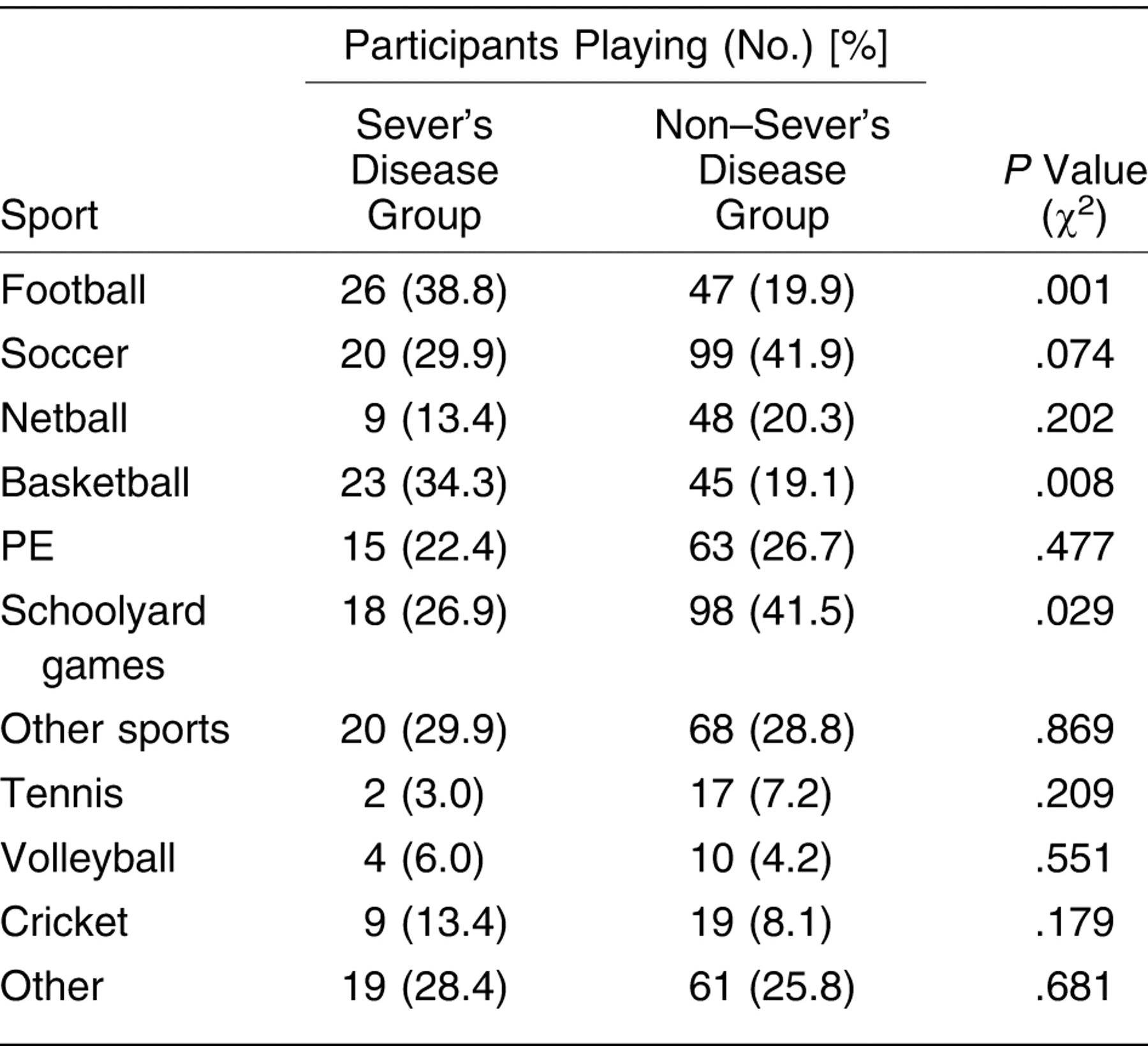
Comparison of the sports played by the two groups demonstrated that significantly more participants in the Sever’s disease group played Australian Rules football and basketball but that significantly fewer participants in the Sever’s disease group participated in schoolyard games (Table 6).
Table 6.
Comparison of Sports Played by Study Group
Onset of Pain in Sever’s Disease Patients
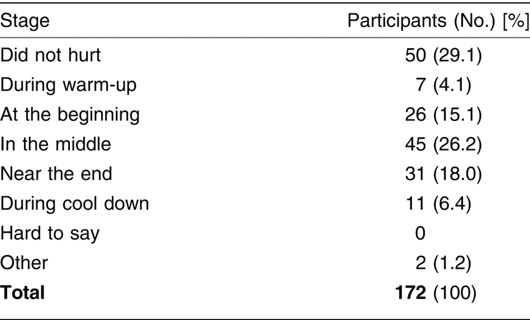
The time when symptoms first appeared for each sport played by patients with Sever’s disease showed a wide spread across the possible categories, with more than a quarter of the recorded sports not causing pain for patients with Sever’s disease (Table 7). The sport categories “schoolyard games,” “other sports,” “PE,” and “other” showed the highest percentage of responses of “did not hurt.”
Table 7.
The Stage of the Activity When the Heels Started to Hurt in the Sever’s Disease Group
Discussion
This study aimed to determine whether there were significant risk factors for the formation of Sever’s disease by comparing a cohort of children with Sever’s disease and a cohort without Sever’s disease. The results suggest that children with Sever’s disease may exhibit some differences in physical characteristics compared with their non–Sever’s disease counterparts and differences from other Sever’s disease groups studied.
Age and Sex
The mean age of the Sever’s disease group in this study was similar to that of patients with Sever’s disease in most other studies, [6,7,15,31,32] although in two studies [8,33] the mean age was a year to 18 months older. More boys (71.6%) than girls were affected by Sever’s disease in this study. Previous literature [6,7,34] suggests that Sever’s disease is more common in males, although Kvist and Heinonen [8] found only a 54% male predominance. Rozenblat and Bauchot [15] had a higher female proportion in their study cohort due to recruitment from dance studios. The higher proportion of boys in the Sever’s disease group of this study is not likely to have been affected by sampling bias toward Australian Rules football and soccer clubs because the small number of participants recruited through this medium could not have caused this difference. The results suggest that there may simply be more boys with the condition or who present for treatment.
Heel Distribution
Patients with Sever’s disease demonstrated a near equal distribution of unilateral (44.8%) and bilateral (55.2%) cases. This distribution is similar to that in some of the literature, [7,14,35] although other larger studies have not reported a consistent distribution pattern. Kvist and Heinonen [8] reported a 15% bilateral prevalence, and Volpon and de Carvalho Filho [36] had only unilateral presentations in their 69 patients with Sever’s disease. McKenzie et al [6] and Rozenblat and Bauchot [15] had a much higher bilateral incidence. If Sever’s disease is an overuse condition, one would surmise that a unilateral appearance would occur on the dominant side of the participant, ie, the foot that is used for unilateral push-off or for initial acceleration. Similarly, a bilateral occurrence may occur in sports and individuals who use both feet in high-stress applications. The study by Kvist and Heinonen [8] had a 73% share of patients in track and field, sports that generally involve very measured step and leading foot patterns, whereas the other studies mentioned had a wider distribution of sports played by their study participants, strengthening this argument. There was generally little difference biomechanically between the two feet, as demonstrated in the mean results of this cohort, so unilateral overuse due to a one-sided biomechanical insufficiency is not a likely scenario. A larger cohort of individuals who played only single sports would help to further test this theory.
Body Mass Index Results
The summary data for the body mass index findings in the two groups yielded results that suggest that the groups studied were different in the younger groups. The 8- to 11-year-olds in the Sever’s disease group seemed to be overweight, as determined by body mass index definition, or very close to it, compared with children without Sever’s disease of the same age. However, this can only be postulated at this stage because there are insufficient numbers in some groups for analysis. There is also insufficient information in the age equivalence data (lack of standard deviations) of Cole et al [30] to run further tests to see whether the age groups from this study were significantly different compared with the age equivalence body mass index data from Cole et al. [30] The binary logistic regression of the Sever’s disease and non–Sever’s disease groups suggest that this is not a risk factor of consequence. The overweight nature of younger patients with Sever’s disease may be due to a lower level of activity in these age groups, which increases at the 12-year-old level, or an adjustment of weight to height ratio with the onset of puberty.
Weight and height were not analyzed separately as the age group studied is prone to large changes in proportions with the onset of puberty. [37] When looked at in isolation, the individual parameters may show trends that should be tempered by inclusion of the other parameter. Current literature also focuses on using body mass index as a weight assessment rather than weight in isolation. [38,39]
Some authors [4,31,40,41] postulated that weight might have been a factor in the development of Sever’s disease. The lack of a significant risk factor being identified in this study suggests that the earlier authors may have been basing this on a small or quite young population. No explanation of the assessment of the overweight status was provided in these articles, and no mention of body mass index was made.
Traditional Foot Measurements
The only measures that produced statistically significant risk factors were forefoot position and left-sided ankle joint dorsiflexion assessment. The forefoot position result suggests that there is a difference in biomechanical malalignment between the groups, but the lack of a significant risk factor between the resting and neutral calcaneal stance positions weakens this association. The “Root” theory [21,42] suggests that this difference should be reflected here as well as for most types of forefoot malalignments because forefoot deviation should result in a compensated resting calcaneal stance position. There is insufficient evidence to definitively say that there is more malalignment in the Sever’s disease group, although the forefoot result suggests that this is an area worthy of further investigation. The continuing problem with reliability and validity of these measurements also weakens any findings that can be extrapolated from the data. However, given the lack of a better measurement system that is used in the podiatric medical community, the results are put forward as the best possible under the circumstances.
Many authors [4,6,7,11,12,41,43-48] have hinted at the presence of a biomechanical malalignment in children with Sever’s disease. However, none of these studies have used a systematic measurement tool or have tested the reliability of the rater(s) involved. The results of this study strengthen the anecdotal findings of the earlier studies and suggest that further investigation into this area as a causative factor and, hence, treatment aimed at this area may be beneficial.
The left ankle joint binary logistic regression result suggests that this is a risk factor. However, the odds ratio of 0.932 also suggests that this difference is small and, although statistically significant, likely to be clinically nonsignificant. The fact that the mean result of both groups was greater than 10° dorsiflexion suggests that ankle equinus is not a major issue for either group in this study. Although ankle equinus is mentioned as a biomechanical finding in many articles, only the study by Szames et al [14] quantified the percentage of participants with equinus (82%). They qualified this by stating that this was a retrospective study with many raters assessing the condition and giving only a “yes” or “no” response for equinus, with no details of measurement or reliability. The present study is the first to reliably measure the amount of ankle joint dorsiflexion. These results suggest that authors [7,9,10,12-14,41,48-58] who have suggested a consistent link between equinus and Sever’s disease may have to more accurately assess the degree of dorsiflexion available given that the mean for the Sever’s disease group was greater than 11°.
Total Activity Hours and Type of Sports Played
Analysis of the two groups did not suggest that total activity hours was a risk factor. This suggests that earlier authors may have overstated this aspect of the risk factor profile for these patients. This may be due to often selecting patients from dedicated sporting clinics and by lack of comparison with a nonsymptomatic group from the same population. [4,6,8,9,31,34,48,51,56,59,60]
Patients with Sever’s disease were found to engage in Australian Rules football and basketball more frequently than were participants without Sever’s disease and were less likely to be involved in schoolyard activities, such as chasey (tag) and handball. Previous studies [6-9,15,34] have found a variety of sports taking predominance in patients with Sever’s disease, especially track and field and soccer. However, because these were studies performed in different countries, and often involved patients recruited from sports injury clinics, comparison with Australian conditions and a more open approach to sampling is difficult. The analysis was not able to take into account comparison of the level of sports played, eg, competition or recreation. Many participants were engaged in single sports at multiple levels, which weakened any overall associations that could be drawn. The significantly greater percentage of patients with Sever’s disease playing Australian Rules football and basketball may, in part, be explained simply by the higher percentage of boys in the Sever’s disease group. This finding was reflected in a survey of activities played by year 7 boys conducted in Adelaide in 1996–1997, [61] which showed that the sports most commonly participated in were Australian Rules football and basketball. The significantly lower percentage of patients with Sever’s disease participating in the schoolyard games category suggests that the Sever’s disease group participated in more classifiable sports, as explained in the data analysis section, leaving this grouping less likely to be filled.
Foot Posture Index Criteria
The Foot Posture Index was originally investigated as an alternative foot measure to the traditional Root et al–type measures because of their well-documented issues with reliability and validity (Redmond AC, unpublished booklet, 2000), as commented on previously herein. The lack of validation of the Foot Posture Index for small movements places caveats on its use as a measurement tool.
Analysis of the Foot Posture Index data from the two groups produced no significant risk factors when assessed as a summated score, but the differences noted in comparison of two of the individual criteria, talar head palpation and shape of the medial longitudinal arch, suggest that some of the criteria are more sensitive than others to change. This was noted in the validation experiment performed by members of this group, [22] where talar head palpation was the only criterion that displayed any logical progression from a supinated to a resting to a pronated position. The fact that the values produced effectively negated each other, ie, the talar head palpation score was approximately twice as likely to be greater in the Sever’s disease group and the shape of the medial longitudinal arch score was approximately twice as likely to be smaller in the Sever’s disease group, also suggests a lack of consistency in the scoring algorithm for all types of feet. This negation of factors suggests that there is no significant difference in biomechanical malalignment between the two groups, which disagrees with the forefoot position findings from the traditional measures. Note, however, that the forefoot measures also have no validation completed for them. Further refinement of the Foot Posture Index and comparison with more traditional foot measures is recommended for further studies. Yates and White, [62] Burns et al, [63] and Burns and Crosbie [64] have used the Foot Posture Index in the assessment of feet but have not compared their results with the traditional foot measures used in this study. They also have not mentioned validity testing of this tool or the cumulative problems discussed by Evans et al. [19] As stated previously, in our opinion the Foot Posture Index should be used only with relative confidence as a measure of gross states until more rigorous testing is performed.
Onset of Pain in Patients with Sever’s Disease
Onset of pain in patients with Sever’s disease when playing a sport was diffuse, with no identified pattern. It was reported that 29.1% of sports undertaken by the Sever’s disease group produced no pain at all and that the remainder was spread out across the time played, with the largest concentration (26.2%) occurring in the middle of the activity. No other studies have reported on this aspect of the condition; thus, no extrapolation can satisfactorily be drawn to time of onset of pain in patients with Sever’s disease. The sports that showed the highest percentages of no pain during their undertaking, such as schoolyard games, other sports, and PE, probably reflect the nature of some of these sports. For example, walking or horseback riding and the level that they were engaged in was mainly for fun or at the PE level, reducing the exertion level of the participant and, hence presumably, the pain level.
Implications for Clinical Practice
The results of this study show that a significant risk exists for the formation of Sever’s disease in adolescents with forefoot to rearfoot malalignment. Although this is not reinforced by a significant risk factor being found between resting and neutral calcaneal stance position differences, measurement of forefoot to rearfoot alignment may potentially provide a diagnostic tool to reinforce results from the squeeze test of the apophyseal area and direct research into appropriate treatment of underlying etiologic factors. No clear evidence was produced in this study to suggest that equinus of the ankle is a consistent finding in Sever’s disease; hence, its measurement as part of a diagnostic process is not supported by this study.
Limitations of the Study
The foot measures used in this study are acknowledged as not being validated. In the case of the traditional foot measures, the continued use of an indefinable neutral position of the subtalar joint will make validation unachievable in the short term. Although the intrarater reliability of the traditional measures used was shown to be greater than 0.75 for the investigator, the generally poor interrater reliability results in the reliability study, [19] and previous literature weaken the strength of these findings when extrapolating them to other raters. The use of a single rater, however, reduces the effect of interrater reliability on the results.
The activity questionnaire was produced from a larger reliable and validated survey [16] using selection of questions thought to be relevant to the information desired for analysis. This survey was also designed to be used as a nonsupervised questionnaire and was used in an interview format. These alterations from the original questionnaire format are acknowledged as having a possible impact on the validity and reliability of the results produced. Additional acknowledgments are made of the introduction of bias by having a nonindependent investigator administer the questionnaires and analyze the data with previous knowledge of the participant’s Sever’s disease status. However, use of a single rater and administration in as consistent a manner as possible should reduce the impact of this possible bias.
Recommendations for Further Study
The development of a valid and reliable tool that requires less tactile and, hence, subjective input from the investigator would greatly enhance the testing of this condition, as its current diagnosis is subject to individual and patient interpretation. Verification of inflammatory processes via histologic analysis, blood testing, or imaging processes and isolation of predictive factors for this condition would enhance its acceptance as a recognizable, treatable condition rather than relying purely on patient feedback.
The development of a valid and reliable foot measurement tool that provided researchers with a difference between the actual and an ideal measurement would greatly strengthen the results of this type of study rather than simple static measures such as the Foot Posture Index. The use of foot modeling, as outlined in the article by Redmond et al, [23] may provide a window into a more reliable dynamic measure of the foot, but owing to the lack of application of this technique to a clinical setting, due to cost and complexity, the results of this type of study may be of less clinical relevance to practitioners. Random sampling of subjects in the Sever’s disease and non–Sever’s disease groups would be the ideal situation to increase the clinical relevance and strength of the inferences, which could be drawn from the results, but this is an unlikely possibility due to the difficulty of recruitment of participants with Sever’s disease. If there was a larger pool of individuals without Sever’s disease recruited, age and sex matching would eliminate the lack of homogeneity between the two groups in this area. Recruitment of individuals without Sever’s disease subjects from a wider age range, possibly 8 to 14 years, would also lessen the likelihood of age differences.
The malalignment hypothesis has not been strongly answered in this study, and a bigger study with a more reliable and valid measurement system that can be used easily clinically, should one come to light, may provide stronger evidence for answering this hypothesis. Ankle joint dorsiflexion and body mass index data may also provide more conclusive results with a larger, random sample, ideally in both groups but at least in the asymptomatic group. Treatment of the biomechanical factors should also be investigated in a controlled setting to determine whether this path produces superior results compared with other methods of treatment.
Conclusions
The results of this study suggest that there is no evidence to support that weight or activity levels are risk factors in the formation of Sever’s disease. There is a weak association on the left side of patients with Sever’s disease, with decreased ankle joint dorsiflexion and a stronger indication that forefoot to rearfoot malalignment is likely to be present in children with Sever’s disease, suggesting that biomechanical malalignment is the area that should be pursued for further investigation.
Financial Disclosure
None reported.
Conflicts of Interest
None reported.
Acknowledgments
The Podiatry Research Group at the University of South Australia for their support throughout this study.
References
- Harding VV: Time schedule for the appearance and fusion of a second accessory center of ossification of the calcaneus.Child Dev23: 181, 1952.
- Hoerr NL, Pyle SI, Francis CC: Radiographic Atlas of Skeletal Development of the Foot and Ankle, Charles C Thomas, Springfield, IL,1962.
- Haglund P: Ueber Fractur des Epiphysenkerns des Calcaneus, nebst allgemeinen Bemerkungen ueber einige aehnliche juvenile Knochenkernverletzungen.Archiv fur klinische Chirurgie82: 922, 1907.
- Sever JW: Apophysitis of the os calcis.NY Med J95: 1025, 1912.
- Scharfbillig RW, Jones S, Scutter SD: Sever’s disease: what does the literature really tell us?JAPMA98: 212, 2008.
- McKenzie DC, Taunton JE, Clement DB, et al: Calcaneal apophysitis in adolescent athletes.Can J Appl Sport Sci6: 123, 1981.
- Micheli LJ, Ireland ML: Prevention and management of calcaneal apophysitis in children: an overuse syndrome.J Pediatr Orthop7: 34, 1987.
- Kvist MH, Heinonen OJ: Calcaneal apophysitis (Sever’s disease): a common cause of heel pain in young athletes.Sci J Med Sci Sport1: 235, 1991.
- Micheli LJ, Fehlandt AF: Overuse injuries to tendons and apophyses in children and adolescents.Clin Sports Med11: 713, 1992.
- Madden CC, Mellion MB: Sever’s disease and other causes of heel pain in adolescents.Am Fam Phys54: 1995, 1996.
- Lewin P: Apophysitis of the os calcis.Surg Gynecol Obstet41: 578, 1925.
- Hauser EDW: Diseases of the Foot, p 172, WB Saunders, Philadelphia,1939.
- Krantz MK: Calcaneal apophysitis: a clinical and roentgenologic study.JAPMA55: 801, 1965.
- Szames SE, Forman WH, Oster J, et al: Sever’s disease and its relationship to equinus: a statistical analysis.Clin Podiatr Med Surg7: 377, 1990.
- Rozenblat M, Bauchot G: Sever’s disease: a new therapeutic approach in a series of 68 athletes.J Traumatol Sport11: 90, 1994.
- Grimmer K, Williams J, Pitt M: Reliability of adolescents’ self-report of recent recreational injury.J Adolesc Health27: 273, 2000.
- Jones D, Louw Q, Grimmer K: Recreational and sporting injury to the adolescent knee and ankle: prevalence and causes.Aust J Physiother46: 179, 2000.
- Scharfbillig R, Scutter SD: Measurement of foot dorsiflexion: a modified Lidcombe template.JAPMA94: 573, 2004.
- Evans AM, Copper AW, Scharfbillig RW, et al: Reliability of the Foot Posture Index and traditional measures of foot position.JAPMA93: 203, 2003.
- Menz HB: Clinical hindfoot measurement: a critical review of the literature.Foot5: 57, 1995.
- Root ML, Orien WP, Weed JH, et al: Biomechanical Examination of the Foot, Vol I, Clinical Biomechanics Corp, Los Angeles,1971.
- Scharfbillig R, Evans AM, Copper AW, et al: Criterion validation of four criteria of the Foot Posture Index.JAPMA94: 31, 2004.
- Redmond AC, Crosbie J, Ouvrier RA: Development and validation of a novel rating system for scoring standing foot posture: the Foot Posture Index.Clin Biomech21: 89, 2006.
- Menz HB, Munteanu SE: Validity of 3 clinical techniques for the measurement of static foot posture in older people.J Orthop Sports Phys Ther35: 179, 2005.
- Walters SJ, Campbell MJ, Lall R: Design and analysis of trials with quality of life as an outcome: a practical guide.J Biopharm Stat11: 155, 2001.
- Cohen ME: Analysis of ordinal dental data: evaluation of conflicting recommendations.J Dental Res80: 309, 2001.
- Sullivan LM, D’Agostino RB Sr: Robustness and power of analysis of covariance applied to ordinal scaled data as arising in randomized controlled trials.Stat Med22: 1317, 2003.
- Menz HB: Two feet, or one person? problems associated with statistical analysis of paired data in foot and ankle medicine.Foot14: 2, 2004.
- Hosmer DW, Lemeshow S: Applied Logistic Regression, John Wiley & Sons Inc, New York,1989.
- Cole TJ, Bellizzi MC, Flegal KM, et al: Establishing a standard definition for child overweight and obesity worldwide: international survey.BMJ320: 1, 2000.
- Meyerding HW, Stuck WG: Painful heels among children (apophysitis).JAMA102: 1658, 1934.
- Simko MV: Heel apophysitis: three case reports.JAPA55: 670, 1965.
- Fehlandt AF, Thabit G, Micheli LJ: Achilles tendonitis and calcaneal apophysitis in adolescent athletes.Med Sci Sports Exerc24: S38, 1992.
- Orava S, Puranen J: Exertion injuries in adolescent athletes.Br J Sports Med12: 4, 1978.
- Liberson A, Lieberson S, Mendes DG, et al: Remodelling of the calcaneus apophysis in the growing child.J Pediatr Orthop B4: 74, 1995.
- Volpon JB, de Carvalho Filho G: Calcaneal apophysitis: a quantitative radiographic evaluation of the secondary ossification center.Arch Orthop Trauma Surg122: 338, 2002.
- Asmussen E, Heeboll-Nielsen K: Physical performance and growth in children: influence of sex, age and intelligence.J App Physiol8: 371, 1956.
- Stettler N, Stallings VA, Troxel AB, et al: Weight gain in the first week of life and overweight in adulthood: a cohort study of European American subjects fed infant formula.Circulation111: 1897, 2005.
- Malecka-Tendera E, Klimek K, Matusik P, et al: Obesity and overweight prevalence in Polish 7- to 9-year-old children.Obes Res13: 964, 2005.
- Trott AW: Children’s foot problems.Orthop Clin North Am13: 641, 1982.
- McCrea JD: Pediatric Orthopaedics of the Lower Extremity, p 249, Futura, New York,1985.
- Root ML, Orien WP, Weed JH: Normal and Abnormal Function of the Foot, Vol II, Clinical Biomechanics Corp, Los Angeles,1977.
- Tax HR: Podopediatrics, 2nd Ed, p 201, Williams & Wilkins, Baltimore,1985.
- Thomson P (ed): Introduction to Podopaediatrics, p 225, WB Saunders, London,1993.
- Valmassey RL (ed): Clinical Biomechanics of the Lower Extremities, p 67, 260, Mosby, St. Louis,1996.
- Dyment PG: Apophyseal injuries.Pediatr Ann26: 28, 1997.
- Topham AE, White JA: Sever’s disease.Phys Ther Case Rep1: 160, 1998.
- Omey ML, Micheli LJ: Foot and ankle problems in the young athlete.Med Sci Sports Exerc31: S470, 1999.
- Stess RM: Persistent calcaneal apophysitis: a case report.JAPA63: 147, 1973.
- Gregg JR, Das M: Foot and ankle problems in the preadolescent and adolescent athlete.Clin Sports Med1: 131, 1982.
- Santopietro FJ: Foot and foot-related injuries in the young athlete.Clin Sports Med7: 563, 1988.
- Subotnik SJ (ed): Sports Medicine of the Lower Extremity, p 264, Churchill-Livingstone, New York,1989.
- Clain MR, Hershman EB: Overuse injuries in children and adolescents.Phys Sports Med17: 111, 1989.
- Dalgleish M: Calcaneal apophysitis (Sever’s disease) clinically based treatment.Sportsmed News15.,1990.
- Garbett L: Calcaneal apophysitis: Sever’s disease.Sportsmed News9.,1991.
- Bartold S: Heel pain in young athletes.Aust Podiatr27: 103, 1993.
- Saperstein AL, Nicholas SJ: Pediatric and adolescent sports medicine.Pediatr Clin North Am43: 1013, 1996.
- Bruns W, Maffulli N: Lower limb injuries in children in sports.Clin Sports Med19: 637, 2000.
- Hetherington BH: Sever’s disease: traction apophysitis of calcaneum [correspondence].N Z J Sports Med13: 58, 1985.
- Crawford AH, Gabriel KR: Foot and ankle problems.Orthop Clin North Am18: 649, 1987.
- Grimmer K: Young People’s Participation in Sports and Recreational Activities and Associated Injury, p 16, University of South Australia, Adelaide,1999.
- Yates B, White S: The incidence and risk factors in the development of medial tibial stress syndrome among naval recruits.Am J Sports Med32: 772, 2004.
- Burns J, Keenan AM, Redmond A: Foot type and overuse injury in triathletes.JAPMA95: 235, 2005.
- Burns J, Crosbie J: Weight bearing ankle dorsiflexion range of motion in idiopathic pes cavus compared to normal and pes planus feet.Foot15: 91, 2005.
© 2011 American Podiatric Medical Association