


Abstract
Introduction: A study of 72 subjects conducted in the European Union and Australia assessed the safety and efficacy of Apligraf (Organogenesis, Inc, Canton, Massachusetts), a bilayered cell therapy composed of living keratinocytes and living fibroblasts in the treatment of non-infected, diabetic foot ulcers (DFU). The design and patient population of this study were similar to a 208-subject United States study (Veves et al., 2001), which led to FDA approval of Apligraf for the treatment of DFU. EU patient outcomes were compared and contrasted to established US-based patient outcome parameters.
Methods: Subjects with a non-infected neuropathic diabetic foot ulcer present for at least two weeks were enrolled in these prospective, multicenter, randomized, controlled, open-label studies that compared Apligraf used in conjunction with standard therapy (sharp debridement, standard wound care, and off-loading) against standard therapy alone.
Results: The design, conduct, and patient populations of the EU and US studies were comparable. Pooling of data was able to be performed because of the similarity and consistency of the two studies. Efficacy and safety results remained consistent across studies independent of mean ulcer duration that was significantly longer in the EU study (21 months, compared to 10 months in the US). Reported adverse events through 12 weeks were comparable across treatment groups in the two studies. Multiple efficacy measures consistently demonstrated superiority of Apligraf treatment over control treated groups in both studies. Combining the data from both studies, 55.2% (80/145) of Apligraf subjects had complete would closure by 12 weeks, compared to 34.3% (46/134) of Control subjects (P = 0.0005; Fisher’s exact test), and Apligraf subjects had a significantly shorter time to complete wound closure (P = 0.0004; log-rank test).
Conclusions: Both the EU and US studies exhibited superior efficacy and comparable safety for subjects treated with Apligraf compared to control treated subjects. The similar outcomes of the two studies provide robust, consistent evidence of the benefit of Apligraf in treating geographically disparate DFU patient populations.
Introduction
Diabetic foot ulcers (DFU) are a major clinical problem and such ulcers may not respond to standard therapeutic procedures. A person-level meta analysis showed that 67% of patients with DFU will fail to heal (Kantor, 2000). The data presented in this poster compared the efficacy and safety of Apligraf (Organogenesis, Inc, Canton, MA) which is composed of living human keratinocytes and fibroblasts, in the treatment of non-infected, DFU as reported in two studies, one conducted in the United States (US) and one conducted in the European Union and Australia (EU).
Both studies were multicenter, randomized controlled trials using intent to treat (ITT) analysis of data. This poster contains results that was designed to test whether data generated by US-based DFU patients, whose outcomes Organogenesis, Inc used for agency approval of Apligraf, could be independently reproduced in a complimentary EU and Australian DFU bridging study.
The EU study compared the efficacy and safety of Apligraf in combination with international standard therapy guidelines (Apelqvist et al., 1999) versus standard therapy alone in the treatment of neuropathic diabetic foot ulcers. The US study compared Apligraf in combination with standard of care at the time (ADA Foot Care Guideline, 1996) with standard of care therapy alone. Efficacy, in both studies, was assessed by time to complete wound healing as well as the incidence of complete wound closure by 12-weeks.
Methods
To determine whether the results of the EU and US studies were comparable, the studies were compared over five major areas which were chosen a priori.
Study design and procedures were compared qualitatively by looking specifically at any differences in inclusion or exclusion criteria and differences in study procedures such as screening, treatment schedule, modalities and follow-up.
The baseline populations in the two studies were compared statistically across multiple characteristics such as age, sex, race, wound size and wound duration and HgbA1c. Appropriate statistical tests were chosen based on the nature of the variable (e.g. continuous, categorical, etc.). The variables chosen were those considered clinically relevant at the time of the design of the two studies and were collected as per study protocols.
Efficacy in the studies was compared on three key measures: 1) time to complete healing as a comparison of the Kaplan-Meier survival functions using the log rank test, 2) incidence of healing at 12 weeks using Fisher’s exact test, 3) time to complete healing with comparison of Cox proportional hazards estimate of treatment effect, after adjusting for pertinent covariates.
Recurrence was compared using a two sided Fisher’s exact test.
Finally, safety was compared in the two studies on the occurrence of three specific events: infection, osteomyelitis, and amputation. Each of these was tested using a two sided Fisher’s exact test. After noting no difference between study groups on these safety variables, data were pooled for analysis by a Cochran-Mantel-Haenszel model using the Breslow-Day test, where possible, to determine if there was evidence of a treatment by study group location interaction.
Results
EU subject recruitment began in 2000 with last follow-up in 2002. Sponsor directed premature closure to enrollment for non-safety related reasons. Initial study design based on US study power analysis was for 240 subject enrollment at 80% power for 2-sided
X2 test with 5% type I error.
EU study patients: 106 subjects; 82 randomized; 72 treated; 33 Apligraf and 39 Control.
EU study specific outcomes:
Apligraf Treatments: Average number of applications was 1.8 during the 12-weeks
Single Treatment: 13/33
Double Treatment: 15/33
Triple Treatment: 5/33
Debridements: Mean was 7.2 and 8.6 in the Apligraf and Controls, respectively.
Apligraf: More subjects did not require debridement following initial or 4-week reapplication (P < 0.001 and 0.0273, respectively).
Adverse Events: 10 SAE’s were reported during the treatment period, with 1 death
Apligraf: 4 subjects, 1 death due to MI, no allergies reported
Control: 5 subjects
Baseline characteristics were highly similar between treated populations (i.e., age, sex, BMI, wound location, number of ulcers at site, wound size and HbA1c). There were, however, three statistically significant exceptions:
Race: US study had a more diverse racial profile than the EU study (P <0.0001)
Ulcer Duration: EU study subjects had a longer period of duration of the treated ulcer compared to the US study by approximately 10 months (P < 0.0001)
Incidence of Charcot: A subset of US study subjects had inactive Charcot’s deformity, whereas none were treated in the EU study (P < 0.0001)
Since the EU study was stopped before full enrollment, results from this study are underpowered for demonstrating statistically significant differences. However, even though the study was halted prematurely, results from the EU study are similar to those of the US study and in particular the EU study showed significant (P = 0.049, Fisher’s exact test) superiority of Apligraf to achieve complete healing at 12-weeks follow-up in comparison to the control group.
In summary, these data show consistent findings for the comparable safety of Apligraf (as compared to Control) in both the US and EU studies, as well as ulcer recurrence, and is a supportive study that bridges the pivotal US study for the EU population.
EU vs. US Study Comparisons
Combining data from both studies, 55.2% (80/145) of Apligraf subjects had complete healing by 12 weeks, compared to 34.3% (46/134) of Control subjects. A two-sided Fisher’s exact test was used to test for a difference between the groups, yielding a P-value of 0.0005. This P-value indicates very strong statistical evidence for the superior performance of Apligraf.

Table 1.
Summary of Standard Therapy Utilized in EU and US Study Procedures

To assess the effect of the treatment while accounting for the study location (i.e., EU or US), a Cochran-Mantel-Haenszel model was employed, controlling for study location as a covariate to predict the effect of treatment group on the time to complete wound closure. After study location was factored into the model, the maximum likelihood P-value for treatment group was 0.0005, once again showing very strong statistical evidence of the superiority of Apligraf.
The Breslow-Day test for homogeneity of odds ratios was used in conjunction with the Cochran-Mantel-Haenszel test to determine if there was supportive evidence of a treatment-by-study-location interaction. This test did not find a statistically significant treatment-by-study-location interaction (P = 0.5721), indicating a lack of statistical evidence supporting the hypothesis that the treatment effects seen in the US and EU studies are different. Thus, the supportive EU bridging study demonstrates the superior results of effectiveness in the Apligraf treatment group, confirming the results from the pivotal US study in an EU population.
Conclusions
Studies conducted in the US and EU show remarkable similarity in: 1) Efficacy of Apligraf in the treatment of DFU, 2) Recurrence of ulceration after treatment, and 3) Safety of Apligraf.
Table 2.
Patient Demographics: Current Study and US Study Populations
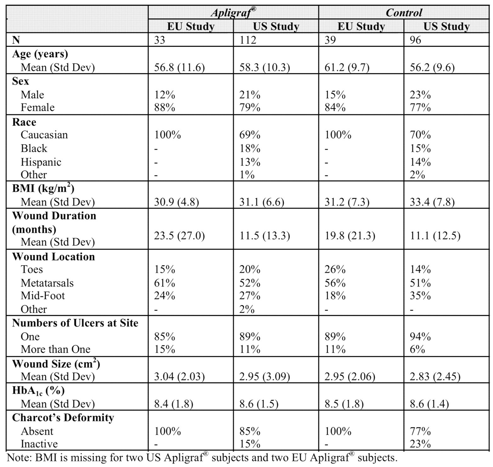
Table 3.
Primary and Secondary Efficacy Outcomes in US and EU Study Populations
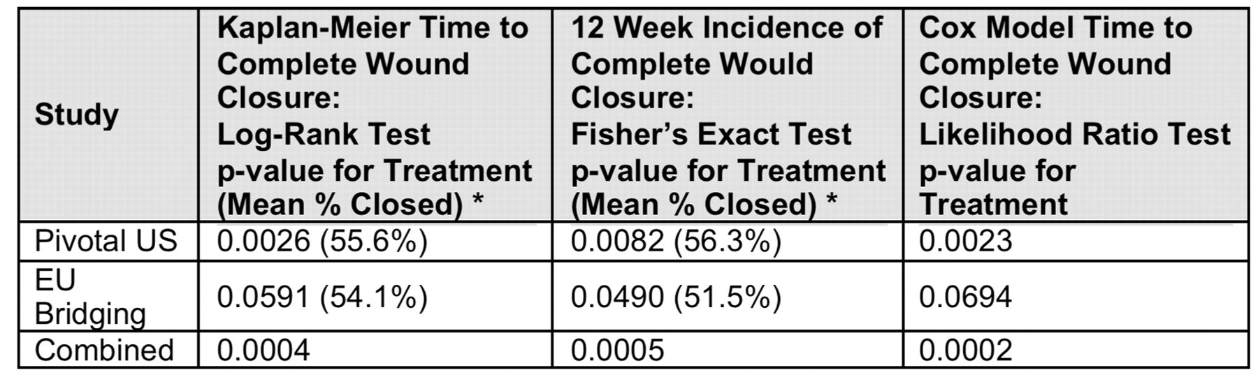
Table 4.
Ulcer Recurrence Rates through 6 Months in US & EU Study Populations

Data from both US and EU studies suggest that the use of Apligraf resulted in a shorter time to wound healing while demonstrating a significant increase in the proportion of healed target diabetic foot ulcers.
The overall results suggest that Apligraf, in combination with debridement, standard wound care, and off-loading should be considered in treating patients with non-healing neuropathic DFU.
US, EU and pooled data provide robust, consistent evidence for the use of Apligraf in geographically disparate DFU populations.
This work was supported by Organogenesis (Canton, MA).
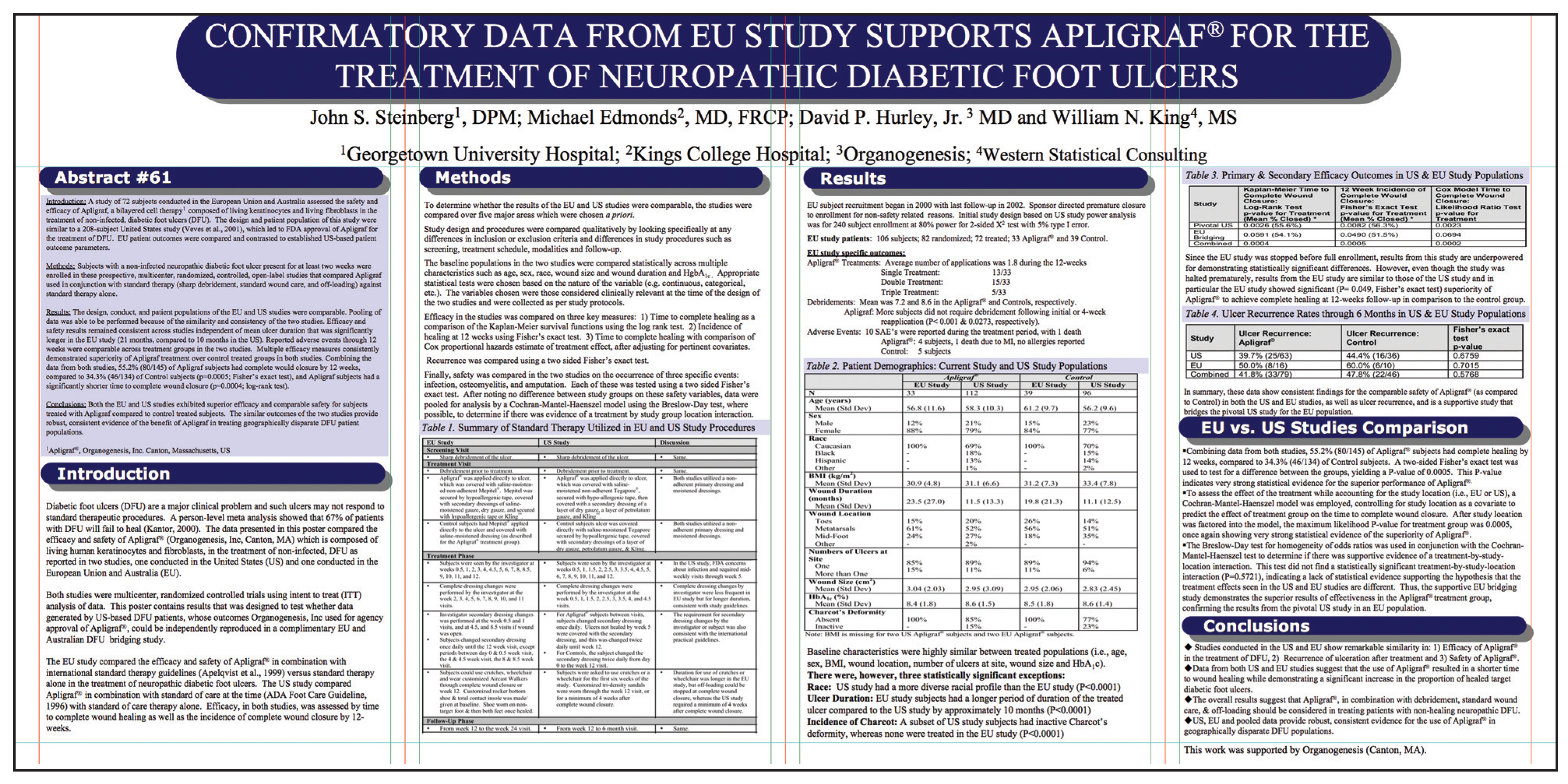
Poster image as presented at the 2009 APMA Annual Scientific Meeting, Toronto.
© 2010 American Podiatric Medical Association