


Abstract
Stress is increasingly recognized as a clinically relevant modifier of tissue repair, yet its effects on wound healing cannot be explained by a single endocrine or immune pathway. Psychological distress, pain, surgical trauma, social adversity, sleep disruption, and chronic disease may converge on the hypothalamic–pituitary–adrenal (HPA) axis, sympathetic–adrenomedullary signaling, local neurocutaneous pathways, immune-cell function, redox balance, angiogenesis, fibroblast activity, extracellular-matrix turnover, and health-related behaviors. This narrative review examines the biopsychosocial mechanisms through which acute and chronic stress can delay wound healing and discusses the implications for surgical wounds, experimental wounds, and chronic non-healing wounds. A structured narrative search of PubMed, Scopus, and Web of Science and citation tracking of key reviews and primary studies was used, with emphasis on peer-reviewed human, translational, and mechanistic evidence relevant to stress biology and wound repair. Human studies consistently demonstrate that caregiving stress, academic examination stress, hostile interpersonal interactions, elevated perceived stress, and preoperative distress are associated with slower healing or dysregulated local wound mediators. Mechanistically, excessive or poorly regulated glucocorticoid signaling can suppress early pro-inflammatory cytokines, impair leukocyte trafficking, fibroblast proliferation, collagen deposition, angiogenesis, and re-epithelialization, whereas sympathetic activation and catecholamines can alter vascular tone and adrenergic signaling within the wound microenvironment. Stress also influences macrophage function, neuropeptide signaling, oxidative stress, pain, sleep, nutrition, treatment adherence, and social behavior. In chronic wounds, these pathways become bidirectional: the wound itself generates pain, odor, exudate, functional impairment, and social isolation, which can sustain psychological distress and neuroimmune dysregulation. Psychological and behavioral interventions, including relaxation with guided imagery, mindfulness-based approaches, expressive writing, and social support, show promising but heterogeneous effects and should be viewed as adjuncts rather than substitutes for evidence-based wound care. Integrating stress assessment and biopsychosocial management into wound-care pathways may improve recovery, particularly in patients with high psychological burden or chronic comorbidity.
1. Introduction
Wounding can be described as disruption of the normal anatomical structure of a tissue with an associated loss of function. Depending on severity, the injury may be limited to disruption of the covering epithelium of the skin or mucosa, or it may extend to deeper structures such as the dermis and subcutaneous tissues, including muscle, tendons, bone, and parenchymal organs [1].
The mechanism of tissue injury may result from acute exposure to different forms of energy, including kinetic energy transmitted during road traffic accidents, falls, and firearm injuries, as well as chemical, thermal, electrical, or ionizing-radiation exposure when the body is unable to tolerate these extreme effects [2,3,4]. Tissue injury may also result from surgical intervention [5]. The severity of the resulting wound varies according to the injuring agent [2].
Injuries may range from minor to life-threatening. Minor injuries generally do not cause serious complications, whereas severe or infected wounds require medical assessment and treatment [5]. According to the triggering mechanism, wounds may result from blunt-force injury, sharp-force injury, or firearm injury [4].
Trauma that occurs suddenly because of violence or an accident produces both local tissue damage and potentially serious systemic consequences. The development of hypovolemic shock, blunt abdominal trauma with internal hemorrhage, or different neurological injuries can initiate comparable processes involving activation of the hypothalamic–pituitary–adrenal axis together with immunological and metabolic responses aimed at restoring homeostasis [6].
A wide range of local and systemic factors can influence healing. Local factors include hypoxia and vascular compromise of arterial, venous, or mixed origin. Systemic factors include age, sex hormones, diabetes, thyroid dysfunction, and obesity, as well as connective-tissue disease, immunodeficiency, and malignancy [4,5]. Additional influences include infection and biofilm formation, the presence of a foreign body, and medications such as corticosteroids, non-steroidal anti-inflammatory drugs, and chemotherapeutic agents [7]. Lifestyle-related factors, including alcohol consumption, smoking, nutrition, and aging, may also affect the healing response, as may stress [4,7,8,9].
1.1. Physiology of Wound Healing
Among living organisms, tissue injury induces a repair response that may proceed through two principal biological outcomes: regeneration and repair. Whereas activation of mitotic processes in affected epithelial, bone, and hepatic tissues characterizes regeneration, tissue or cellular repair is associated with scar formation [5].
Wound healing is an evolutionarily developed, complex, multifactorial, and precisely regulated process through which tissue defects are restored. Injury is the trigger that initiates the subsequent changes directed toward tissue recovery. The natural course of wound healing is dynamic and comprises four sequential, overlapping, and obligatory phases: hemostasis, inflammation, proliferation, and remodeling [10].
1.1.1. Hemostasis
Hemostasis is the phase in which blood loss caused by injury is limited through clot formation. The initial vasoconstriction is short-lived and is soon followed by vasodilation, which permits the influx of leukocytes and additional platelets. Both the extrinsic and intrinsic coagulation pathways are activated [11].
During hemostasis, a temporary fibrin wound matrix is generated [5]. This matrix also provides a temporary barrier that protects underlying tissues from further injury and infection [12]. Platelets and mast cells participate actively in this phase. Activated platelets release multiple signaling molecules that influence and modulate the function of other platelets, leukocytes, and endothelial cells. Subsequently, arachidonic-acid products, prostaglandins, leukotrienes, and thromboxanes mediate events that contribute to the following inflammatory phase. Mast cells release histamine and contribute to the vascular response through vasodilation, increased permeability, and inflammatory-cell infiltration [13].
1.1.2. Inflammation
Inflammation is an integral component of healing and is considered a driving process that activates and facilitates progression of tissue repair [14]. The inflammatory phase is directed toward elimination of invading microorganisms and removal of cellular debris [12].
Neutrophils and macrophages are active participants in this phase. They infiltrate the wound site and release pro-inflammatory mediators [5,12]. Damage-associated molecular patterns (DAMPs) released by necrotic cells and damaged tissue, together with pathogen-associated molecular patterns (PAMPs) derived from bacterial components, serve as initiating signals. These signals activate resident immune cells, including mast cells, Langerhans cells, T cells, and macrophages, as well as downstream inflammatory pathways. Their secreted pro-inflammatory cytokines and chemokines promote further cellular influx [15].
Neutrophils are among the first cells to infiltrate the wound and continue to migrate into the injured tissue during the first 48 h. This active recruitment occurs under the influence of chemotactic factors [13], including interleukin-1 (IL-1), tumor necrosis factor-alpha (TNF-α), and bacterial endotoxins such as lipopolysaccharide (LPS) [13,15].
Neutrophils constitute a first line of defense against infection and foreign antigens through phagocytosis, bactericidal activity, and debridement of devitalized tissue [5]. Their activity is mediated through released reactive oxygen species (ROS), antimicrobial peptides and extracellular traps, eicosanoids, and proteolytic enzymes. Neutrophils have different fates: some remain within the inflammatory exudate, whereas others are removed from the tissue by macrophage efferocytosis, apoptosis, necrosis, or phagocytosis [15].
Monocytes and macrophages are the next major cellular participants. As key components of the immune system, they are actively involved in tissue recovery after injury. Circulating monocytes are recruited to the wound in response to chemotactic signals and enter the injured tissue, where they may transiently persist and produce pro-inflammatory and angiogenic factors [5]. Subsequently, monocytes differentiate into macrophages [15]. Both monocytes and macrophages may exert pro-inflammatory or anti-inflammatory and reparative effects depending on the local wound microenvironment [5].
Macrophages are major effector cells in the wound-healing process [15]. Traditionally, macrophage activation has been described using the M1/M2 classification. However, this represents a simplified conceptual framework, as macrophages in vivo exhibit substantial phenotypic plasticity and exist across a continuum of activation states rather than as two strictly separated populations. Within this simplified framework, classically activated M1-like macrophages predominate during the early inflammatory phase, whereas M2-like reparative phenotypes become increasingly important during the transition toward proliferation and tissue repair [16].
Differentiation toward an M1-like phenotype is promoted by signals associated with pathogens and tissue damage, including pathogen-associated molecular patterns and damage-associated molecular patterns. These macrophages predominate during the early stages of wound repair and contribute to antimicrobial defense and removal of damaged tissue. M1-like macrophages produce reactive oxygen species (ROS) and pro-inflammatory cytokines, including IL-1, IL-6, IL-12, and TNF-α, and may also release growth factors such as vascular endothelial growth factor (VEGF) and platelet-derived growth factor (PDGF). They demonstrate high phagocytic activity, facilitate the clearance of pathogens and devitalized tissue, and participate in efferocytosis [15,16].
Successful clearance of bacteria and tissue debris remains an important determinant of the duration of inflammation and the normal progression of wound healing [13].
As the inflammatory response resolves, the macrophage population progressively shifts toward more reparative M2-like phenotypes, contributing to the transition from inflammation to proliferation and supporting further wound healing. Th2 cytokines, such as IL-4 and IL-13, are important signals promoting alternative macrophage activation [16].
M2-like macrophages perform multiple reparative functions. They can produce anti-inflammatory and pro-resolving mediators, including IL-10 and transforming growth factor-beta (TGF-β), which contribute to suppression and resolution of excessive inflammatory activity. In addition, reparative macrophages promote wound healing through the production of growth factors. VEGF contributes to neoangiogenesis; TGF-β promotes fibroblast differentiation into myofibroblasts and stimulates extracellular-matrix synthesis and scar formation; and epidermal growth factor (EGF) contributes to restoration of the epidermal layer [17]. Through these functions, reparative macrophage phenotypes participate in angiogenesis, fibroblast activation, extracellular-matrix deposition, re-epithelialization, and tissue remodeling.
Lymphocytes are attracted to wound tissue at approximately 72 h after injury [12,13]. Different T-cell populations are involved in the regulation of wound healing, including Th1, Th2 and Th17 cells, cytotoxic effector T cells, regulatory T cells, and memory CD8+ T cells. These lymphocyte subsets can influence inflammatory regulation, immune-cell activity, and the transition toward tissue repair and the proliferative phase [18].
1.1.3. Proliferation
The proliferative phase is directed toward restoration of the tissue defect and reduction in the wounded area through the coordinated formation of new tissue. In healing cutaneous wounds, this phase may extend for approximately three weeks, although its duration depends on wound size, severity, local conditions, and the overall healing response [10,19].
Proliferation is a complex process mediated by the migration and activity of endothelial cells, fibroblasts, myofibroblasts, and epithelial cells. Angiogenesis, formation of granulation tissue, collagen deposition, epithelialization, and wound contraction occur simultaneously during this phase [13].
Angiogenesis is the formation of new blood vessels from microvascular endothelial cells and represents a response to the increased metabolic requirements of proliferating tissue [15]. Neoangiogenesis is mediated by signaling molecules including vascular endothelial growth factor (VEGF) and matrix metalloproteinases (MMPs) [13].
Stimulated fibroblasts participate in extracellular-matrix (ECM) production. The resulting granulation tissue contains hyaluronan, fibronectin, proteoglycans, and collagen, predominantly type III collagen. Progressive matrix deposition promotes the fibroblast-to-myofibroblast transition. Myofibroblasts contribute to wound contraction and also support angiogenesis [13].
Epithelialization is another major event during the proliferative phase. Epithelial cells at the wound margins acquire a proliferative phenotype, migrate, and move across the wound bed, cover denuded surfaces, and restore the epithelial barrier [13]. Cellular migration is regulated in part by matrix metalloproteinases, particularly MMP-1 and MMP-9 [15]. In larger wound defects, substantial wound contraction also contributes to reduction in the wound area [13].
1.1.4. Remodeling
The final stage of wound healing is the longest and may continue for months to years [13]. During this phase, granulation tissue progressively matures into permanent scar tissue, and the extracellular matrix undergoes continuous degradation, resynthesis, and structural reorganization. These processes aim to increase the mechanical stability and tensile strength of the repaired tissue [10,19].
During remodeling, the collagen content and organization of the wound change substantially. Active collagen synthesis continues during the early period of scar formation, after which the type III reticular collagen deposited predominantly during the proliferative phase is progressively replaced by stronger type I fibrillar collagen as the scar matures [10].
At the same time, the cellularity and vascularity of the healing tissue decrease. Many fibroblasts, inflammatory cells, and newly formed blood vessels disappear from the wound through apoptosis, regression, migration, and other mechanisms as the metabolically active granulation tissue develops into a less cellular and less vascular mature scar [19]. Components of the provisional extracellular matrix, including hyaluronan and fibronectin, are also progressively degraded and remodeled through cellular and matrix-metalloproteinase activity.
Together, collagen replacement, fiber reorganization, increased cross-linking, and progressive extracellular-matrix remodeling improve the mechanical strength and functional stability of the newly formed tissue. Nevertheless, scar tissue does not completely regain the tensile strength of uninjured skin; mature scar tissue may reach approximately 80% of the tensile strength of normal skin [10].
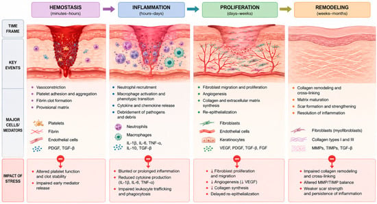
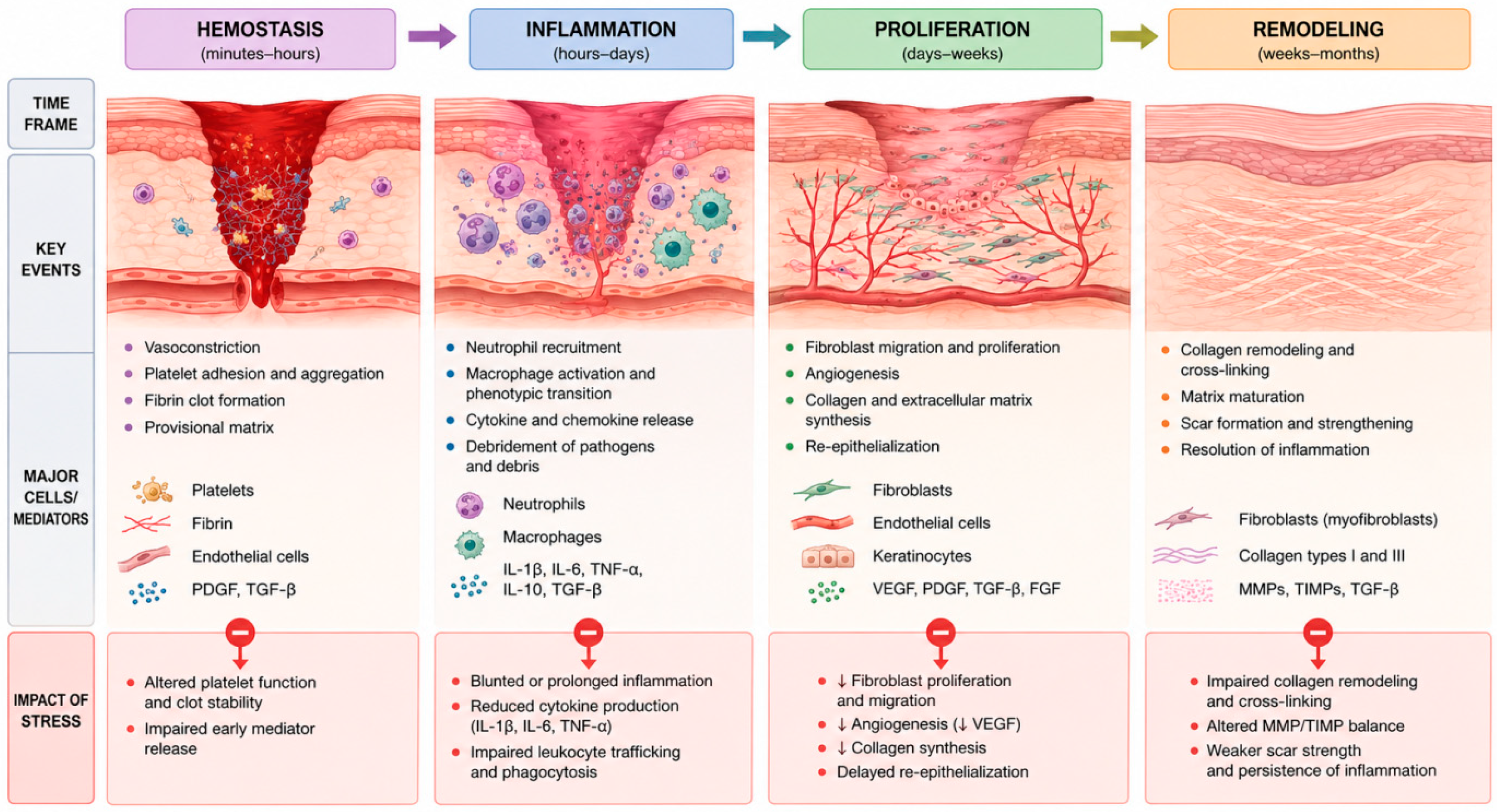
The principal phases of physiological wound healing and the major sites at which stress-related mechanisms may interfere with tissue repair are summarized in Figure 1.
Figure 1.
Phases of wound healing and major sites of stress-related interference. Normal wound repair progresses through overlapping phases of hemostasis, inflammation, proliferation, and remodeling. Stress-related neuroendocrine and immune alterations may interfere with vascular responses, inflammatory signaling, leukocyte function, angiogenesis, fibroblast activity, collagen synthesis, re-epithelialization, and extracellular-matrix remodeling. Note: This figure was generated with the assistance of ChatGPT (GPT-5.6 Sol; OpenAI, San Francisco, CA, USA) and subsequently reviewed and edited by the authors.
1.2. Wounds as a “Silent” Epidemic in Society
The natural course of wound healing is most characteristic of acute wounds [7]. Physiological healing commonly occurs over approximately 4–6 weeks, although maturation continues beyond this period [11].
Any influence that disrupts the finely regulated balance of events across the healing phases can result in delayed repair and contribute to the development of non-healing chronic wounds, thereby increasing both psychological and physiological burden for patients [12,20].
Contemporary living conditions expose individuals to many factors capable of modifying physiological processes, including normal wound healing. Several highly prevalent and socially significant diseases, whose global burden continues to increase, contribute substantially to chronic-wound development. These include diabetes mellitus, obesity, cardiovascular pathology, and stress and distress together with related pathology [21,22,23]. Population aging further contributes to this growing challenge, as age-related changes in tissue repair, immune function, vascular integrity, and the increasing prevalence of chronic comorbidities may collectively predispose older individuals to impaired wound healing and chronic wound development [24].
Economic conditions, the organization of health systems, access to effective healthcare, and impaired living conditions and quality of life are also determining factors [7,23]. Collectively, these factors contribute to the high prevalence and substantial clinical impact of chronic wounds, which have been described by some authors as a “silent” epidemic [7].
Over recent decades, human populations have been exposed to persistent and increasingly intense stress. Stress and distress, together with stress-related disorders, can influence lifestyle, the capacity to adapt to changing environmental conditions, and the effective engagement of regulatory and compensatory mechanisms. Their high global prevalence places stress-related health problems among the major population-level challenges with epidemic characteristics [22,23].
2. Materials and Methods
2.1. Review Design
This article was designed as a narrative review aimed at providing an integrative synthesis of the current evidence regarding the effects of psychological and physiological stress on wound healing. Particular emphasis was placed on the biopsychosocial mechanisms through which stress may interfere with the normal sequence of tissue repair, including neuroendocrine activation, immune and inflammatory dysregulation, altered angiogenesis, extracellular matrix remodeling, oxidative stress, pain, sleep disturbances, health-related behaviors, and social factors.
Given the broad and interdisciplinary nature of the topic, a narrative review methodology was considered appropriate for integrating evidence derived from human observational and experimental studies, clinical investigations, systematic reviews and meta-analyses, as well as relevant animal and in vitro studies addressing specific mechanistic pathways. The review was developed with reference to the methodological principles for high-quality narrative reviews described by the Scale for the Assessment of Narrative Review Articles (SANRA), particularly with regard to the justification of the review, formulation of its objectives, transparency of the literature search, appropriate referencing, and critical interpretation of the available evidence [25].
2.2. Literature Search Strategy
A focused literature search was conducted in PubMed, Scopus, and Web of Science between 15 and 20 July 2026 to identify peer-reviewed publications relevant to the relationship between stress and wound healing. The search included articles available in the databases up to 20 July 2026. No strict lower publication-date limit was applied because several seminal experimental and clinical studies investigating psychophysiological stress and wound repair were published before the most recent two decades and remain fundamental to the development of the current psychoneuroimmunological model of wound healing. Nevertheless, preference was given to more recent publications when contemporary evidence of comparable methodological quality was available.
The search strategy combined terms related to wound repair with terms describing psychological stress, neuroendocrine regulation, inflammation, and psychosocial determinants. Search terms included combinations of the following keywords and phrases: “stress”, “psychological stress”, “chronic stress”, “acute stress”, “distress”, “wound healing”, “wound repair”, “tissue repair”, “cutaneous wound healing”, “chronic wounds”, “surgical wounds”, “psychoneuroimmunology”, “hypothalamic–pituitary–adrenal axis”, “HPA axis”, “cortisol”, “glucocorticoids”, “catecholamines”, “sympathetic nervous system”, “inflammation”, “cytokines”, “IL-1β”, “IL-6”, “TNF-α”, “macrophages”, “angiogenesis”, “VEGF”, “fibroblasts”, “collagen”, “extracellular matrix”, “matrix metalloproteinases”, “oxidative stress”, “reactive oxygen species”, “re-epithelialization”, “neuropeptides”, “substance P”, “pain”, “sleep”, “social support”, “anxiety”, “depression”, and “biopsychosocial”.
Representative Boolean combinations included:
(“stress” OR “psychological stress” OR “chronic stress” OR “distress”) AND (“wound healing” OR “wound repair” OR “tissue repair”)
and
(“wound healing” OR “chronic wound*” OR “surgical wound*”) AND (“cortisol” OR “glucocorticoid*” OR “catecholamine*” OR “HPA axis” OR “inflammation” OR “cytokine*” OR “psychoneuroimmunology”).
Additional focused searches were performed for individual mechanisms and clinically relevant modifiers identified during preparation of the review, including macrophage polarization, angiogenesis, fibroblast activity, collagen deposition, extracellular matrix remodeling, oxidative stress, neuropeptide signaling, wound-related pain, sleep disturbance, depression, anxiety, and social support. Reference lists of key reviews, meta-analyses, and highly relevant primary studies were also manually screened to identify additional publications not retrieved during the initial database searches.
2.3. Study Selection and Evidence Synthesis
Publications were eligible if they provided clinically or biologically relevant evidence on the relationship between stress and wound healing across acute, surgical, traumatic, and chronic wounds. Human studies were prioritized, while animal and in vitro studies were included when they clarified underlying physiological or pathophysiological processes.
Eligible evidence included clinical and experimental studies, observational and controlled investigations, randomized trials, systematic reviews, meta-analyses, and authoritative narrative reviews. Priority was given to studies assessing wound-healing outcomes or stress-related biomarkers directly linked to tissue repair, including inflammatory activity, immune function, angiogenesis, fibroblast activity, collagen and extracellular matrix remodeling, infection risk, and postoperative recovery. Therapeutic glucocorticoid exposure was identified as a potential source of confounding when interpreting the clinical evidence linking psychological stress to wound-healing outcomes.
Studies were excluded if they lacked a relevant connection between stress and tissue repair, focused solely on wound management, or provided insufficient methodological detail. Non-peer-reviewed material, duplicate publications, and sources that could not be adequately evaluated were also excluded. English-language publications were preferentially considered.
Because of substantial heterogeneity in wound type, study populations, stress exposure, biomarkers, and healing outcomes, the evidence was synthesized narratively rather than by meta-analysis. When multiple types of evidence were available for the same question, greater interpretative weight was given to systematic reviews and meta-analyses, followed by randomized or controlled clinical studies, prospective observational studies, and other human investigations. Seminal older studies were retained when they provided foundational evidence that remained directly relevant to the mechanisms under discussion. Animal and in vitro studies were used primarily to support biological plausibility or to clarify mechanisms that could not be adequately investigated in humans.
When findings across studies were inconsistent, differences in study design, population characteristics, wound type, duration and nature of stress exposure, biological compartment assessed, timing of measurements, and outcome definitions were considered before drawing conclusions. Greater emphasis was placed on findings that were reproduced across independent studies or supported by convergent clinical, experimental, and mechanistic evidence. Isolated or conflicting findings were interpreted cautiously and were not treated as definitive evidence.
4. Human and Translational Evidence Linking Stress to Delayed Healing
Evidence from different experimental and clinical settings generally supports an association between psychological stress and impaired wound healing, although substantial methodological heterogeneity exists across studies. In a seminal human study, caregivers of relatives with dementia showed substantially delayed healing of standardized punch-biopsy wounds compared with matched controls, providing early evidence that chronic real-life stress can impair tissue repair [38]. Similarly, a within-person study of dental students found that oral mucosal wounds produced during an examination period healed approximately 40% more slowly than wounds created during vacation, accompanied by markedly reduced IL-1β mRNA expression at the wound site [39].
Subsequent studies further supported a biological link between psychological stress and impaired wound healing. Higher perceived stress was associated with reduced local production of IL-1α and IL-8 in experimentally induced blister wounds and with increased salivary cortisol [40]. In healthy men with standardized punch-biopsy wounds, greater perceived stress and higher morning cortisol responses were also associated with slower healing, supporting a role for HPA-axis activation beyond behavioral influences alone [41].
Interpersonal stress produces similar effects. In a crossover study of married couples, blister wounds healed more slowly after a marital-conflict interaction than after a supportive interaction; local wound production of IL-6, TNF-α, and IL-1β was lower after conflict, and couples characterized by consistently hostile behavior healed at only about 60% of the rate of low-hostility couples [42].
Clinical studies have extended these observations to surgery and chronic wounds. Greater preoperative stress in patients undergoing inguinal-hernia repair predicted lower IL-1 concentrations in postoperative wound fluid, indicating impaired early local repair signaling [43]. In patients with naturally occurring chronic wounds, anxiety and depressive symptoms have been associated with delayed healing, although chronic-wound studies are more heterogeneous and more vulnerable to confounding by disease severity, pain, mobility, vascular status, and treatment burden [44,45]. Therapeutic corticosteroid use may independently affect wound healing and should therefore be considered when interpreting associations between psychological stress and delayed tissue repair. This is particularly relevant in surgical patients and individuals with chronic wounds, where concurrent medication use and underlying comorbidities may complicate causal interpretation [46,47].
A meta-analysis of human studies reported a moderate association between psychological stress and delayed wound healing (r ≈ −0.42) [48]. Subsequent reviews confirmed this relationship, although evidence remains stronger for experimental and surgical wounds than for chronic wounds [28,33]. Representative human studies supporting this association are summarized in Table 1.
Table 1.
Representative human studies investigating psychosocial stress and wound-healing outcomes: methodological characteristics and principal findings.
Although these studies collectively support an association between psychological stress and impaired wound healing, their findings should be interpreted in the context of substantial methodological heterogeneity. The investigated populations range from healthy volunteers and students to caregivers exposed to chronic stress, surgical patients, and individuals with chronic wounds. Likewise, the wound models include oral mucosal wounds, punch biopsies, suction blisters, surgical wounds, and naturally occurring chronic wounds, while the assessed outcomes vary from time to wound closure and re-epithelialization to local cytokine concentrations, cortisol responses, and other biological markers of repair. Stress exposure and assessment also differ considerably across studies, encompassing chronic caregiving stress, academic examination stress, interpersonal conflict, perceived stress, and preoperative distress. Consequently, direct quantitative comparison between individual studies is limited, and the magnitude of the observed effects should not be assumed to be equivalent across wound types or clinical populations. Nevertheless, the recurrence of broadly similar stress-related impairments across distinct experimental and clinical contexts strengthens the overall qualitative evidence that psychological stress can act as a relevant modifier of tissue repair.
5. Neuroendocrine Mechanisms
5.1. HPA-Axis Activation and Glucocorticoid Signaling
Perception of a stressor activates corticotropin-releasing hormone (CRH) neurons in the hypothalamus, followed by pituitary adrenocorticotropic hormone (ACTH) secretion and adrenal glucocorticoid release. Cortisol is essential for metabolic adaptation and prevents uncontrolled inflammation; however, sustained or inappropriately timed glucocorticoid exposure can delay repair. Experimental and clinical evidence indicates that glucocorticoids can suppress inflammatory-cell recruitment and cytokine production, reduce fibroblast proliferation, inhibit collagen synthesis, impair angiogenesis, delay re-epithelialization, and weaken wound contraction [34,49].
The timing of stress exposure is critical. Early wound healing requires a controlled inflammatory response involving cytokines, chemokines, and growth factors. Stress-related cortisol elevation may suppress this response, reducing immune-cell recruitment and subsequent progression to the proliferative phase, as supported by human wound-fluid and blister studies [40,43]. Experimental evidence further implicates glucocorticoid signaling, including local activation by 11β-hydroxysteroid dehydrogenase type 1 (11β-HSD1), as inhibition of these pathways can attenuate stress-induced delays in healing [50].
Chronic stress does not necessarily result in sustained hypercortisolemia, but may instead alter diurnal cortisol rhythms, feedback sensitivity, or glucocorticoid responsiveness [51]. In wound healing, both excessive glucocorticoid signaling, which may suppress early inflammation, and reduced glucocorticoid control of persistent inflammation can impair repair. This dual dysregulation may explain seemingly contradictory findings in stress immunology [35,52]. An important distinction should be made between endogenous stress-related glucocorticoids and therapeutically administered corticosteroids. Endogenous cortisol secretion is regulated by the HPA axis and follows circadian rhythms, with transient elevations supporting physiological adaptation, whereas prolonged or dysregulated exposure may impair inflammatory signaling and tissue repair. Local cortisol availability is additionally modulated by cutaneous 11β-HSD1 activity [53,54]. In contrast, the effects of exogenous glucocorticoids depend on dose, duration, route of administration, and treatment timing. Although prolonged systemic corticosteroid therapy may impair fibroblast activity, collagen synthesis, and wound healing, short-term administration does not necessarily produce comparable effects [46,47].
5.2. Sympathetic–Adrenomedullary Activation and Catecholamines
Stress also activates the sympathetic nervous system and adrenal medulla, increasing norepinephrine and epinephrine. Catecholamines influence healing through vascular, immune, and cellular mechanisms. Sympathetic vasoconstriction can reduce cutaneous blood flow and oxygen delivery, particularly when baseline perfusion is already compromised by peripheral arterial disease, smoking, edema, or diabetes. Adequate oxygen tension is required for oxidative bacterial killing, collagen hydroxylation, fibroblast function, and angiogenesis; therefore, stress-related alterations in perfusion can amplify other local causes of hypoxia [30,55].
Adrenergic receptors are expressed on keratinocytes, fibroblasts, endothelial cells, and immune cells. In restraint-stressed mice, blockade of α-adrenergic receptors attenuated impairment in wound closure and contraction, whereas β-adrenergic blockade did not, indicating an important α-adrenergic contribution in that model [56]. Catecholamine effects are context-dependent, however, and acute adrenergic activation should not be assumed to be uniformly detrimental. The relevant problem in chronic stress is sustained or repeated signaling that distorts the coordinated timing of perfusion, inflammation, and repair [57].
5.3. The Local Cutaneous Stress System
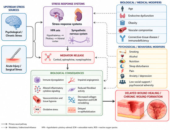
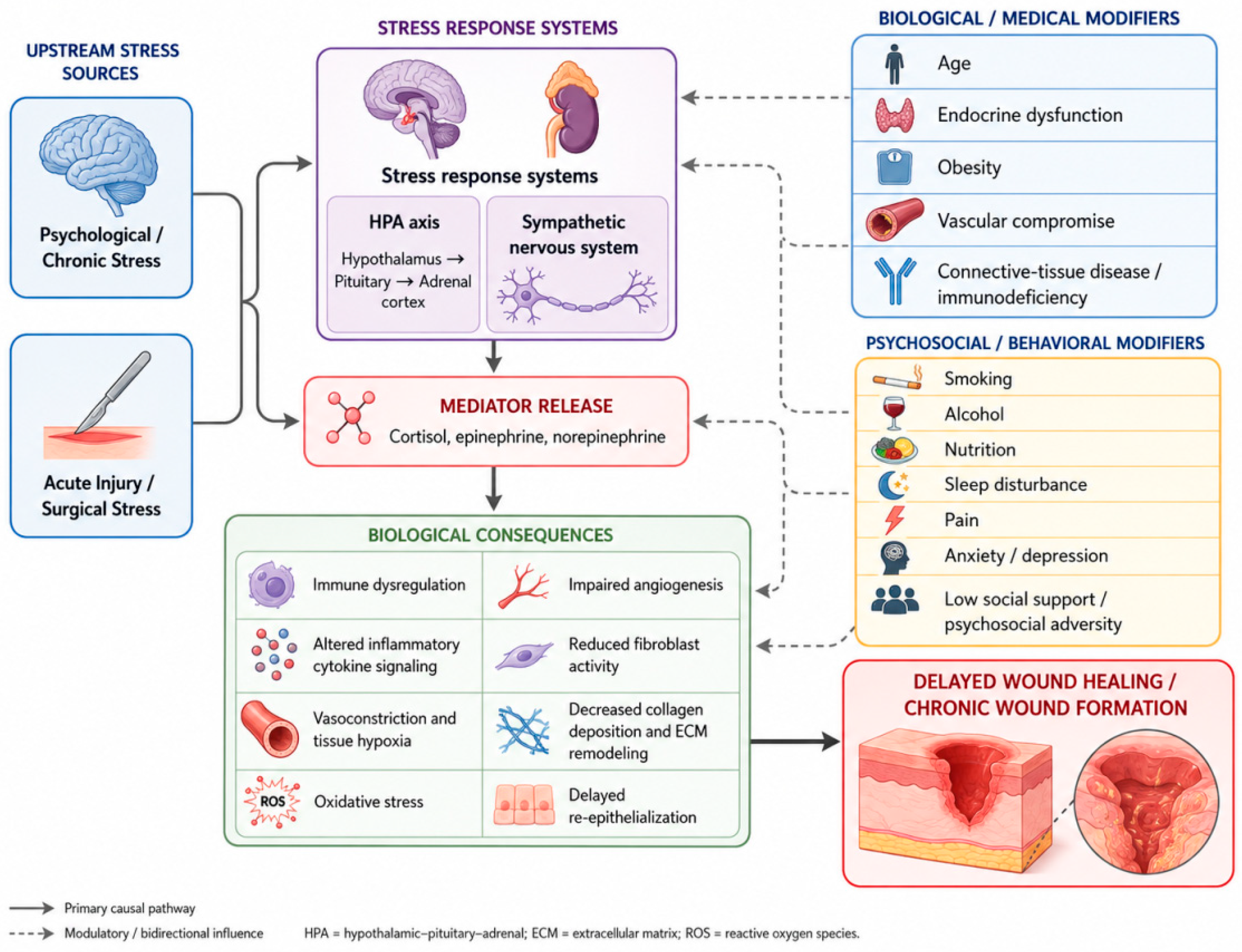
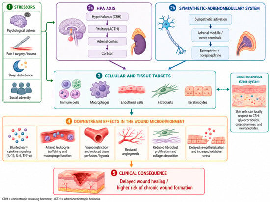
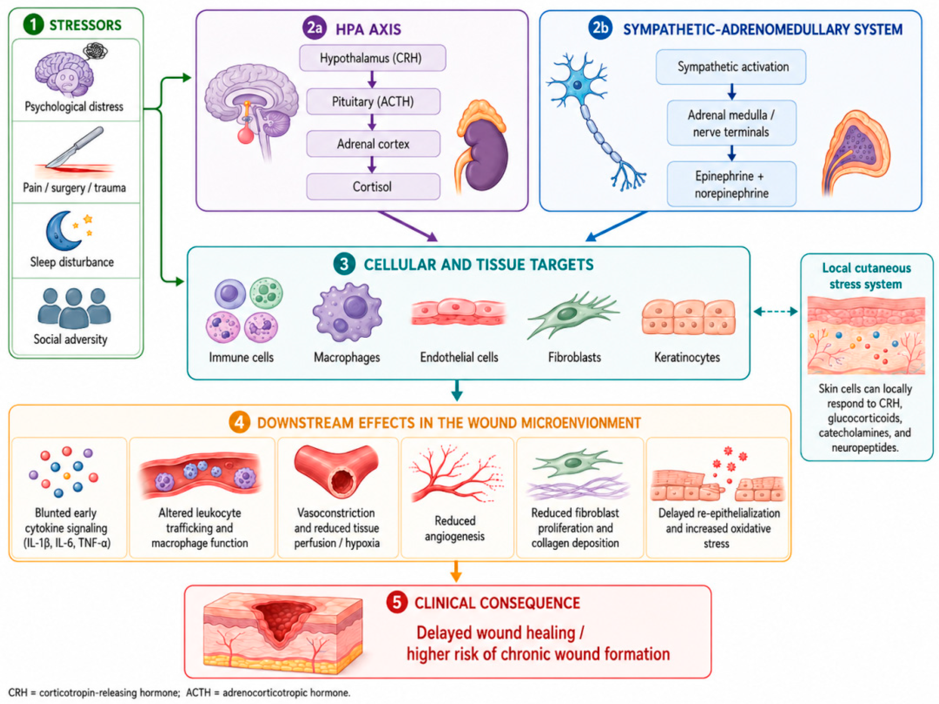
The skin is not merely a passive target of systemic stress hormones. Keratinocytes, fibroblasts, immune cells, endothelial cells, adnexal structures, and peripheral nerves participate in a local neuroendocrine network capable of producing and responding to CRH, ACTH-related peptides, glucocorticoids, catecholamines, neurotrophins, and neuropeptides. This local “brain–skin” or neurocutaneous axis allows psychological and systemic stress signals to be translated directly into changes in barrier function, vascular responses, inflammation, and repair [58,59]. The principal neuroendocrine pathways linking stress to impaired wound repair are summarized in Figure 3.
Figure 3.
Neuroendocrine pathways linking stress to impaired wound repair. Psychological and physiological stressors activate the hypothalamic–pituitary–adrenal (HPA) axis and sympathetic–adrenomedullary system, resulting in glucocorticoid and catecholamine signaling. These systemic pathways, together with the local cutaneous stress system, influence immune cells, macrophages, endothelial cells, fibroblasts, and keratinocytes, thereby altering inflammatory signaling, leukocyte trafficking, tissue perfusion, angiogenesis, collagen deposition, re-epithelialization, and oxidative balance. Note: This figure was generated with the assistance of ChatGPT (GPT-5.6 Sol; OpenAI, San Francisco, CA, USA) and subsequently reviewed and edited by the authors.
6. Immune and Inflammatory Dysregulation
6.1. Cytokine Timing Rather than Simple “Suppression”
A central insight from psychoneuroimmunology is that successful healing requires correctly timed inflammation rather than maximal inflammation. Stress can reduce local production of IL-1α, IL-1β, IL-6, IL-8, and TNF-α during the early wound response, delaying neutrophil recruitment and downstream activation of fibroblasts, keratinocytes, and endothelial cells [39,40,42]. Yet chronic systemic stress can also promote elevated circulating inflammatory markers. The apparent contradiction is explained by compartment, timing, and receptor sensitivity, whereby early local immune activity at the wound site is suppressed despite a concurrent systemic pro-inflammatory state [60].
Failure of early inflammatory signaling can postpone the transition to proliferation [54]. Conversely, failure to terminate inflammation after pathogen and debris clearance can prolong protease activity, oxidative stress, and tissue destruction [61]. Chronic non-healing wounds often exhibit this persistent inflammatory state, with prolonged neutrophil activity, macrophage dysfunction, excessive proteases, microbial burden, and cellular senescence [20]. Stress may aggravate either pattern depending on wound type, comorbidities, and healing stage.
6.2. Neutrophils, Macrophages, and Antigen-Presenting Cells
Stress-induced changes occur before wounding as well as after injury. Pre-existing immunological state of the skin may determine the quality of the later repair response. In human skin sampled before experimental wounding, higher stress has been associated with fewer macrophages and lower immune-cell activation; lower activation and fewer Langerhans cells were subsequently associated with slower healing [62].
Macrophage plasticity is central to wound healing. Early pro-inflammatory macrophages support debridement and antimicrobial defense, whereas later reparative phenotypes promote angiogenesis, fibroplasia, and resolution [63]. Experimental and mechanistic evidence suggests that stress-related neuroendocrine signaling can influence monocyte trafficking and immune-cell function [64]. However, direct evidence demonstrating stress-induced alterations in macrophage phenotype transitions within human wounds remains limited. The proposed contribution of impaired transition toward reparative macrophage states to stress-related healing impairment is therefore based predominantly on experimental stress models and on broader mechanistic evidence regarding macrophage plasticity in wound healing [63,64,65].
The increasing understanding of macrophage plasticity has also created opportunities for therapeutic modulation of the wound inflammatory microenvironment. Experimental biomaterial-based approaches have sought to shift persistent pro-inflammatory macrophage activity toward more reparative phenotypes by targeting intracellular signaling pathways involved in macrophage activation. For example, Wang et al. demonstrated that a chlorogenic-acid-loaded, pH/ROS-responsive nanocomposite hydrogel modulated macrophage phenotype through NF-κB/JAK-STAT-related signaling, while simultaneously providing antioxidant, antibacterial, and pro-angiogenic effects in diabetic wound models. Such findings support the concept that macrophage-directed interventions may help restore the transition from persistent inflammation toward tissue repair, although their clinical efficacy and optimal timing remain to be established [66].
6.3. Host Defense, Infection, and Biofilm
Psychological stress can impair innate and adaptive immune function and may reduce resistance to infection. In wounds, impaired neutrophil function, altered cytokine production, and reduced tissue oxygenation can compromise bacterial clearance. Infection and biofilm then become additional biological stressors that sustain inflammation, increase protease activity, and reinforce delayed healing. This creates a self-perpetuating loop in which stress impairs antimicrobial defense, microbial persistence intensifies inflammation and pain, and the worsening wound increases psychological distress [67,68,69].
7. Angiogenesis, Fibroblast Function, ECM Remodeling, and Re-Epithelialization
The proliferative phase is particularly vulnerable to stress because it depends on coordinated activity of endothelial cells, fibroblasts, myofibroblasts, and keratinocytes. Glucocorticoids can inhibit fibroblast proliferation and collagen production, whereas altered adrenergic signaling can influence cell migration and vascular responses [70]. Reduced expression or effectiveness of VEGF and other growth factors can limit angiogenesis, especially in hypoxic or metabolically compromised tissues [71]. Stress-related suppression of early cytokines also has downstream effects because cytokines and growth factors form interdependent signaling networks that initiate granulation tissue formation [37].
Fibroblasts require adequate oxygen, nutrients, growth-factor signaling, and a permissive inflammatory microenvironment to synthesize ECM. Prolonged glucocorticoid exposure decreases collagen synthesis and can interfere with the balance of MMPs and tissue inhibitors of metalloproteinases. Excessive MMP activity degrades ECM and growth factors; insufficient remodeling, conversely, produces disorganized matrix [72]. In chronic wounds, protease imbalance is a hallmark of a microenvironment that destroys newly formed matrix faster than it can be replaced [20].
Re-epithelialization depends on keratinocyte activation, proliferation, and migration across a viable matrix [73]. Experimental and mechanistic evidence further suggests that endocrine stress mediators and local adrenergic and neuropeptide signaling may influence keratinocyte behavior and re-epithelialization [58,59,70]. However, direct human evidence linking psychological stress to specific keratinocyte responses during wound repair remains limited, and these mechanisms should therefore be interpreted primarily as biologically plausible pathways rather than as definitively established clinical effects. Recent reviews also emphasize the potential influence of stress on neocollagenesis and scar maturation, suggesting that psychobiological context may affect not only time to closure but also later scar quality [69].
8. Oxidative Stress and Stress-Impaired Regeneration
ROS are necessary signaling molecules during normal wound repair. At controlled concentrations they support platelet activation, antimicrobial defense, angiogenic signaling, and cell migration. When ROS production exceeds antioxidant capacity, however, oxidative stress damages lipids, proteins, DNA, mitochondria, and ECM and sustains inflammatory signaling. Diabetes, obesity, smoking, ischemia, aging, infection, and chronic stress can all increase oxidative burden [74].
Stress-induced sympathetic activation, glucocorticoid signaling, sleep disruption, and inflammatory dysregulation may all impact mitochondrial and redox pathways. Recent experimental work in chronic social stress has demonstrated delayed excisional-wound healing accompanied by reduced cytoprotective and regenerative markers and increased markers of apoptosis and impaired autophagy, supporting a link between persistent psychosocial stress and altered cellular resilience [75]. Although animal findings cannot be directly extrapolated to human clinical wounds, they identify testable molecular targets for future translational research (Table 2).
Table 2.
Mechanistic pathways through which stress may impair wound repair.
Mitochondrial dysfunction and impaired autophagic regulation are also emerging as potential therapeutic targets in chronic and diabetic wounds. Recent preclinical work in diabetic foot ulcer models has demonstrated that interventions targeting interconnected complement, mitochondrial, and autophagy-related pathways may restore mitochondrial membrane potential and ATP production, enhance autophagic activity, reduce excessive oxidative stress, and improve tissue regeneration. In particular, an engineered exosome-based hydrogel system acting through complement 1q binding protein-related signaling was reported to modulate the complement pathway while improving mitochondrial function and autophagy, resulting in accelerated wound closure in experimental diabetic foot ulcers. These findings illustrate the therapeutic potential of targeting redox and mitochondrial homeostasis together with immune regulation, although translation to clinical wound care will require further validation [76].
10. Comorbidities and Populations at Particular Risk
Stress-related effects are likely to be most clinically important when they occur in patients who already have limited physiological reserve. Diabetes combines hyperglycemia, neuropathy, impaired immunity, microvascular and macrovascular disease, oxidative stress, and altered growth-factor signaling. Obesity contributes to chronic low-grade inflammation and mechanical stress. Cardiovascular and peripheral vascular disease reduce perfusion. Aging is associated with structural skin changes, fibroblast senescence, altered inflammation, and slower progression through healing phases [24]. In these contexts, even a moderate additional stress-related impairment may cross a threshold from delayed healing to chronic non-healing.
The same principle applies to patients receiving systemic corticosteroids, chemotherapy, radiotherapy, or immunosuppressive treatment and to those with malnutrition, smoking, alcohol misuse, or severe sleep disturbance. These factors can interact multiplicatively rather than additively. Consequently, clinical assessment should identify clusters of risk rather than focusing on psychological stress in isolation [83,84,85].
12. Research Gaps and Future Directions
First, chronic-wound research should move beyond cross-sectional associations. Prospective studies should repeatedly measure perceived stress, depression, anxiety, pain, sleep, social isolation, wound area, perfusion, microbial burden, and treatment adherence together with endocrine and immune biomarkers. This would clarify whether stress predicts healing independently of wound severity and whether changes in stress precede changes in repair.
Second, stress should be characterized multidimensionally. Single cortisol samples are insufficient because HPA-axis function includes diurnal slope, awakening response, reactivity, recovery, and receptor sensitivity. Autonomic markers, including heart-rate variability, may complement endocrine measures by capturing regulatory flexibility, although their specific predictive value for wound healing remains to be established in adequately designed clinical studies.
Third, studies should integrate wound-fluid cytokines, proteases, growth factors, immune-cell phenotyping, tissue oxygenation, neuropeptides, redox markers, and omics approaches. Recent experimental work points to autophagy, apoptosis, cytoprotection, neuroregeneration, and nerve–immune interactions as underexplored stress-sensitive pathways [59,75].
Fourth, future studies should test moderators including age, sex and sex hormones, diabetes, obesity, vascular disease, smoking, social deprivation, psychiatric symptoms, and wound type. A stress effect observed in a healthy punch-biopsy model may not have the same magnitude or mechanism in an ischemic diabetic foot ulcer. Future clinical studies should also help define which patient groups derive the greatest benefit from routine psychosocial assessment. Particular attention may be warranted in patients with chronic or non-healing wounds, diabetic foot ulcers, vascular or ischemic wounds, persistent wound-related pain, recurrent infection, major medical comorbidity, prolonged treatment courses, or substantial functional impairment. Patients reporting sleep disturbance, anxiety or depressive symptoms, social isolation, poor treatment adherence, or high perceived stress may also represent priority groups for targeted assessment. Prospective studies should determine whether systematic screening for psychological stress, sleep problems, pain, and emotional distress in these populations improves treatment adherence, quality of life, and objective wound-healing outcomes.
Fifth, intervention research needs larger preregistered trials using objective healing outcomes. Psychological interventions should be tested as adjuncts to standardized evidence-based wound care, with explicit attention to mechanism, adherence, timing, dose, and cost-effectiveness. Digital delivery, remote coaching, sleep interventions, pain-focused approaches, and integrated behavioral-health models may be especially relevant for patients requiring prolonged outpatient care.
Finally, studies should investigate scar quality and tissue function in addition to time to closure. Stress may influence collagen organization, remodeling, pigmentation, pruritus, and the long-term experience of scars, outcomes that are clinically meaningful but underrepresented in the classic stress–healing literature.
13. Limitations of the Current Evidence
Several limitations constrain interpretation of the field. Many foundational studies have small sample sizes and use standardized experimental wounds in healthy volunteers. Such models offer strong internal control but limited external validity for complex chronic wounds. Stress measurement varies across studies and often relies on self-report, while endocrine measures are frequently sampled at only one or a few time points. Wound-healing outcomes also differ substantially, including time to closure, barrier recovery, cytokines, hydroxyproline deposition, wound area, and clinician-rated healing. These methodological differences contribute to heterogeneity and limit quantitative synthesis.
Animal models provide valuable mechanistic evidence for glucocorticoid, adrenergic, neuropeptide, and oxidative pathways, but species differences in skin architecture, contraction-dominant healing, stress paradigms, and endocrine physiology limit direct translation. In chronic wounds, confounding by diabetes, ischemia, infection, neuropathy, edema, medications, and socioeconomic factors is difficult to eliminate. Consequently, psychological stress should be regarded as a plausible and clinically relevant modifier of healing rather than a single primary cause of non-healing.
A further limitation of the present review is the preferential inclusion of English-language publications. This approach may have introduced language selection bias by reducing the likelihood that relevant findings published in other languages were identified and considered. Consequently, some region-specific or otherwise relevant evidence may not have been represented in the present synthesis.
14. Conclusions
Wound healing depends on coordinated hemostatic, inflammatory, proliferative, vascular, neural, endocrine, and remodeling processes. Stress can disrupt this coordination through neuroendocrine activation, altered immune and inflammatory signaling, oxidative stress, impaired angiogenesis and fibroplasia, and behavioral factors.
Available evidence indicates that both chronic and short-term psychological stress can delay wound healing. The impact may be greatest in patients with chronic wounds or reduced physiological reserve, where stress interacts with diabetes, vascular disease, aging, infection, pain, and social disadvantage, potentially creating a bidirectional cycle of distress and impaired repair.
A biopsychosocial approach therefore complements standard wound management by addressing distress, pain, sleep disturbance, and social barriers alongside local wound care. Further clinical studies are needed to identify the most relevant stress-related pathways and determine whether targeted interventions can improve healing, reduce complications, and enhance quality of life.
Supplementary Materials
The following supporting information can be downloaded at: https://www.mdpi.com/article/10.3390/pathophysiology33040085/s1, Supplementary File S1: Declaration of Generative AI Use and Detailed Figure-Generation Prompts for Figure 1, Figure 2 and Figure 3.
Author Contributions
Conceptualization, G.G.P. and M.H.; methodology, G.G.P. and M.H.; investigation, G.G.P., A.H. and M.H.; resources, G.G.P., A.H. and M.H.; data curation, G.G.P. and A.H.; writing—original draft preparation, G.G.P. and M.H.; writing—review and editing, G.G.P., A.H. and M.H.; visualization, G.G.P.; supervision, M.H.; project administration, G.G.P. and M.H. All authors have read and agreed to the published version of the manuscript.
Funding
This research is funded by the Medical University of Varna, Bulgaria.
Institutional Review Board Statement
Not applicable.
Informed Consent Statement
Not applicable.
Data Availability Statement
No new data were created or analyzed in this study. Data sharing is not applicable to this article.
Acknowledgments
During the preparation of this manuscript, the authors used ChatGPT (GPT-5.6 Sol; OpenAI, San Francisco, CA, USA) for the generation of the scientific illustrations presented in Figure 1, Figure 2 and Figure 3 and graphic abstract. The tool was used to create conceptual scientific diagrams illustrating the physiological phases of wound healing, the biopsychosocial mechanisms underlying stress-related impairment of tissue repair, and the neuroendocrine pathways linking psychological and physiological stress to delayed wound healing. The prompts used for figure generation are provided in the Supplementary Material. The authors reviewed and edited all generated outputs to ensure scientific accuracy, conceptual consistency, and alignment with the literature discussed in the manuscript and take full responsibility for the content of this publication.
Conflicts of Interest
The authors declare no conflicts of interest.
Abbreviations
The following abbreviations are used in this manuscript:
| 11β-HSD1 | 11β-hydroxysteroid dehydrogenase type 1 |
| ACTH | Adrenocorticotropic hormone |
| ATP | Adenosine triphosphate |
| CD8+ | Cluster of differentiation 8 positive |
| CGRP | Calcitonin gene-related peptide |
| CRH | Corticotropin-releasing hormone |
| DAMPs | Damage-associated molecular patterns |
| DNA | Deoxyribonucleic acid |
| ECM | Extracellular matrix |
| EGF | Epidermal growth factor |
| JAK | Janus kinase |
| HPA | Hypothalamic–pituitary–adrenal |
| IL | Interleukin |
| LPS | Lipopolysaccharide |
| MBSR | Mindfulness-based stress reduction |
| MMPs | Matrix metalloproteinases |
| mRNA | Messenger RNA |
| NF-κB | Nuclear factor kappa B |
| NPY | Neuropeptide Y |
| PAMPs | Pathogen-associated molecular patterns |
| PDGF | Platelet-derived growth factor |
| RCT | Randomized controlled trial |
| ROS | Reactive oxygen species |
| SANRA | Scale for the Assessment of Narrative Review Articles |
| SP | Substance P |
| STAT | Signal transducer and activator of transcription |
| TGF-β | Transforming growth factor beta |
| Th1 | T helper 1 |
| Th2 | T helper 2 |
| Th17 | T helper 17 |
| TNF-α | Tumor necrosis factor alpha |
| VEGF | Vascular endothelial growth factor |
| VIP | Vasoactive intestinal peptide |
References
- Velnar, T.; Bailey, T.; Smrkolj, V. The wound healing process: An overview of the cellular and molecular mechanisms. J. Int. Med. Res. 2009, 37, 1528–1542. [Google Scholar] [CrossRef] [Scilit]
- Lupton, J.R.; Davis-O’Reilly, C.; Jungbauer, R.M.; Newgard, C.D.; Fallat, M.E.; Brown, J.B.; Mann, N.C.; Jurkovich, G.J.; Bulger, E.; Gestring, M.L.; et al. Mechanism of injury and special considerations as predictive of serious injury: A systematic review. Acad. Emerg. Med. 2022, 29, 1106–1117. [Google Scholar] [CrossRef] [Scilit]
- Grande, C.M. Mechanisms and patterns of injury: The key to anticipation in trauma management. Crit. Care Clin. 1990, 6, 25–35. [Google Scholar] [CrossRef] [Scilit]
- Chhabra, S.; Chhabra, N.; Kaur, A.; Gupta, N. Wound Healing Concepts in Clinical Practice of OMFS. J. Maxillofac. Oral Surg. 2017, 16, 403–423. [Google Scholar] [CrossRef] [Scilit]
- Sulakhiya, K.; Soni, P.; Tembhre, M.K.; Kungumaraj, H.J.; Paliwal, R.; Kumar, S. 2-Physiology and pharmacology of wounds. In Nanotechnological Aspects for Next-Generation Wound Management; Solanki, P.R., Kumar, A., Pratap Singh, R., Singh, J., Singh, K.R.B., Eds.; Academic Press: Cambridge, MA, USA, 2024; pp. 21–54. [Google Scholar] [CrossRef] [Scilit]
- Dumovich, J.; Singh, P. Physiology, Trauma. In StatPearls; StatPearls Publishing: Treasure Island, FL, USA, 2022. Available online: http://www.ncbi.nlm.nih.gov/books/NBK538478/ (accessed on 21 August 2026).
- Eming, S.A.; Martin, P.; Tomic-Canic, M. Wound repair and regeneration: Mechanisms, signaling, and translation. Sci. Transl. Med. 2014, 6, 265sr6. [Google Scholar] [CrossRef] [Scilit]
- Ravshanova, I.E. Health and its determining factors. JournalNX—A Multidiscip. Peer Rev. J. 2023, 9, 383–390. [Google Scholar]
- Ogrodnik, M. Aging: The wound that never starts healing. Nat. Commun. 2025, 16, 8732. [Google Scholar] [CrossRef] [Scilit]
- Almadani, Y.H.; Vorstenbosch, J.; Davison, P.G.; Murphy, A.M. Wound Healing: A Comprehensive Review. Semin. Plast. Surg. 2021, 35, 141–144. [Google Scholar] [CrossRef] [Scilit]
- Wallace, H.A.; Basehore, B.M.; Zito, P.M. Wound Healing Phases. In StatPearls; StatPearls Publishing: Treasure Island, FL, USA, 2023. Available online: http://www.ncbi.nlm.nih.gov/books/NBK470443/ (accessed on 21 August 2026).
- Han, X.; Ju, L.S.; Irudayaraj, J. Oxygenated Wound Dressings for Hypoxia Mitigation and Enhanced Wound Healing. Mol. Pharm. 2023, 20, 3338–3355. [Google Scholar] [CrossRef] [Scilit]
- Singh, S.; Young, A.; McNaught, C.-E. The physiology of wound healing. Surg.—Oxf. Int. Ed. 2017, 35, 473–477. [Google Scholar] [CrossRef] [Scilit]
- Sawaya, A.P.; Stone, R.C.; Brooks, S.R.; Pastar, I.; Jozic, I.; Hasneen, K.; O’Neill, K.; Mehdizadeh, S.; Head, C.R.; Strbo, N.; et al. Deregulated immune cell recruitment orchestrated by FOXM1 impairs human diabetic wound healing. Nat. Commun. 2020, 11, 4678. [Google Scholar] [CrossRef] [Scilit]
- Wilkinson, H.N.; Hardman, M.J. Wound healing: Cellular mechanisms and pathological outcomes. Open Biol. 2020, 10, 200223. [Google Scholar] [CrossRef] [Scilit]
- Chen, C.; Liu, T.; Tang, Y.; Luo, G.; Liang, G.; He, W. Epigenetic regulation of macrophage polarization in wound healing. Burns Trauma 2023, 11, tkac057. [Google Scholar] [CrossRef] [Scilit]
- Ren, J. Epigenetic remodeling of M2 macrophage polarization in skin wound healing: Mechanisms and therapeutic implications. J. Radiat. Res. Appl. Sci. 2025, 18, 102072. [Google Scholar] [CrossRef] [Scilit]
- Peter, S.E.; Riaz, F.; Li, Y.; Zhao, X.; Pan, F. Immunomodulatory roles of regulatory T cells in cutaneous wound healing: Mechanisms and therapeutic opportunities. Front. Immunol. 2026, 17, 1737438. [Google Scholar] [CrossRef] [Scilit]
- Gonzalez, A.C.d.O.; Costa, T.F.; Andrade, Z.d.A.; Medrado, A.R.A.P. Wound healing—A literature review. An. Bras. Dermatol. 2016, 91, 614–620. [Google Scholar] [CrossRef] [Scilit]
- Pastar, I.; Balukoff, N.C.; Marjanovic, J.; Chen, V.Y.; Stone, R.C.; Tomic-Canic, M. Molecular Pathophysiology of Chronic Wounds: Current State and Future Directions. Cold Spring Harb. Perspect. Biol. 2023, 15, a041243. [Google Scholar] [CrossRef] [Scilit]
- O’Connor, P.D. Pathology of Lifestyle Diseases: Obesity, Diabetes, and Cardiovascular Disorders. J. West 2026, 65, 239–242. [Google Scholar]
- Piao, X.; Xie, J.; Managi, S. Continuous worsening of population emotional stress globally: Universality and variations. BMC Public Health 2024, 24, 3576. [Google Scholar] [CrossRef] [Scilit]
- Canaletti, E.F.; Lun, P.; Stutzman, L.D.; Chan, M.; Cheung, F. Rising tide of stress: Global trends and structural predictors over 18 years. Wellbeing Space Soc. 2026, 10, 100319. [Google Scholar] [CrossRef] [Scilit]
- Rabin, D.A.; Kim, T.; Kaniuk, J.K.; Lopez, M.P.; Green, S.; Chaudhry, A.S.; Kozlowski, K.; Griffin, D.; Adam, T.H.; Mustoe, T.A.; et al. Mechanisms and Clinical Manifestations of Age-Related Impairment in Wound Healing. Int. Wound J. 2026, 23, e70968. [Google Scholar] [CrossRef] [Scilit]
- Baethge, C.; Goldbeck-Wood, S.; Mertens, S. SANRA-a scale for the quality assessment of narrative review articles. Res. Integr. Peer Rev. 2019, 4, 5. [Google Scholar] [CrossRef] [Scilit]
- Dhabhar, F.S. The Short-Term Stress Response—Mother Nature’s Mechanism for Enhancing Protection and Performance Under Conditions of Threat, Challenge, and Opportunity. Front. Neuroendocrinol. 2018, 49, 175–192. [Google Scholar] [CrossRef] [Scilit]
- Krugers, H.J.; Joëls, M. The Stress Response Is Adaptive in a Context- and State-Dependent Manner. Cells 2025, 14, 1957. [Google Scholar] [CrossRef] [Scilit]
- Gouin, J.-P.; Kiecolt-Glaser, J.K. The Impact of Psychological Stress on Wound Healing: Methods and Mechanisms. Immunol. Allergy Clin. N. Am. 2011, 31, 81–93. [Google Scholar] [CrossRef] [Scilit]
- Bolton, D. A revitalized biopsychosocial model: Core theory, research paradigms, and clinical implications. Psychol. Med. 2023, 53, 7504–7511. [Google Scholar] [CrossRef] [Scilit]
- Chu, B.; Marwaha, K.; Sanvictores, T.; Awosika, A.O.; Ayers, D. Physiology, Stress Reaction. In StatPearls; StatPearls Publishing: Treasure Island, FL, USA, 2026. Available online: http://www.ncbi.nlm.nih.gov/books/NBK541120/ (accessed on 22 August 2026).
- McLoughlin, E.; Arnold, R.; Moore, L.J. The tendency to appraise stressful situations as more of a threat is associated with poorer health and well-being. Stress Health 2024, 40, e3358. [Google Scholar] [CrossRef] [Scilit]
- Kirkbride, J.B.; Anglin, D.M.; Colman, I.; Dykxhoorn, J.; Jones, P.B.; Patalay, P.; Pitman, A.; Soneson, E.; Steare, T.; Wright, T.; et al. The social determinants of mental health and disorder: Evidence, prevention and recommendations. World Psychiatry 2024, 23, 58–90. [Google Scholar] [CrossRef] [Scilit]
- Basu, S.; Goswami, A.G.; David, L.E.; Mudge, E. Psychological Stress on Wound Healing: A Silent Player in a Complex Background. Int. J. Low. Extrem. Wounds 2024, 23, 365–371. [Google Scholar] [CrossRef] [Scilit]
- Christian, L.M.; Graham, J.E.; Padgett, D.A.; Glaser, R.; Kiecolt-Glaser, J.K. Stress and Wound Healing. Neuroimmunomodulation 2006, 13, 337–346. [Google Scholar] [CrossRef] [Scilit]
- Slominski, A.T.; Zmijewski, M.A. Glucocorticoids Inhibit Wound Healing: Novel Mechanism of Action. J. Investig. Dermatol. 2017, 137, 1012–1014. [Google Scholar] [CrossRef] [Scilit]
- Cohen, S.; Janicki-Deverts, D.; Doyle, W.J.; Miller, G.E.; Frank, E.; Rabin, B.S.; Turner, R.B. Chronic stress, glucocorticoid receptor resistance, inflammation, and disease risk. Proc. Natl. Acad. Sci. USA 2012, 109, 5995–5999. [Google Scholar] [CrossRef] [Scilit]
- Balcerowska, M.; Kwaśnik, P. The multifaceted impact of stress on immune function. Mol. Biol. Rep. 2025, 52, 1008. [Google Scholar] [CrossRef] [Scilit]
- Kiecolt-Glaser, J.K.; Marucha, P.T.; Malarkey, W.B.; Mercado, A.M.; Glaser, R. Slowing of wound healing by psychological stress. Lancet 1995, 346, 1194–1196. [Google Scholar] [CrossRef] [Scilit]
- Marucha, P.T.; Kiecolt-Glaser, J.K.; Favagehi, M. Mucosal wound healing is impaired by examination stress. Psychosom. Med. 1998, 60, 362–365. [Google Scholar] [CrossRef] [Scilit]
- Glaser, R.; Kiecolt-Glaser, J.K.; Marucha, P.T.; MacCallum, R.C.; Laskowski, B.F.; Malarkey, W.B. Stress-related changes in proinflammatory cytokine production in wounds. Arch. Gen. Psychiatry 1999, 56, 450–456. [Google Scholar] [CrossRef] [Scilit]
- Ebrecht, M.; Hextall, J.; Kirtley, L.-G.; Taylor, A.; Dyson, M.; Weinman, J. Perceived stress and cortisol levels predict speed of wound healing in healthy male adults. Psychoneuroendocrinology 2004, 29, 798–809. [Google Scholar] [CrossRef] [Scilit]
- Kiecolt-Glaser, J.K.; Loving, T.J.; Stowell, J.R.; Malarkey, W.B.; Lemeshow, S.; Dickinson, S.L.; Glaser, R. Hostile marital interactions, proinflammatory cytokine production, and wound healing. Arch. Gen. Psychiatry 2005, 62, 1377–1384. [Google Scholar] [CrossRef] [Scilit]
- Broadbent, E.; Petrie, K.J.; Alley, P.G.; Booth, R.J. Psychological stress impairs early wound repair following surgery. Psychosom. Med. 2003, 65, 865–869. [Google Scholar] [CrossRef] [Scilit]
- Cole-King, A.; Harding, K.G. Psychological factors and delayed healing in chronic wounds. Psychosom. Med. 2001, 63, 216–220. [Google Scholar] [CrossRef] [Scilit]
- Vileikyte, L. Stress and wound healing. Clin. Dermatol. 2007, 25, 49–55. [Google Scholar] [CrossRef] [Scilit]
- Wang, A.S.; Armstrong, E.J.; Armstrong, A.W. Corticosteroids and wound healing: Clinical considerations in the perioperative period. Am. J. Surg. 2013, 206, 410–417. [Google Scholar] [CrossRef] [Scilit]
- Appoo, A.; Christensen, B.L.; Somayaji, R. Examining the Association between Immunosuppressants and Wound Healing: A Narrative Review. Adv. Skin Wound Care 2024, 37, 261–267. [Google Scholar] [CrossRef] [Scilit]
- Walburn, J.; Vedhara, K.; Hankins, M.; Rixon, L.; Weinman, J. Psychological stress and wound healing in humans: A systematic review and meta-analysis. J. Psychosom. Res. 2009, 67, 253–271. [Google Scholar] [CrossRef] [Scilit]
- Anstead, G.M. Steroids, retinoids, and wound healing. Adv. Wound Care 1998, 11, 277–285. [Google Scholar]
- Youm, J.-K.; Park, K.; Uchida, Y.; Chan, A.; Mauro, T.M.; Holleran, W.M.; Elias, P.M. Local blockade of glucocorticoid activation reverses stress- and glucocorticoid-induced delays in cutaneous wound healing. Wound Repair Regen. 2013, 21, 715–722. [Google Scholar] [CrossRef] [Scilit]
- Knezevic, E.; Nenic, K.; Milanovic, V.; Knezevic, N.N. The Role of Cortisol in Chronic Stress, Neurodegenerative Diseases, and Psychological Disorders. Cells 2023, 12, 2726. [Google Scholar] [CrossRef] [Scilit]
- de Almeida, T.F.; de Castro Pires, T.; Monte-Alto-Costa, A. Blockade of glucocorticoid receptors improves cutaneous wound healing in stressed mice. Exp. Biol. Med. 2016, 241, 353–358. [Google Scholar] [CrossRef] [Scilit]
- Hall, L.; Hart, R. Role of corticosteroids in skin physiology and therapeutic potential of an 11β-HSD1 inhibitor: A review. Int. J. Dermatol. 2024, 63, 443–454. [Google Scholar] [CrossRef] [Scilit]
- Tsai, K.H.-Y.; Shi, H.; Parungao, R.J.; Naficy, S.; Ding, X.; Ding, X.; Hew, J.J.; Wang, X.; Chrzanowski, W.; Lavery, G.G.; et al. Skin 11β-hydroxysteroid dehydrogenase type 1 enzyme expression regulates burn wound healing and can be targeted to modify scar characteristics. Burns Trauma 2023, 11, tkac052. [Google Scholar] [CrossRef] [Scilit]
- Kvetnansky, R.; Lu, X.; Ziegler, M.G. Stress-triggered changes in peripheral catecholaminergic systems. Adv. Pharmacol. 2013, 68, 359–397. [Google Scholar] [CrossRef] [Scilit]
- Eijkelkamp, N.; Engeland, C.G.; Gajendrareddy, P.K.; Marucha, P.T. Restraint stress impairs early wound healing in mice via alpha-adrenergic but not beta-adrenergic receptors. Brain Behav. Immun. 2007, 21, 409–412. [Google Scholar] [CrossRef] [Scilit]
- Kim, M.-H.; Gorouhi, F.; Ramirez, S.; Granick, J.L.; Byrne, B.A.; Soulika, A.M.; Simon, S.I.; Rivkah Isseroff, R. Catecholamine Stress Alters Neutrophil Trafficking and Impairs Wound Healing by β2-Adrenergic Receptor–Mediated Upregulation of IL-6. J. Investig. Dermatol. 2014, 134, 809–817. [Google Scholar] [CrossRef] [Scilit]
- Paus, R.; Theoharides, T.C.; Arck, P.C. Neuroimmunoendocrine circuitry of the “brain-skin connection”. Trends Immunol. 2006, 27, 32–39. [Google Scholar] [CrossRef] [Scilit]
- Zhou, Y.; Yang, J.; Chen, L.; Yu, Q.; Yang, Y. The neuro-cutaneous axis: The role of nerve cells in wound healing. Biochem. Biophys. Res. Commun. 2025, 793, 153038. [Google Scholar] [CrossRef] [Scilit]
- Larouche, J.; Sheoran, S.; Maruyama, K.; Martino, M.M. Immune Regulation of Skin Wound Healing: Mechanisms and Novel Therapeutic Targets. Adv. Wound Care 2018, 7, 209–231. [Google Scholar] [CrossRef] [Scilit]
- Zhang, Y.; Guo, D.; Song, L.; Su, G.; Lin, Z.; Zhang, H.; Wan, H.; Zhang, W.; Zou, Z. Inflammation and wound healing: A comprehensive overview of mechanisms, therapeutic strategies, and translational perspectives. Biomark. Res. 2026, 14, 75. [Google Scholar] [CrossRef] [Scilit]
- Koschwanez, H.; Vurnek, M.; Weinman, J.; Tarlton, J.; Whiting, C.; Amirapu, S.; Colgan, S.; Long, D.; Jarrett, P.; Broadbent, E. Stress-related changes to immune cells in the skin prior to wounding may impair subsequent healing. Brain Behav. Immun. 2015, 50, 47–51. [Google Scholar] [CrossRef] [Scilit]
- Yan, L.; Wang, J.; Cai, X.; Liou, Y.; Shen, H.; Hao, J.; Huang, C.; Luo, G.; He, W. Macrophage plasticity: Signaling pathways, tissue repair, and regeneration. MedComm 2024, 5, e658. [Google Scholar] [CrossRef] [Scilit]
- Wohleb, E.S.; McKim, D.B.; Sheridan, J.F.; Godbout, J.P. Monocyte trafficking to the brain with stress and inflammation: A novel axis of immune-to-brain communication that influences mood and behavior. Front. Neurosci. 2015, 8, 447. [Google Scholar] [CrossRef] [Scilit]
- Koh, T.J.; DiPietro, L.A. Inflammation and wound healing: The role of the macrophage. Expert Rev. Mol. Med. 2011, 13, e23. [Google Scholar] [CrossRef] [Scilit]
- Wang, S.; Liu, Y.; Wang, X.; Chen, L.; Huang, W.; Xiong, T.; Wang, N.; Guo, J.; Gao, Z.; Jin, M. Modulating macrophage phenotype for accelerated wound healing with chlorogenic acid-loaded nanocomposite hydrogel. J. Control. Release 2024, 369, 420–443. [Google Scholar] [CrossRef] [Scilit]
- Obeagu, E.I. Stress, neutrophils, and immunity: A dynamic interplay. Ann. Med. Surg. 2025, 87, 3573–3585. [Google Scholar] [CrossRef] [Scilit]
- Alotiby, A. Immunology of Stress: A Review Article. J. Clin. Med. 2024, 13, 6394. [Google Scholar] [CrossRef] [Scilit]
- Mochel, K.; Bronte, J.; Kasaba, M.; Vempati, A.; Tam, C.; Hazany, S. The Impact of Psychological Stress on Wound Healing: Implications for Neocollagenesis and Scar Treatment Efficacy. Clin. Cosmet. Investig. Dermatol. 2025, 18, 1625–1637. [Google Scholar] [CrossRef] [Scilit]
- Smith, J.; Rai, V. Novel Factors Regulating Proliferation, Migration, and Differentiation of Fibroblasts, Keratinocytes, and Vascular Smooth Muscle Cells during Wound Healing. Biomedicines 2024, 12, 1939. [Google Scholar] [CrossRef] [Scilit]
- Lee, C.; Kim, M.-J.; Kumar, A.; Lee, H.-W.; Yang, Y.; Kim, Y. Vascular endothelial growth factor signaling in health and disease: From molecular mechanisms to therapeutic perspectives. Signal Transduct. Target. Ther. 2025, 10, 170. [Google Scholar] [CrossRef] [Scilit]
- Plikus, M.V.; Wang, X.; Sinha, S.; Forte, E.; Thompson, S.M.; Herzog, E.L.; Driskell, R.R.; Rosenthal, N.; Biernaskie, J.; Horsley, V. Fibroblasts: Origins, definitions, and functions in health and disease. Cell 2021, 184, 3852–3872. [Google Scholar] [CrossRef] [Scilit]
- Garlick, J.A.; Parks, W.C.; Welgus, H.G.; Taichman, L.B. Re-epithelialization of human oral keratinocytes in vitro. J. Dent. Res. 1996, 75, 912–918. [Google Scholar] [CrossRef] [Scilit]
- Ukaegbu, K.; Allen, E.; Svoboda, K.K.H. Reactive Oxygen Species and Antioxidants in Wound Healing: Mechanisms and Therapeutic Potential. Int. Wound J. 2025, 22, e70330. [Google Scholar] [CrossRef] [Scilit]
- Makyeyeva, L.; Belenichev, I.; Aliyeva, O.; Frolov, O.; Petakh, P.; Kamyshnyi, O. Impact of chronic social stress on molecular markers of skin regeneration during experimental excisional wounding. Front. Immunol. 2025, 16, 1656214. [Google Scholar] [CrossRef] [Scilit]
- Wang, S.-Q.; Jin, M.-J.; Guo, Z.-K.; Shen, D.-R.; Jin, L.-N.; Cheng, F.; Zhao, Y.-R.; Liu, T.; Li, Y.-C.; Wang, N.-Y.; et al. Trace element-dictated exosome modules and self-adaptive dual-network hydrogel orchestrate diabetic foot regeneration through complement-mitochondria-autophagy circuitry. Mil. Med. Res. 2025, 12, 71. [Google Scholar] [CrossRef] [Scilit]
- Das, A.; Gupta, R.; Huda, F.; Kumar, N.; Krishnan, V.; Basu, S. Effect of sleep quality on wound healing among patients undergoing emergency laparotomy: An observational study. J. Clin. Sleep Med. 2025, 21, 503–512. [Google Scholar] [CrossRef] [Scilit]
- McGuire, L.; Heffner, K.; Glaser, R.; Needleman, B.; Malarkey, W.; Dickinson, S.; Lemeshow, S.; Cook, C.; Muscarella, P.; Melvin, W.S.; et al. Pain and wound healing in surgical patients. Ann. Behav. Med. 2006, 31, 165–172. [Google Scholar] [CrossRef] [Scilit]
- Seiger, A.N.; Penzel, T.; Fietze, I. Chronic pain management and sleep disorders. Cell Rep. Med. 2024, 5, 101761. [Google Scholar] [CrossRef] [Scilit]
- Werdin, F.; Tennenhaus, M.; Schaller, H.-E.; Rennekampff, H.-O. Evidence-based Management Strategies for Treatment of Chronic Wounds. Eplasty 2009, 9, e19. [Google Scholar]
- Detillion, C.E.; Craft, T.K.S.; Glasper, E.R.; Prendergast, B.J.; DeVries, A.C. Social facilitation of wound healing. Psychoneuroendocrinology 2004, 29, 1004–1011. [Google Scholar] [CrossRef] [Scilit]
- Klein, T.M.; Andrees, V.; Kirsten, N.; Protz, K.; Augustin, M.; Blome, C. Social participation of people with chronic wounds: A systematic review. Int. Wound J. 2020, 18, 287–311. [Google Scholar] [CrossRef] [Scilit]
- Fountoukidis, G.; Schiza, A.; Smith, D.; Othman, M.; Bergman, M.; Ahlgren, J.; Lambe, M.; Irenaeus, S.; Valachis, A. Effect of alcohol consumption on oncological treatment effectiveness and toxicity in patients with cancer: A systematic review and meta-analysis. BMC Cancer 2025, 25, 246. [Google Scholar] [CrossRef] [Scilit]
- Cotogni, P.; Pedrazzoli, P.; De Waele, E.; Aprile, G.; Farina, G.; Stragliotto, S.; De Lorenzo, F.; Caccialanza, R. Nutritional Therapy in Cancer Patients Receiving Chemoradiotherapy: Should We Need Stronger Recommendations to Act for Improving Outcomes? J. Cancer 2019, 10, 4318–4325. [Google Scholar] [CrossRef] [Scilit]
- García-Estrada, J.; Luquin, S.; Pesqueda-Cendejas, K.; Ruiz-Ballesteros, A.I.; Campos-López, B.; Meza-Meza, M.R.; Parra-Rojas, I.; González-Castañeda, R.E.; Ramos-Lopez, O.; De la Cruz-Mosso, U. Malnutrition in Substance Use Disorders: A Critical Issue in Their Treatment and Recovery. Healthcare 2025, 13, 868. [Google Scholar] [CrossRef] [Scilit]
- Wu, Y.-W.; Wang, C.-Y.; Cheng, N.-C.; Lin, H.-J.; Huang, H.-L.; Huang, J.-H.; Chen, C.-C.; Lee, J.-K.; Chen, P.-L.; Hsu, P.-C.; et al. 2024 TSOC/TSPS Joint Consensus: Strategies for Advanced Vascular Wound Management in Arterial and Venous Diseases. Acta Cardiol. Sin. 2024, 40, 1–44. [Google Scholar] [CrossRef] [Scilit]
- Broadbent, E.; Kahokehr, A.; Booth, R.J.; Thomas, J.; Windsor, J.A.; Buchanan, C.M.; Wheeler, B.R.L.; Sammour, T.; Hill, A.G. A brief relaxation intervention reduces stress and improves surgical wound healing response: A randomised trial. Brain Behav. Immun. 2012, 26, 212–217. [Google Scholar] [CrossRef] [Scilit]
- Robinson, H.; Norton, S.; Jarrett, P.; Broadbent, E. The effects of psychological interventions on wound healing: A systematic review of randomized trials. Br. J. Health Psychol. 2017, 22, 805–835. [Google Scholar] [CrossRef] [Scilit]
- Meesters, A.; Den Bosch-Meevissen, Y.M.C.I.; Weijzen, C.A.H.; Buurman, W.A.; Losen, M.; Schepers, J.; Thissen, M.R.T.M.; Alberts, H.J.E.M.; Schalkwijk, C.G.; Peters, M.L. The effect of Mindfulness-Based Stress Reduction on wound healing: A preliminary study. J. Behav. Med. 2018, 41, 385–397. [Google Scholar] [CrossRef] [Scilit]
- Ferreira, G.; Faria, S.; Carvalho, A.; Pereira, M.G. Relaxation intervention to improve diabetic foot ulcer healing: Results from a pilot randomized controlled study. Wound Repair Regen. 2023, 31, 528–541. [Google Scholar] [CrossRef] [Scilit]
- Robinson, H.; Jarrett, P.; Vedhara, K.; Broadbent, E. The effects of expressive writing before or after punch biopsy on wound healing. Brain Behav. Immun. 2017, 61, 217–227. [Google Scholar] [CrossRef] [Scilit]

Disclaimer/Publisher’s Note: The statements, opinions and data contained in all publications are solely those of the individual author(s) and contributor(s) and not of MDPI and/or the editor(s). MDPI and/or the editor(s) disclaim responsibility for any injury to people or property resulting from any ideas, methods, instructions or products referred to in the content. |
© 2026 by the authors. Licensee MDPI, Basel, Switzerland. This article is an open access article distributed under the terms and conditions of the Creative Commons Attribution (CC BY) license.