


Abstract
Background/Objectives: Lung cancer tumors are the most frequent malignant tumors and the primary cause of cancer-related deaths. Despite the improvement in survival related to the implementation of immunotherapy, a significant proportion of patients fail to demonstrate satisfactory response. As a result, the need for new biomarkers, which will predict patient response, as well as for novel therapeutic targets, is urgent. SHP-2 is an intracellular signal transduction molecule, modifying signal transduction of numerous intracellular cascades, acting primarily as an oncogene. In this study we analyzed the expression pattern of SHP-2 in a cohort of non-small-cell lung cancer cases and attempted to correlate it with available clinicopathological parameters. Methods: We performed immunohistochemistry for SHP-2 detection in a cohort of 258 NSCLC cases. H-score was estimated and statistical analyses correlated it with survival and other clinicopathological and molecular parameters. Results: SHP-2 expression was observed in 60 cases (23.3%), with an H-score ranging from 150 to 300. Positive samples were distributed among the three major histological subtypes, without statistically significant differences. Statistical analysis revealed significant positive correlation of SHP-2 expression with smoking, PD-L1 levels, and KRAS mutations. Moreover, positive SHP-2 immunohistochemistry was associated with better clinical outcome in both the total cohort and in the group of patients who received immunotherapy. Finally, the simultaneous presence of SHP-2 expression and KRAS mutation was correlated with prolonged survival. Conclusions: Our findings indicate that SHP-2 could be a useful prognostic factor and a very reliable biomarker in predicting response to immune checkpoint inhibitors.
1. Introduction
Lung cancer is the most common malignancy and the most frequent cause of cancer-related deaths on a global scale, equally affecting men and women and showing a strong correlation with smoking [1,2,3]. It is characterized by high histological and molecular heterogeneity. From a histological perspective, it has been classically segregated into small-cell and non-small-cell lung cancer (SCLC and NSCLC), with the former category representing aggressive tumors with neuroendocrine differentiation and the latter encompassing both adeno- and squamous cell malignancies. NSCLC accounts for the vast majority of disease cases [4,5,6]. Squamous cell tumors arise characteristically in large airways, originating from squamous metaplasia of the bronchial epithelium, and are primarily driven by TP53 inactivating mutations [7,8]. On the other hand, the majority of adenocarcinomas occur in peripheral locations, with type II pneumocytes considered as their cell of origin, and display much higher histological and molecular variability [9]. While EGFR and KRAS gain-of-function mutations represent the most prevalent genetic alterations, additional genes have been identified as major factors in disease pathogenesis, such as ALK, ROS1, and NTRK [10,11]. In general, KRAS-activating somatic alterations are associated with dismal prognosis and worse PFS and OS, as observed in a meta-analysis of 43 studies [12]. On the other hand, ALK translocations correlate with better PFS and OS compared to those lacking ALK genetic alterations, underlying the role of ALK as a favorable prognostic factor [13]. Besides offering a deeper insight into the biology, these genomic alterations have a profound impact on the treatment of lung carcinomas as they generate actionable molecules, which serve as targets for therapeutic intervention.
Lung cancer is one of the first malignancies where immune checkpoint inhibitors (ICIs), especially anti-PD-1/anti PD-L1 and anti-CTLA-4 agents, were implemented with promising results. These new therapeutic approaches have been incorporated into everyday clinical practice and are usually used in combination with traditional surgical, chemotherapeutic, and radiotherapy treatments [14,15]. Despite the promising results observed in a proportion of cases, the majority of lung carcinoma patients fail to demonstrate satisfactory response [16,17]. As a result, there is an urgent need for the establishment of standardized criteria that will allow the selection of patients who are more likely to experience substantial survival benefit from immunotherapy. The expression levels of PD-L1 by both neoplastic and immune cells is, at the moment, the only biomarker recommended by the National Comprehensive Cancer Network (NCCN) and regularly used to determine patient suitability [18,19,20]. However, experience so far has proved that the presence of PD-L1 is a necessary but not sufficient condition for the successful implementation of immune checkpoint blockade, as a lot of factors interfere with the interaction of neoplastic cells with the host immune system and consequently affect the development of immune response. Tumor mutational burden (TMB), reflecting the abundance of cancer cells, neoantigens, and genetic alterations with a direct impact on the recruitment and activation of immune cells in the tumor microenvironment, are two additional parameters associated with the response to ICIs [21]. An extensive meta-analysis conducted on 28 studies, including a total of about 3500 patients, compared the efficacy of PD-1/PD-L1 treatment either as monotherapy or combined with anti-CTLA4 agents with chemotherapy in cases of low or high TMB. As expected, ICIs were associated with prolonged OS and PFS in patients with high TMB, while in the low TMB group, chemotherapy was associated with better PFS and OS did not show a significant difference between the two treatment approaches [22]. Despite the robust findings supporting the predictive role of TMB in immunotherapy response, it has not been incorporated in routine clinical practice yet. A better understanding of tumor–immune system interactions is therefore required so that reliable markers of ICI efficacy can be identified. In this setting, future research efforts should aim to decipher the exact mechanisms that tumor cells implement in order to avoid immune surveillance.
SHP-2 is an intracellular signal transduction molecule encoded by the PTPN11 gene. It serves as a tyrosine phosphatase and contains a catalytic PTP domain, which exerts phosphate group cleavage activity, and two SH2 domains, which controls its interactions with other cellular proteins. SHP-2 is recruited by multiple tyrosine kinase receptors (RTKs) and modifies the signal transduction of numerous intracellular cascades, acting primarily as an oncogene [23,24]. While its most well-characterized partner is RAS and the most thoroughly studied pathway it interferes with is RAS-RAF-MEK-ERK, it has been found to enhance the downstream signaling of PI3K/Akt and JAK/STAT as well, in a tumor-promoting manner [25,26,27,28]. It also plays a crucial role in immune response by positively modulating PD-1 signaling, leading to attenuation of the CD3/ZAP70 cascade, and consequently facilitating suppression of adaptive immune cell activation. This pivotal immunomodulatory role of SHP-2 renders it a promising target, which could be utilized in combination treatment schemes, along with ICIs, with the potential to boost immunotherapy efficacy [29,30].
In this study, we analyzed the expression pattern of SHP-2 in a cohort of NSCLC specimens and correlated it with several clinicopathological parameters, including patient survival, providing innovative data on its potential role as a biomarker and therapeutic target.
2. Materials and Methods
2.1. Study Cohort
The present study was carried out in the First Department of Pathology, National and Kapodistrian University of Athens School of Medicine, Laiko General Hospital (NKUA), and was approved by the Ethics Committee of NKUA (Protocol No. 1718004921). Our cohort consisted of 258 patients, for whom formalin-fixed paraffin-embedded (FFPE) tissues were available in the archive of the department, derived primarily from biopsies or fine needle aspirates from thoracic sites (lung, lymph nodes, pleura), as well as from surgical specimens. A small number of samples consisted of brain or bone metastasis biopsies. All patients included in our study had a histologically confirmed diagnosis of NSCLC, which had been established based on criteria set by the Tumor Histology International Association for the study of Lung Cancer/American Thoracic Society/European Respiratory Society International Multidisciplinary Classification of Lung Adenocarcinoma/2011, WHO 5th edition 2021 [31].
Demographic, clinical, and pathologic characteristics were also available, including age, gender, history of smoking, tumor histology, administration of immunotherapy, progression-free-survival (PFS), and overall survival (OS). Follow-up data were collected via clinical and radiological evaluation every 3 to 6 months, according to institutional protocols. For both PFS and OS estimation, treatment initiation was used as a starting point. PFS was defined as the period until documented disease progression, while OS was calculated until the date of death or last follow-up.
Moreover, numerous immunohistochemical and molecular analyses had taken place in our cohort samples, including immunohistochemistry for PD-L1, immunohistochemistry and FISH for ALK, immunohistochemistry and CISH for ROS1, as well as genome sequencing for the detection of KRAS, EGFR, and BRAF mutations. For a detailed analysis of the protocols implemented for these analyses, please look at our previous publication [32].
2.2. SHP-2 Immunohistochemistry
SHP-2 immunohistochemistry was performed on 4 μm thick FFPE sections, using the SHP-2 (D50F2) rabbit monoclonal antibody from Cell Signaling Technology. Staining was evaluated by two experienced pathologists (PK and SS) and the H-score was estimated for each sample, in a range of 0–300, by multiplying the percentage of positive cells (0–100) with the intensity of staining (0: no staining, 1: mild intensity, 2: moderate intensity, 3: strong intensity) [33]. Based on a previous paper, we considered as positive the samples with a H-score over 150 [34].
2.3. Statistical Analysis
Statistical analysis was performed in order to correlate SHP-2 expression with the available clinicopathological, immunohistochemical, and molecular data, using Pearson’s chi-square test with continuity correction or Fisher’s exact test, as appropriate. All tests were two-sided, and the level of statistical significance was set at 0.05. Differences in progressive-free survival (PFS) and overall survival (OS) between groups were estimated by the log-rank test and represented by Kaplan–Meier curves.
3. Results
3.1. Epidemiological and Clinical Characteristics
Our cohort consisted of 258 patients, including 184 (71.3%) men and 74 (28.7%) women, with a median age of 66 years old (range: 34–88). The majority of them, namely 200 patients (77.5%), had a history of smoking. Regarding the histological profile of tumors, adenocarcinoma was the most common subtype, accounting for 219 cases (84.9%), with squamous cell carcinoma coming second with 28 cases (10.9%). Ten patients’ neoplasms (3.9%) had been identified as NSCLC not otherwise specified (NSCLC-NOS), while one case of a well-differentiated neuroendocrine tumor (lung carcinoid) completed the study population. Concerning the clinical outcome of our cases, their median OS was 11 months (range: 1–50), while their PFS was just 3 months (range: 0–36). Immunotherapy in the form of immune checkpoint inhibitors was included in the therapeutic protocol of 136 patients (52.7%). The main molecules utilized were inhibitors of PD-1/PD-L1. Table 1 summarizes epidemiological and clinical features of our cohort.
Table 1.
Epidemiological and clinical characteristics of our study population.
3.2. Molecular and Immunohistochemical Characteristics
As already mentioned, sequencing analyses for the identification of EGFR, KRAS, and BRAF mutations, in situ hybridization for the detection of ALK amplification, and immunohistochemical assessment of ROS1 and PD-L1 expression were also available.
All EGFR, KRAS, and BRAF mutations were encountered in adenocarcinomas (Table 2). The most commonly mutated gene was KRAS, with gain-of-function alterations identified in 70 specimens (27.1%), followed by EGFR, which displayed activating mutations in 24 cases (9.3%). The most prevalent EGFR genetic perturbation was exon 19 deletion (Ex19del), detected in 15 cases (5.8%), followed by exon 20 insertion (Ex20ins) and exon 21 point-mutation (L858R) observed in five (1.9%) and four (1.6%) tumors, respectively. BRAF changes were encountered in only five adenocarcinomas of our cohort (1.9%), while amplification of the ALK locus was detected in nine lung adenocarcinomas (3.5%).
Table 2.
Molecular alterations of our specimens.
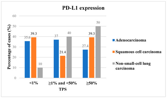
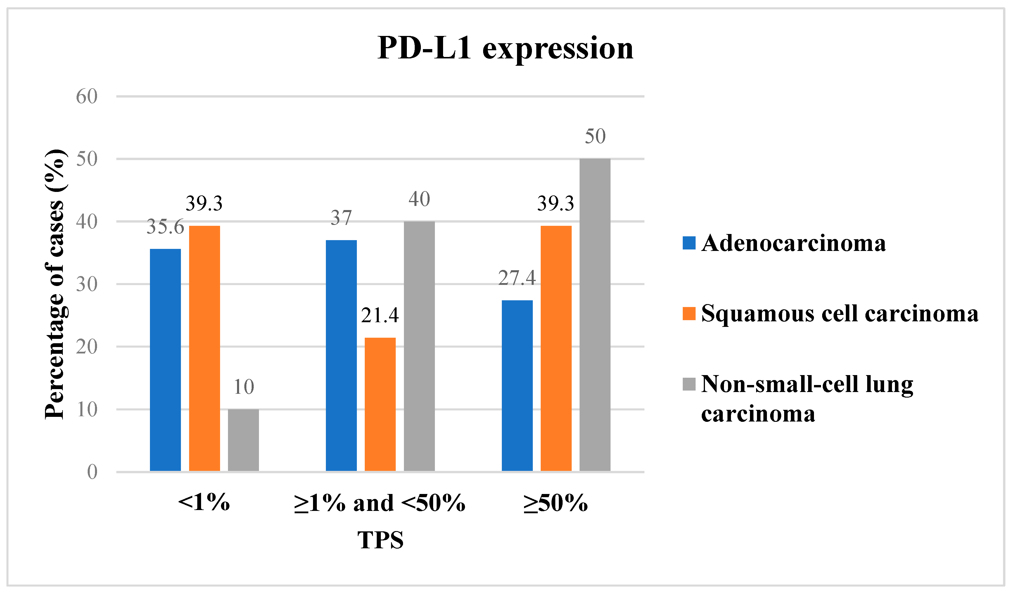
Concerning immunohistochemical markers, ROS1 expression was observed in 21 specimens (8.1%) with a H-score ranging from 120 to 250. Twenty of the 21 positive tumors were adenocarcinomas, and one of them was a NSCLC-NOS. PD-L1 expression was observed in 189 samples, with the median percentage of positive cancer cells (TPS) standing at 5% (range: 0–95%). The staining levels of PD-L1 demonstrated significant variation, with 90 specimens (34.9%) showing a TPS of less than 1%, 92 lying between 1% and 50% (35.7%), and the remaining 76 cases (29.4%) displaying >50% positive tumor cells (Figure 1). In contrast to ROS1 expression, PD-L1 was more homogeneously distributed among different histological subtypes, with positive staining observed in 160/219 (73.1%) adenocarcinomas, 19/28 (67.9%) squamous cell carcinomas, and 9/10 (90%) NSCLC-NOS.
Figure 1.
PD-L1 expression levels among the three different histological subtypes of lung tumors.
3.3. SHP-2 Expression and Statistical Correlations with Other Clinicopathological Parameters
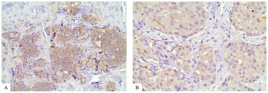
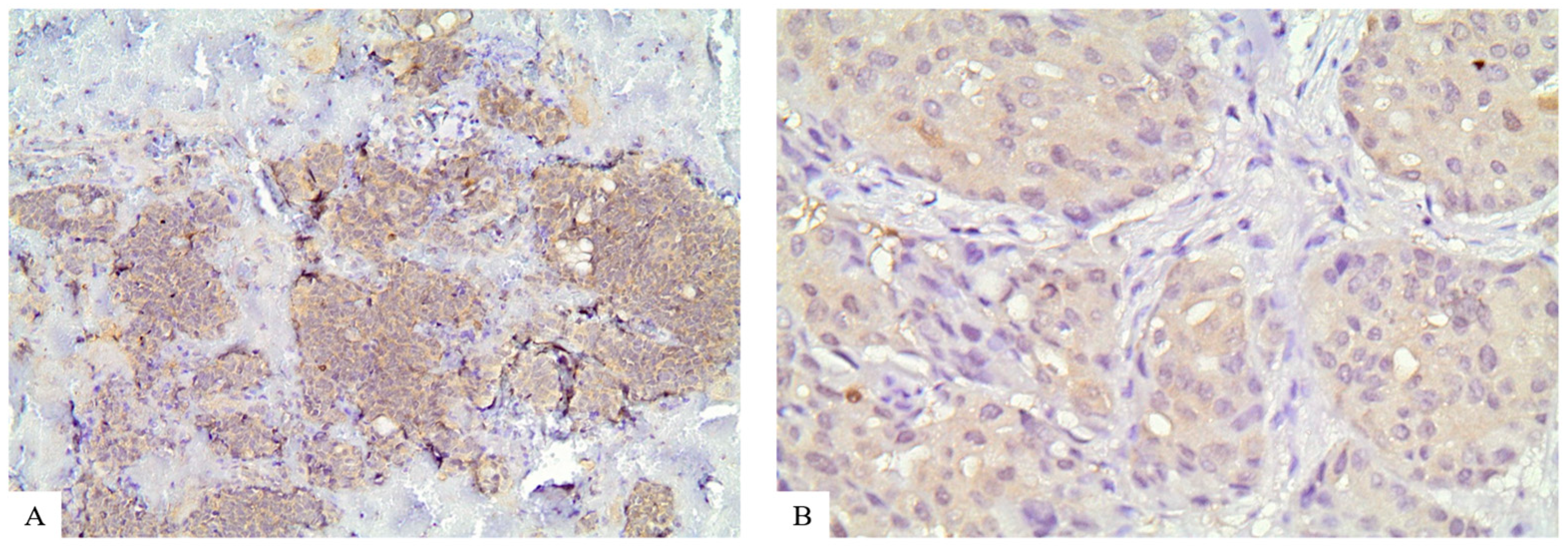
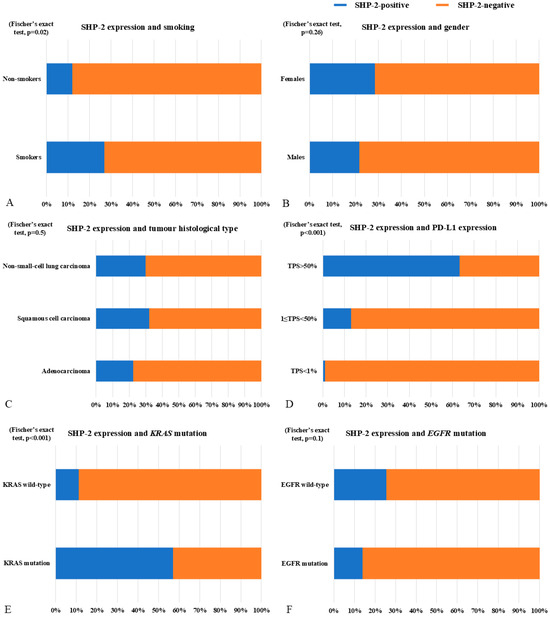
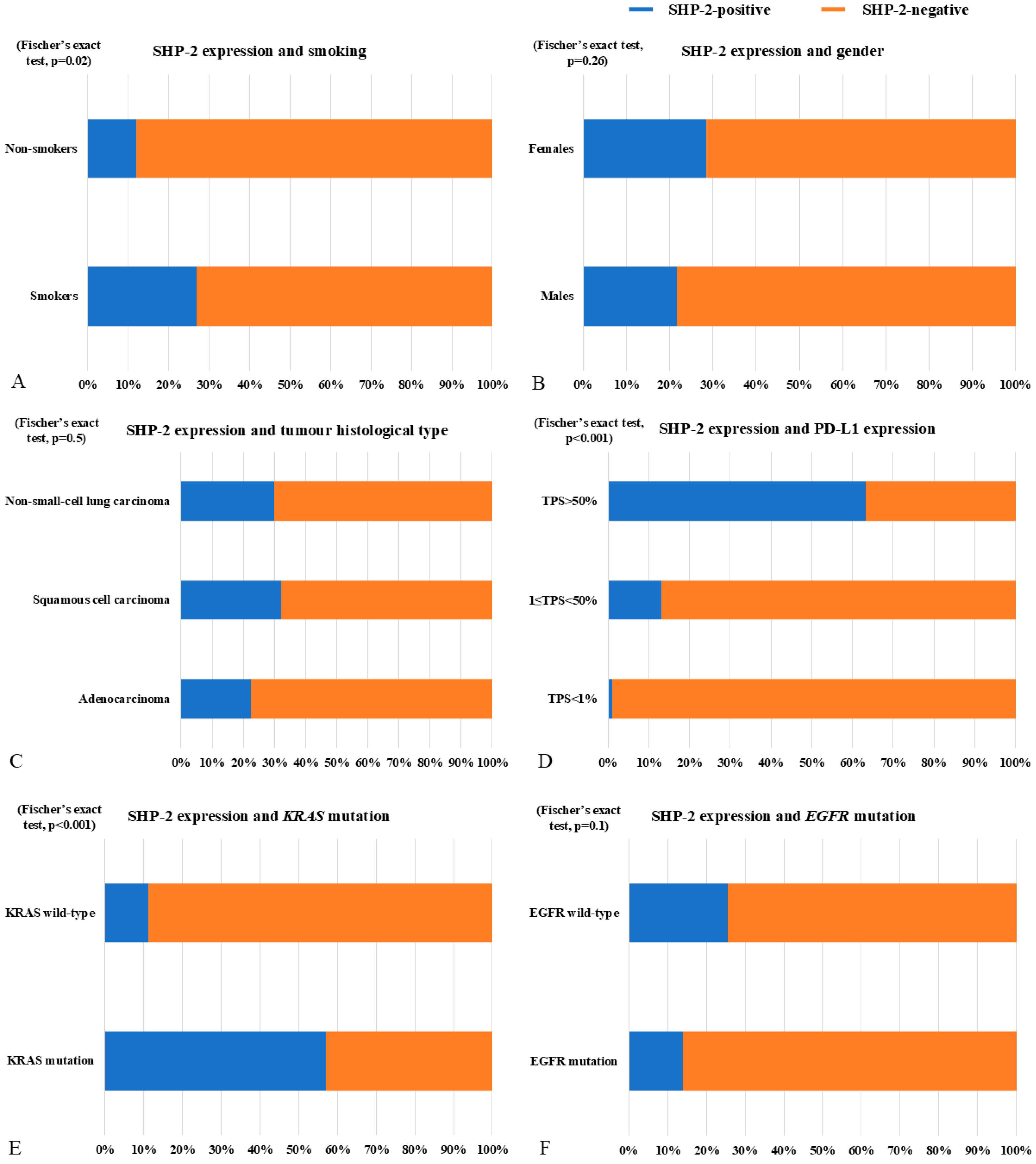
SHP-2 staining was considered positive in 60 samples (23.3%), with exclusively cytoplasmic localization (Figure 2), and distributed among the three major histological subtypes without statistically significant differences (Fisher’s exact test, p = 0.5) (Figure 3). The H-score of positive cases ranged between 150 and 300. While adenocarcinomas accounted for the majority of positive specimens, namely 49 (22.4%), the highest positivity frequency was detected in tumors of squamous differentiation, with nine of them (32.1%) displaying SHP-2 expression. The two remaining positive neoplasms were NSCLC-NOS (20%) (Table 3).
Figure 2.
Cytoplasmic expression of SHP-2 in a squamous cell lung carcinoma: (A) ×200; (B) ×400.
Figure 3.
Correlation of SHP-2 expression with multiple clinicopathological and molecular parameters: (A) smoking (Fisher’s exact test, p = 0.02), (B) gender (Fisher’s exact test, p = 0.26, (C) tumor histological type (Fisher’s exact test, p = 0.5), (D) PD-L1 expression (Fisher’s exact test, p < 0.001), (E) KRAS mutation (Fisher’s exact test, p < 0.001), and (F) EGFR mutation (Fisher’s exact test, p = 0.1).
Table 3.
ROS1 and SHP-2 immunohistochemical expression.
Additional statistical analyses revealed a very strong correlation of SHP-2 staining with PD-L1 expression levels (Fisher’s exact test, p < 0.001) and with the presence of KRAS mutations (Fisher’s exact test, p < 0.001) (Figure 3). More specifically, moving from low (<1%) to moderate (≥1%, <50%) and to high (≥50%) PD-L1 expression, the frequency of SHP-2 positivity progressively increased, rising from 1/90 (1.1%) to 12/92 (13%) and reaching a peak of 48/76 (43.2%) in the high PD-L1 expressors group (Figure 3). Regarding KRAS genetic alterations, they were detected in 40/70 (57.1%) SHP-2-positive cases compared to 21/189 (11.2%) SHP-2-negative cases. A statistically significant association was also identified between SHP-2 expression and smoking status (Fisher’s exact test, p = 0.02), as a higher SHP-2 positivity frequency was encountered in smokers (40/184, 21.7%) compared to non-smokers (7/58, 12.1%) (Figure 3). No statistically significant correlation was observed between SHP-2 staining and gender, EGFR mutational status, as well as with ALK and ROS1 expression (Fisher’s exact test, p > 0.05) (Figure 3).
3.4. Survival Analyses
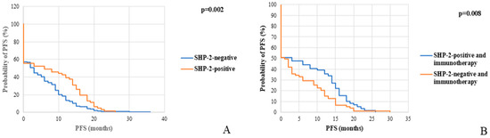
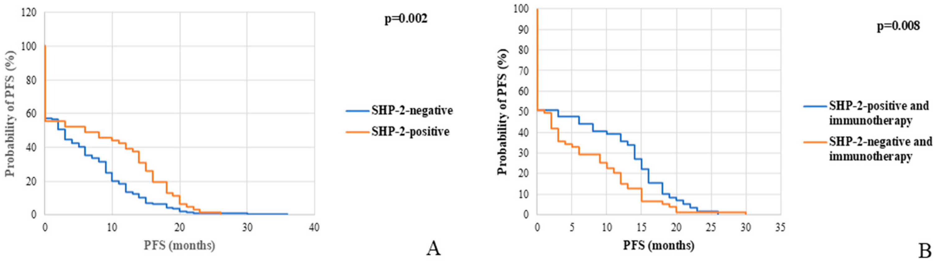
Expression levels of SHP-2 showcased a statistically significant correlation with clinical outcomes (OS and PFS). Positive SHP-2 immunohistochemical staining was associated with prolonged PFS (log-rank test, p = 0.002) in the total cohort (Figure 4). A mildly weaker association (log-rank test, p = 0.008) was observed among patients who received immunotherapy, as cases with SHP-2 expression showcased longer PFS compared to those lacking SHP-2 (Figure 4).
Figure 4.
Kaplan–Meier curves correlating SHP-2 expression with survival in the whole cohort ((A), log-rank test, p = 0.002) and among the patients who received immunotherapy ((B), log-rank test, p = 0.008).
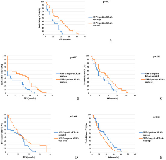
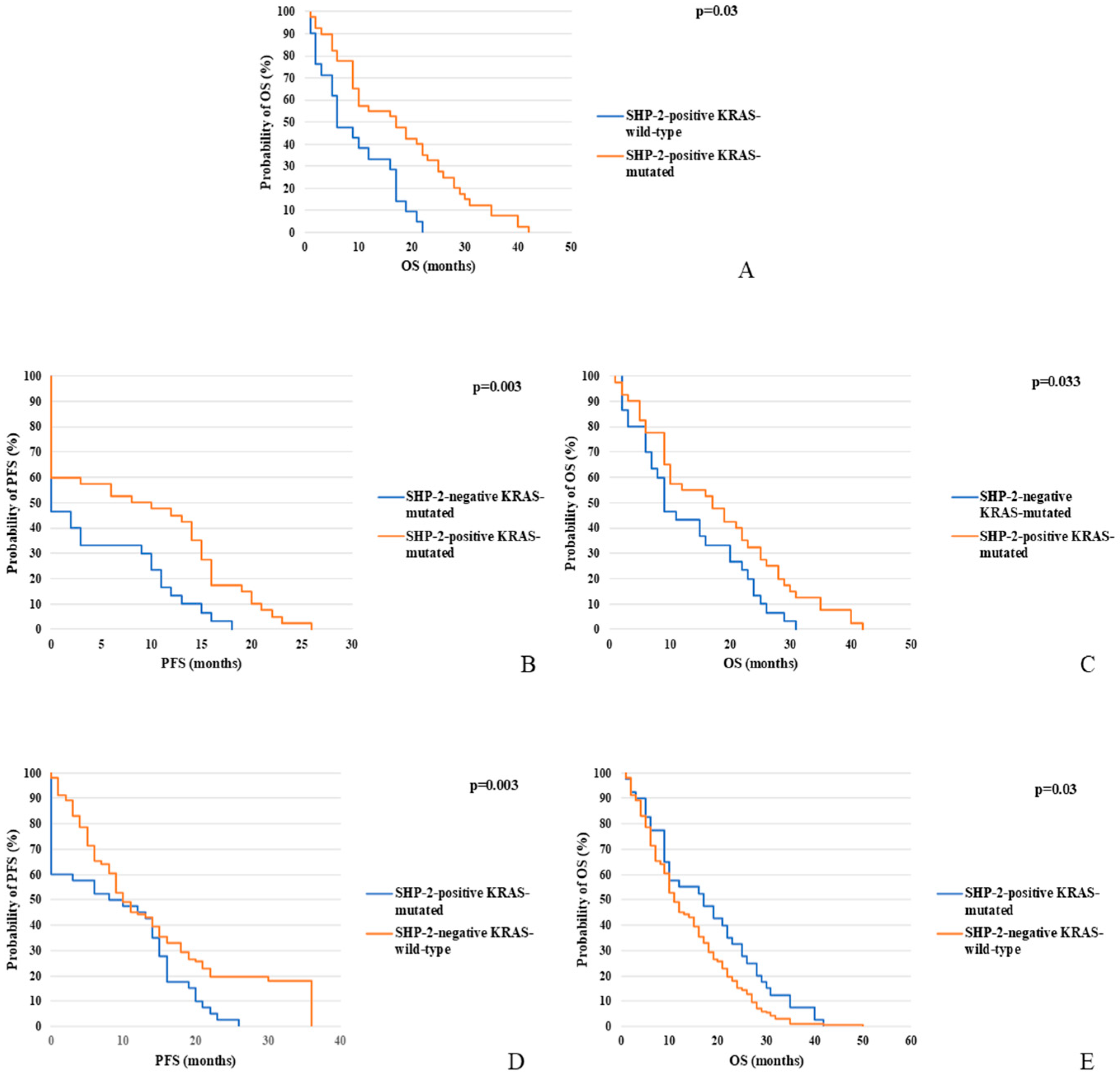
Interesting findings were also extracted from the multiple different combinations of SHP-2 expression and KRAS mutational status (Figure 5). First of all, in SHP-2-positive cases, the presence of KRAS gain-of-function mutations was correlated with prolonged OS (log-rank test, p = 0.03). On the other hand, in patients with KRAS-mutant neoplasms, SHP-2 expression was correlated with improved PFS (log-rank test, p = 0.003) and OS (log-rank test, p = 0.033). Finally, the concurrent presence of SHP-2 immunohistochemical staining and KRAS mutation was also associated with better PFS (log-rank test, p = 0.003) and OS (log-rank test, p = 0.03).
Figure 5.
Kaplan–Meier curves correlating SHP-2 expression and KRAS mutational status with survival. (A) Correlation of KRAS mutation with OS in SHP-2-positive cases (log-rank test, p = 0.03). (B,C) Correlation of SHP-2 expression with PFS (log-rank test, p = 0.03) and OS (log-rank test, p = 0.033) among KRAS-mutant cases. (D,E) Correlation of simultaneous SHP-2 expression and KRAS mutation with PFS (log-rank test, p = 0.003) and OS (log-rank test, p = 0.03).
4. Discussion
In this study we analyzed the expression pattern of SHP-2 using immunohistochemistry in a large cohort of NSCLC cases and attempted to correlate it with available clinicopathological and molecular data.
This is, to our knowledge, the first comprehensive review of SHP-2 expression in such an extensive pool of NSCLCs. Overall, SHP-2 staining was considered positive in 60 of the 258 samples (23.3%) and showed statistically significant correlation with smoking; the positive cases were distributed among different histological subtypes, without statistically significant variations. More specifically, SHP-2 expression was observed in 49/219 (22.4%) adenocarcinomas, 9/29 (32.1%) squamous cell carcinomas, and 2/10 (20%) NSCLCs. SHP-2 levels were also positively correlated with smoking, the presence of KRAS mutation, and PD-L1 expression. Another interesting finding concerns the role of SHP-2 as a prognostic and predictive marker capable of estimating a patient’s survival and response to immunotherapy. In more detail, SHP-2 expression was associated with prolonged PFS in both the total patient cohort (log-rank test, p = 0.002) and specifically in those who received immunotherapy with checkpoint inhibitors (log-rank test, p = 0.008). Especially in the group of patients who received immunotherapy schemes, the most straightforward explanation could be the increased levels of PD-L1 encountered in SHP-2-positive specimens. Significant correlations regarding patient survival were also identified in relation to KRAS mutational status. In general, the co-existence of SHP-2 expression and KRAS gain-of-function mutation was associated with better PFS and OS, regardless of the treatment protocol implemented.
A number of other studies have analyzed the role of SHP-2 in lung tumors via multiple methods, from protein expression evaluation by immunohistochemistry to molecular detection and experimental approaches. One of them utilized a tissue microarray of 80 NSCLC cases to assess SHP-2 expression and revealed increased immunohistochemical staining in neoplastic compared to tumor-adjacent tissue. Moreover, a statistically significant correlation was observed between SHP-2 expression levels and the presence of lymph node metastases [35]. Another study analyzed the expression pattern of KRAS and PTPN11 genes via The Cancer Genome Atlas (TCGA) database and revealed a very strong positive statistical correlation, while SHP-2 levels were lower in KRAS-wild-type compared to KRAS-mutant samples. In addition, lower SHP-2 was associated with improved prognosis and better overall survival [36]. In addition to the assessment of SHP-2 expression levels, mutations in different foci of the gene locus have been identified. Bentires Alj et al. analyzed 65 different NSCLC cell lines and found two distinct alterations in the PTPN11 sequence (V45L, N85S) [37].
Despite the limited and preliminary data on the role of SHP-2 in lung carcinomas, a number of studies have already evaluated its potential as a therapeutic target via both in vitro and in vivo experiments. A genetically engineered mouse model evaluated the oncogenic capacity of SHP-2 via a doxycycline (Dox)-inducible gain-of-function SHP-2 mutation, activated exclusively in Clara cells, by a Clara cell secretory protein (CCSP)-reverse tetracycline transactivator (rtTA). Mice developed both bronchiolar adenomas and lung adenocarcinomas, which regressed after Dox withdrawal. Moreover, all lung tumors, both benign and malignant, demonstrated strong pERK1/2 immunohistochemical staining, implying a pivotal role of the MAPK signaling cascade in SHP-2 tumorigenic function [38]. Another study tested the effect of a SHP-2 inhibitor, termed embelin, in KRAS-driven lung adenocarcinoma cell lines. Embelin is a naturally occurring para-benzoquinone, blocking the SHP-2 catalytic domain. Indeed, it was found to reduce proliferation and induce apoptosis of NSCLC cells. It also suppressed their migratory capacity and invasive potential, an effect that was accompanied by a downregulation in epithelial-to-mesenchymal transition (EMT) factors. This multidimensional anti-tumor activity was also verified in lung cancer heterotopic xenograft models [36]. Another natural compound which has displayed a tumoricidal effect against KRAS-mutant NSCLC cell lines is 3-acetoxylteuvincenone G (3-AG), which is an abietane diterpenoid extracted from the whole plants of Ajuga ovalifolia var. calantha. It has been shown to bind to the catalytic domain of SHP-2, blocking its phosphorylation capacity, leading to the suppression of cell proliferation and invasion and induction of A549 cell line apoptosis [39]. Another SHP-2 inhibitor showed little effect when tested on its own in a lung cancer cell line, while it significantly amplified the cytotoxic effect of MEK inhibitors when evaluated in combination schemes. This finding was also confirmed genetically: the CRISPR-associated protein 9 (Cas9)-mediated deletion of PTPN11 increased sensitivity to MEK blockade [40]. In contrast to the above findings, in vivo experiments showcased a robust anti-tumor effect of PTPN11 knock-out even in the absence of MEK inhibition [40]. SHP-2 blockade is also a promising approach in overcoming resistance to tyrosine kinase inhibition. TNO55 is a SHP-2-targeting molecule which has been evaluated in the setting of combination schemes, along with EGFR and KRAS inhibitors in lung cancer cell lines, preventing the emergence of acquired resistance and enhancing their tumoricidal effect [41,42]. Another interesting study conducted in broadly used lung adenocarcinoma cell lines revealed that ERK phosphorylation upon EGF and TNFa stimulation was suppressed in the presence of allosteric SHP-2 inhibitors SHP099 and TNO155. SHP-2 blockade also lead to a similar inhibitory effect on TNFa-induced EphA2 activation, which was also accompanied by diminished mobility and invasive potential of cells in wound-healing assays [43].
Besides the direct effect on cancer cells, SHP-2 is also crucial in the crosstalk between the tumor and immune microenvironments, rendering its targeting as a promising strategy in improving the response to immunotherapy with checkpoint inhibitors. Experiments in an EGFR-mutant lung adenocarcinoma cell line identified SHP-2 as a key driver of interferon-γ (IFN-γ) signaling suppression in tumor cells. EGFR gain-of-function mutations led to increased levels of IFN receptors (IFNG1, IFNG2), which was paradoxically accompanied by blunted pathway activation. The alleviation of IFN-γ signaling was associated with increased STAT1 de-phosphorylation, which was mediated by SHP-2, as the genetic deletion of PTPN11 in the same cell line reversed this effect. In further support of this finding, combination of a PD-1 inhibitor and a SHP-2-targeting molecule led to a synergistic therapeutic effect in a heterotopic xenograft mouse model of lung adenocarcinoma, implying a role in increasing tumor responsiveness to immune checkpoint inhibitors (ICBs) [44]. In contrast to these results, and in agreement with our findings, a study correlating SHP-2 expression patterns with response to ICIs in a cohort of NSCLC cases revealed that higher SHP-2 levels were associated with better response in KRAS-mutant neoplasms [45]. It is therefore crucial to take into consideration the context, especially the genetic background of tumors, in order to predict the possible effect of SHP-2 in immune response and immunotherapy efficacy. It is also important to always consider the limitations of each method, both immunohistochemical and in vivo/in vitro experimental studies.
Interesting findings of distinct SHP-2 levels in tumor cells compared to non-neoplastic tissues have also been described in a variety of other solid malignancies of heterogeneous histogenesis [46]. The most characteristic examples include neoplasms of the gastrointestinal tract as well as of pancreatic origin. In pancreatic and gastric cancer, a similar pattern to NSCLCs of increased SHP-2 expression in neoplastic cells, compared to non-neoplastic cells, was encountered [47,48]. On the contrary, a downregulation of SHP-2 was detected in esophageal squamous cell carcinomas in comparison to normal tissue [49]. Studies on colorectal lesions in both pre-invasive (adenomas) and invasive stages point towards a more intricate role of the SHP-2 protein in tumorigenesis. Adenomas and early invasive (stage I) lesions showed higher SHP-2 expression than more advanced tumors [50]. In addition to gastrointestinal malignancies, SHP-2 has been studied in thyroid, oral and laryngeal, and prostate and gynecological system neoplasms, with heterogeneous and sometimes contradictory results [46]. In thyroid carcinomas and oral and laryngeal squamous cell tumors, SHP-2 demonstrated a similar profile as in the lung, pancreas and stomach, with increased expression in tumor cells compared to non-neoplastic tissue. Moreover, it showcased a positive correlation with advanced tumor stage [51,52,53]. In prostate cancer, conflicting data have been derived with one study demonstrating SHP-2 overexpression, while another one revealed that reduced cytoplasmic staining is associated with tumor development and progression [54,55]. The heterogenous patterns of SHP-2 expression observed in different tumor subtypes clearly showcase the context-dependent role it exerts in tumorigenesis.
5. Conclusions
The existing data are mainly preliminary in order to justify an immediate incorporation of SHP-2 targeting in routine treatment protocols of NSCLC. However, our study provides the first analysis of its expression pattern in such an extended cohort of NSCLC cases, giving some initial insight on its potential role as a prognostic and/or predictive biomarker. Based on our findings, it could serve as a very reliable predictor of response to ICIs, even though additional studies are required in order to validate our results.
Author Contributions
Conceptualization, S.S. and K.S.; methodology, S.S. and P.K.; formal analysis, K.S. and S.S.; investigation, K.S., S.S. and G.P.; resources, G.P. and E.K.; data curation, K.S., G.P. and E.K.; writing—original draft preparation, K.S.; writing—review and editing, S.S.; supervision, S.S. and P.K. All authors have read and agreed to the published version of the manuscript.
Funding
This research received no external funding.
Institutional Review Board Statement
The study was conducted in accordance with the Declaration of Helsinki, and approved by the Ethics Committee of NKUA (Protocol No. 1718004921, date of approval 31 October 2019).
Informed Consent Statement
Informed consent was obtained from all patients who participated in the study.
Data Availability Statement
The original contributions presented in this study are included in the article. Further inquiries can be directed to the corresponding author.
Conflicts of Interest
The authors declare no conflicts of interest.
Abbreviations
The following abbreviations are used in this manuscript:
| NSCLC | Non-small-cell lung cancer |
| SCLC | Small-cell lung cancer |
| PFS | Progression-free survival |
| OS | Overall survival |
| NOS | Not otherwise specified |
| ICIs | Immune checkpoint inhibitors |
| NCCN | National Comprehensive Cancer Network |
| NKUA | National and Kapodistrian University of Athens |
| TCGA | The Cancer Genome Atlas |
| RTK | Tyrosine Kinase Receptor |
| CCSP | Clara cell secretory protein |
| rtTA | Reverse tetracycline transactivator |
| EMT | Epithelial-to-mesenchymal transition |
| 3-AG | 3-acetoxylteuvincenone G |
| Cas9 | CRISPR-associated protein 9 |
References
- Smolarz, B.; Łukasiewicz, H.; Samulak, D.; Piekarska, E.; Kołaciński, R.; Romanowicz, H. Lung Cancer—Epidemiology, Pathogenesis, Treatment and Molecular Aspect (Review of Literature). Int. J. Mol. Sci. 2025, 26, 2049. [Google Scholar] [CrossRef] [Scilit] [PubMed]
- Thandra, K.C.; Barsouk, A.; Saginala, K.; Aluru, J.S.; Barsouk, A. Epidemiology of Lung Cancer. Contemp. Oncol. 2021, 25, 45–52. [Google Scholar] [CrossRef] [Scilit] [PubMed]
- Alberg, A.J.; Ford, J.G.; Samet, J.M. Epidemiology of Lung Cancer: ACCP Evidence-Based Clinical Practice Guidelines (2nd Edition). Chest 2007, 132, 29S–55S. [Google Scholar] [CrossRef] [Scilit] [PubMed]
- Lightner-Ferrer, C.; Burke, L. Pathology of Lung Tumours: WHO 2021 Updates and Resection Status. Diagn. Histopathol. 2026, 32, 92–95. [Google Scholar] [CrossRef] [Scilit]
- Roșu, S.M.; Biciușcă, V.; Stan, S.I.; Mitroi, D.M.; Mahler, B.; Trașcă, D.M.; Popescu, M.; Pădureanu, M.M.; Stoica, A.Ș.; Pencea, T.I.; et al. Non-Small-Cell Lung Cancer: From Histopathological Classification to Precision Oncology—A Narrative Review. J. Clin. Med. 2026, 15, 3042. [Google Scholar] [CrossRef] [Scilit] [PubMed]
- Nicholson, A.G.; Tsao, M.S.; Beasley, M.B.; Borczuk, A.C.; Brambilla, E.; Cooper, W.A.; Dacic, S.; Jain, D.; Kerr, K.M.; Lantuejoul, S.; et al. The 2021 WHO Classification of Lung Tumors: Impact of Advances Since 2015. J. Thorac. Oncol. 2022, 17, 362–387. [Google Scholar] [CrossRef] [Scilit] [PubMed]
- Joshi, A.; Mishra, R.; Desai, S.; Chandrani, P.; Kore, H.; Sunder, R.; Hait, S.; Iyer, P.; Trivedi, V.; Choughule, A.; et al. Molecular Characterization of Lung Squamous Cell Carcinoma Tumors Reveals Therapeutically Relevant Alterations. Oncotarget 2021, 12, 578. [Google Scholar] [CrossRef] [Scilit] [PubMed]
- Gómez-López, S.; Whiteman, Z.E.; Janes, S.M. Mapping Lung Squamous Cell Carcinoma Pathogenesis through In Vitro and In Vivo Models. Commun. Biol. 2021, 4, 937. [Google Scholar] [CrossRef] [Scilit] [PubMed]
- Seguin, L.; Durandy, M.; Feral, C.C. Lung Adenocarcinoma Tumor Origin: A Guide for Personalized Medicine. Cancers 2022, 14, 1759. [Google Scholar] [CrossRef] [Scilit] [PubMed]
- Staaf, J.; Aine, M.; Nacer, D.F.; Planck, M.; Arbajian, E. Molecular Characteristics of Lung Adenocarcinoma with Respect to Patient Age at Diagnosis. Int. J. Cancer 2023, 153, 197–209. [Google Scholar] [CrossRef] [Scilit] [PubMed]
- Devarakonda, S.; Morgensztern, D.; Govindan, R. Genomic Alterations in Lung Adenocarcinoma. Lancet Oncol. 2015, 16, e342–e351. [Google Scholar] [CrossRef] [Scilit] [PubMed]
- Marabese, M.; Ganzinelli, M.; Garassino, M.C.; Shepherd, F.A.; Piva, S.; Caiola, E.; Macerelli, M.; Bettini, A.; Lauricella, C.; Floriani, I.; et al. KRAS Mutations Affect Prognosis of Non-Small-Cell Lung Cancer Patients Treated with First-Line Platinum Containing Chemotherapy. Oncotarget 2015, 6, 34014–34022. [Google Scholar] [CrossRef] [Scilit] [PubMed]
- Zhang, H.; Shan, G.; Cai, B.; Bi, G.; Chen, Z.; Liang, J.; Besskaya, V.; Zheng, Y.; Guo, W.; Wang, L.; et al. Clinicopathological and Prognostic Implications of ALK Rearrangement in Patients with Completely Surgically Resected Lung Adenocarcinoma. Thorac. Cancer 2021, 12, 3011–3018. [Google Scholar] [CrossRef] [Scilit] [PubMed]
- Pardoll, D.M. The Blockade of Immune Checkpoints in Cancer Immunotherapy. Nat. Rev. Cancer 2012, 12, 252–264. [Google Scholar] [CrossRef] [Scilit] [PubMed]
- Zamani, M.R.; Šácha, P. Immune Checkpoint Inhibitors in Cancer Therapy: What Lies beyond Monoclonal Antibodies? Med. Oncol. 2025, 42, 273. [Google Scholar] [CrossRef] [Scilit] [PubMed]
- Mamdani, H.; Matosevic, S.; Khalid, A.B.; Durm, G.; Jalal, S.I. Immunotherapy in Lung Cancer: Current Landscape and Future Directions. Front. Immunol. 2022, 13, 823618. [Google Scholar] [CrossRef] [Scilit] [PubMed]
- Capella, M.P.; Pang, S.A.; Magalhaes, M.A.; Esfahani, K. A Review of Immunotherapy in Non-Small-Cell Lung Cancer. Curr. Oncol. 2024, 31, 3495–3512. [Google Scholar] [CrossRef] [Scilit] [PubMed]
- Younis, A.; Gribben, J. Immune Checkpoint Inhibitors: Fundamental Mechanisms, Current Status and Future Directions. Immuno 2024, 4, 186–210. [Google Scholar] [CrossRef] [Scilit]
- Davis, A.A.; Patel, V.G. The Role of PD-L1 Expression as a Predictive Biomarker: An Analysis of All US Food and Drug Administration (FDA) Approvals of Immune Checkpoint Inhibitors. J. Immunother. Cancer 2019, 7, 278. [Google Scholar] [CrossRef] [Scilit] [PubMed]
- Patel, S.P.; Kurzrock, R. PD-L1 Expression as a Predictive Biomarker in Cancer Immunotherapy. Mol. Cancer Ther. 2015, 14, 847–856. [Google Scholar] [CrossRef] [Scilit] [PubMed]
- Sun, Q.; Hong, Z.; Zhang, C.; Wang, L.; Han, Z.; Ma, D. Immune Checkpoint Therapy for Solid Tumours: Clinical Dilemmas and Future Trends. Signal Transduct. Target. Ther. 2023, 8, 320. [Google Scholar] [CrossRef] [Scilit] [PubMed]
- Meng, G.; Liu, X.; Ma, T.; Lv, D.; Sun, G. Predictive Value of Tumor Mutational Burden for Immunotherapy in Non-Small Cell Lung Cancer: A Systematic Review and Meta-Analysis. PLoS ONE 2022, 17, e0263629. [Google Scholar] [CrossRef] [Scilit] [PubMed]
- Imbody, D.; Arce, K.; Solanki, H.S.; Haura, E.B.; Pellini, B. Targeting SHP2 Signaling in Lung Cancer. J. Thorac. Oncol. 2024, 19, 18–24. [Google Scholar] [CrossRef] [Scilit] [PubMed]
- Lin, C.-C.; Wieteska, L.; Suen, K.M.; Kalverda, A.P.; Ahmed, Z.; Ladbury, J.E. Grb2 Binding Induces Phosphorylation-Independent Activation of Shp2. Commun. Biol. 2021, 4, 437. [Google Scholar] [CrossRef] [Scilit] [PubMed]
- Zehender, A.; Huang, J.; Györfi, A.-H.; Matei, A.-E.; Trinh-Minh, T.; Xu, X.; Li, Y.-N.; Chen, C.-W.; Lin, J.; Dees, C.; et al. The Tyrosine Phosphatase SHP2 Controls TGFβ-Induced STAT3 Signaling to Regulate Fibroblast Activation and Fibrosis. Nat. Commun. 2018, 9, 3259. [Google Scholar] [CrossRef] [Scilit] [PubMed]
- Ruess, D.A.; Heynen, G.J.; Ciecielski, K.J.; Ai, J.; Berninger, A.; Kabacaoglu, D.; Görgülü, K.; Dantes, Z.; Wörmann, S.M.; Diakopoulos, K.N.; et al. Mutant KRAS-Driven Cancers Depend on PTPN11/SHP2 Phosphatase. Nat. Med. 2018, 24, 954–960. [Google Scholar] [CrossRef] [Scilit] [PubMed]
- Ryan, M.B.; de la Cruz, F.F.; Phat, S.; Myers, D.T.; Wong, E.; Shahzade, H.A.; Hong, C.B.; Corcoran, R.B. Vertical Pathway Inhibition Overcomes Adaptive Feedback Resistance to KrasG12C Inhibition. Clin. Cancer Res. 2020, 26, 1633–1643. [Google Scholar] [CrossRef] [Scilit] [PubMed]
- López Moreno, S.F.; Lenz, S.A.; Casso-Chapa, B.; Paniagua-Bojorges, A.; Kim, J.H.; Palaskas, N.L.; Nead, K.T.; Samanthapudi, V.S.K.; Mejia, G.; Hoang, O.; et al. SHP2: A Redox-Sensitive Regulator Linking Immune Checkpoint Inhibitor Therapy to Cancer Treatment and Vascular Risk. Antioxidants 2025, 14, 1388. [Google Scholar] [CrossRef] [Scilit] [PubMed]
- Chen, D.; Barsoumian, H.B.; Yang, L.; Younes, A.I.; Verma, V.; Hu, Y.; Menon, H.; Wasley, M.; Masropour, F.; Mosaffa, S.; et al. SHP-2 and PD-L1 Inhibition Combined with Radiotherapy Enhances Systemic Antitumor Effects in an Anti-PD-1-Resistant Model of Non-Small Cell Lung Cancer. Cancer Immunol. Res. 2020, 8, 883–894. [Google Scholar] [CrossRef] [Scilit] [PubMed]
- Sheppard, K.-A.; Fitz, L.J.; Lee, J.M.; Benander, C.; George, J.A.; Wooters, J.; Qiu, Y.; Jussif, J.M.; Carter, L.L.; Wood, C.R.; et al. PD-1 Inhibits T-Cell Receptor Induced Phosphorylation of the ZAP70/CD3ζ Signalosome and Downstream Signaling to PKCθ. FEBS Lett. 2004, 574, 37–41. [Google Scholar] [CrossRef] [Scilit] [PubMed]
- WHO Classification of Tumours Editorial Board. Thoracic Tumours, 5th ed.; International Agency for Research on Cancer: Lyon, France, 2021; Volume 5. [Google Scholar]
- Karatrasoglou, E.A.; Chatziandreou, I.; Sakellariou, S.; Stamopoulos, K.; Kavantzas, N.; Lazaris, A.C.; Korkolopoulou, P.; Saetta, A.A. Association between PD-L1 Expression and Driver Gene Mutations in Non-Small Cell Lung Cancer Patients: Correlation with Clinical Data. Virchows Arch. 2020, 477, 207–217. [Google Scholar] [CrossRef] [Scilit] [PubMed]
- Erfani, E.; Roudi, R.; Rakhshan, A.; Sabet, M.N.; Shariftabrizi, A.; Madjd, Z. Comparative Expression Analysis of Putative Cancer Stem Cell Markers CD44 and ALDH1A1 in Various Skin Cancer Subtypes. Int. J. Biol. Markers 2016, 31, e53–e61. [Google Scholar] [CrossRef] [Scilit] [PubMed]
- Chen, M.-J.; Wang, Y.-C.; Wu, D.-W.; Chen, C.-Y.; Lee, H. Association of Nuclear Localization of SHP2 and YAP1 with Unfavorable Prognosis in Non-Small Cell Lung Cancer. Pathol. Res. Pract. 2019, 215, 801–806. [Google Scholar] [CrossRef] [Scilit] [PubMed]
- Tang, C.; Luo, D.; Yang, H.; Wang, Q.; Zhang, R.; Liu, G.; Zhou, X. Expression of SHP2 and Related Markers in Non-Small Cell Lung Cancer: A Tissue Microarray Study of 80 Cases. Appl. Immunohistochem. Mol. Morphol. 2013, 21, 386–394. [Google Scholar] [CrossRef] [Scilit] [PubMed]
- Fu, N.J.; Sheng, Y.W.; Fan, Z.; Wu, Z.; Li, L.Y.; Xi, R.Y.; Shi, X.K.; Zhang, G.L.; Wang, F. Synthetic Lethality of SHP2 and XIAP Suppresses Proliferation and Metastasis in KRAS-Mutant Nonsmall Cell Lung Cancer. Adv. Sci. 2025, 12, 2411642. [Google Scholar] [CrossRef] [Scilit] [PubMed]
- Bentires-Alj, M.; Paez, J.G.; David, F.S.; Keilhack, H.; Halmos, B.; Naoki, K.; Maris, J.M.; Richardson, A.; Bardelli, A.; Sugarbaker, D.J.; et al. Activating Mutations of the Noonan Syndrome-Associated SHP2/PTPN11 Gene in Human Solid Tumors and Adult Acute Myelogenous Leukemia. Cancer Res. 2004, 64, 8816–8820. [Google Scholar] [CrossRef] [Scilit] [PubMed]
- Schneeberger, V.E.; Luetteke, N.; Ren, Y.; Berns, H.; Chen, L.; Foroutan, P.; Martinez, G.V.; Haura, E.B.; Chen, J.; Coppola, D.; et al. SHP2E76K Mutant Promotes Lung Tumorigenesis in Transgenic Mice. Carcinogenesis 2014, 35, 1717–1725. [Google Scholar] [CrossRef] [Scilit] [PubMed]
- Xu, W.-Q.; Qi, S.-Z.; Zhao, J.-F.; Li, L.-P.; Ding, C.-H.; Liu, W.-S. Discovery of 1H-Pyrazolo [3,4-b]Pyrazine Derivatives as Selective Allosteric Inhibitor of Protein Tyrosine Phosphatase SHP2 for the Treatment of KRASG12C-Mutant Non-Small Cell Lung Cancer. J. Biomol. Struct. Dyn. 2025, 43, 5465–5473. [Google Scholar] [CrossRef] [Scilit] [PubMed]
- Mainardi, S.; Mulero-Sánchez, A.; Prahallad, A.; Germano, G.; Bosma, A.; Krimpenfort, P.; Lieftink, C.; Steinberg, J.D.; De Wit, N.; Gonçalves-Ribeiro, S.; et al. SHP2 Is Required for Growth of KRAS-Mutant Non-Small-Cell Lung Cancer in Vivo. Nat. Med. 2018, 24, 961–967. [Google Scholar] [CrossRef] [Scilit] [PubMed]
- Liu, C.; Lu, H.; Wang, H.; Loo, A.; Zhang, X.; Yang, G.; Kowal, C.; Delach, S.; Wang, Y.; Goldoni, S.; et al. Combinations with Allosteric SHP2 Inhibitor TNO155 to Block Receptor Tyrosine Kinase Signaling. Clin. Cancer Res. 2021, 27, 342–354. [Google Scholar] [CrossRef] [Scilit] [PubMed]
- Ho, C.S.L.; Tüns, A.I.; Schildhaus, H.-U.; Wiesweg, M.; Grüner, B.M.; Hegedus, B.; Schuler, M.; Schramm, A.; Oeck, S. HER2 Mediates Clinical Resistance to the KRASG12C Inhibitor Sotorasib, Which Is Overcome by Co-Targeting SHP2. Eur. J. Cancer 2021, 159, 16–23. [Google Scholar] [CrossRef] [Scilit] [PubMed]
- Nguyen, K.; Zhou, Y.; Yokoyama, S.; Sasaki, S.-I.; Yano, S.; Hayashi, R.; Koizumi, K.; Hayakawa, Y.; Sakurai, H. Allosteric SHP2 Inhibitors Suppress Lung Cancer Cell Migration by Inhibiting Non-Canonical Activation of EphA2 via the ERK-RSK Signaling Pathway. Sci. Rep. 2025, 15, 36926. [Google Scholar] [CrossRef] [Scilit] [PubMed]
- Zhuang, W.-T.; Pang, L.-L.; Hu, L.-Y.; Liao, J.; Zhan, J.-H.; Li, T.; Chen, R.-X.; Zheng, J.-N.; Li, A.-L.; Yu, W.-Y.; et al. EGFR Activation Disrupts Immunotherapy Response via SHP2-Mediated Suppression of Tumor-Intrinsic Response to IFN-γ. J. Clin. Investig. 2026, 136, e194377. [Google Scholar] [CrossRef] [Scilit] [PubMed]
- Feng, H.-B.; Chen, Y.; Xie, Z.; Jiang, J.; Zhong, Y.M.; Guo, W.-B.; Yan, W.Q.; Lv, Z.Y.; Lu, D.X.; Liang, H.L.; et al. High SHP2 Expression Determines the Efficacy of PD-1/PD-L1 Inhibitors in Advanced KRAS Mutant Non-Small Cell Lung Cancer. Thorac. Cancer 2021, 12, 2564–2573. [Google Scholar] [CrossRef] [Scilit] [PubMed]
- Chen, X.; Keller, S.J.; Hafner, P.; Alrawashdeh, A.Y.; Avery, T.Y.; Norona, J.; Zhou, J.; Ruess, D.A. Tyrosine Phosphatase PTPN11/SHP2 in Solid Tumors-Bull’s Eye for Targeted Therapy? Front. Immunol. 2024, 15, 1340726. [Google Scholar] [CrossRef] [Scilit] [PubMed]
- Zheng, J.; Huang, S.; Huang, Y.; Song, L.; Yin, Y.; Kong, W.; Chen, X.; Ouyang, X. Expression and Prognosis Value of SHP2 in Patients with Pancreatic Ductal Adenocarcinoma. Tumor Biol. 2016, 37, 16207–16213. [Google Scholar] [CrossRef] [Scilit] [PubMed]
- Dong, S.; Li, F.-Q.; Zhang, Q.; Lv, K.-Z.; Yang, H.-L.; Gao, Y.; Yu, J.-R. Expression and Clinical Significance of SHP2 in Gastric Cancer. J. Int. Med. Res. 2012, 40, 2083–2089. [Google Scholar] [CrossRef] [Scilit] [PubMed]
- Qi, C.; Han, T.; Tang, H.; Huang, K.; Min, J.; Li, J.; Ding, X.; Xu, Z. Shp2 Inhibits Proliferation of Esophageal Squamous Cell Cancer via Dephosphorylation of Stat3. Int. J. Mol. Sci. 2017, 18, 134. [Google Scholar] [CrossRef] [Scilit] [PubMed]
- Gagné-Sansfaçon, J.; Coulombe, G.; Langlois, M.-J.; Langlois, A.; Paquet, M.; Carrier, J.; Feng, G.-S.; Qu, C.-K.; Rivard, N. SHP-2 Phosphatase Contributes to KRAS-Driven Intestinal Oncogenesis but Prevents Colitis-Associated Cancer Development. Oncotarget 2016, 7, 65676. [Google Scholar] [CrossRef] [Scilit] [PubMed]
- Cao, J.; Huang, Y.-Q.; Sun, J.; Lan, X.-B.; Ge, M.-H. Clinicopathological and Prognostic Significance of SHP2 and Hook1 Expression in Patients with Thyroid Carcinoma. Hum. Pathol. 2018, 81, 105–112. [Google Scholar] [CrossRef] [Scilit] [PubMed]
- Hu, Z.Q.; Ma, R.; Zhang, C.M.; Li, J.; Li, L.; Hu, Z.T.; Gao, Q.; Li, W.M. Expression and Clinical Significance of Tyrosine Phosphatase SHP2 in Thyroid Carcinoma. Oncol. Lett. 2015, 10, 1507. [Google Scholar] [CrossRef] [Scilit] [PubMed]
- Wang, J.; Ouyang, S.; Zhang, X.; Zhao, S.; Cheng, M.; Fan, X.; Cai, Y.; Liao, L. Deregulation of Hsa_circ_0001971/MiR-186 and Hsa_circ_0001874/MiR-296 Signaling Pathways Promotes the Proliferation of Oral Squamous Carcinoma Cells by Synergistically Activating SHP2/PLK1 Signals. Sci. Rep. 2021, 11, 20561. [Google Scholar] [CrossRef] [Scilit] [PubMed]
- Zhang, K.; Zhao, H.; Ji, Z.; Zhang, C.; Zhou, P.; Wang, L.; Chen, Q.; Wang, J.; Zhang, P.; Chen, Z.; et al. Shp2 Promotes Metastasis of Prostate Cancer by Attenuating the PAR3/PAR6/APKC Polarity Protein Complex and Enhancing Epithelial-to-Mesenchymal Transition. Oncogene 2016, 35, 1271–1282. [Google Scholar] [CrossRef] [Scilit] [PubMed]
- Tassidis, H.; Brokken, L.J.S.; Jirström, K.; Bjartell, A.; Ulmert, D.; Härkönen, P.; Wingren, A.G. Low Expression of SHP-2 Is Associated with Less Favorable Prostate Cancer Outcomes. Tumour Biol. 2013, 34, 637–642. [Google Scholar] [CrossRef] [Scilit] [PubMed]

Disclaimer/Publisher’s Note: The statements, opinions and data contained in all publications are solely those of the individual author(s) and contributor(s) and not of MDPI and/or the editor(s). MDPI and/or the editor(s) disclaim responsibility for any injury to people or property resulting from any ideas, methods, instructions or products referred to in the content. |
© 2026 by the authors. Licensee MDPI, Basel, Switzerland. This article is an open access article distributed under the terms and conditions of the Creative Commons Attribution (CC BY) license.