


Simple Summary
Prostate cancer is one of the most common cancers in men, and most patients eventually develop resistance to standard hormone-blocking treatments—a stage called castration-resistant prostate cancer. Predicting which patients will progress quickly remains a major clinical challenge. In this study, we analyzed tumor DNA from 92 prostate cancer patients using next-generation sequencing, a technology that reads thousands of genes simultaneously to identify genetic changes that could predict faster progression to castration resistance. We found that alterations in the KMT2C gene—which normally acts as a molecular brake on cancer cell transformation—were the strongest predictor of early resistance, with affected patients progressing nearly four times faster than those without this alteration. Patients with defects in mismatch repair genes, while not progressing faster, had significantly shorter survival, highlighting their candidacy for immunotherapy. These findings support incorporating genetic tumor profiling at the time of diagnosis to guide personalized treatment decisions.
Abstract
The prognostic significance of next-generation sequencing (NGS) findings during the hormone-sensitive phase of prostate cancer remains incompletely characterized. This retrospective cohort study included 92 patients who underwent NGS analysis on tumor tissue between 2019 and 2025. The primary endpoint was time to castration-resistant prostate cancer (CRPC) from androgen deprivation therapy (ADT) initiation; secondary endpoints were overall survival from ADT initiation (OS-ADT) and from diagnosis. Kaplan-Meier and Cox regression analyses were performed. CRPC developed in 66 patients (71.7%) at a median of 21.1 months. The most frequently altered genes were ATR (35.9%), PTEN (28.3%), TP53 (26.1%), and BRCA2 (15.2%). KMT2C alteration (5.4%) was the strongest independent genomic predictor of shorter time to CRPC (HR = 6.804, p = 0.003) and OS-ADT (HR = 4.730, p = 0.019). TP53 alteration independently predicted shorter OS-ADT (HR = 1.810, p = 0.038). High genomic burden independently predicted shorter time to CRPC (HR = 1.917, p = 0.032). Homologous recombination repair deficiency was not associated with outcomes, attributable to high ATR alteration frequency introducing pathway heterogeneity. Mismatch repair deficiency showed a borderline association with shorter OS-ADT (20.7 vs. 44.0 months; p = 0.060). An exploratory composite risk score stratified patients into three prognostic groups with markedly different outcomes (HR = 7.904, p = 0.001). NGS analysis during the hormone-sensitive phase identifies independent predictors of castration resistance, supporting its integration at ADT initiation for risk stratification and biomarker-guided treatment planning.
1. Introduction
Globally, prostate cancer (PCa) ranks as the second-most frequently diagnosed malignancy in men, accounting for approximately 1.4 million new cases in 2022 [1]. In parallel with this global burden, the incidence of PCa in Türkiye was documented at 40.3 per 100,000 in 2018 [2]. Androgen deprivation therapy (ADT) remains the standard of care for metastatic hormone-sensitive prostate cancer (mHSPC); however, most patients progress to castration-resistant prostate cancer (CRPC) within 18–24 months [3,4]. This transition—defined by prostate-specific antigen (PSA) and/or radiological progression despite castrate serum testosterone levels (<50 ng/dL)—carries a poor prognosis even in the contemporary treatment era [4,5].
Classical prognostic parameters, including PSA kinetics and Gleason grade, are useful in clinical practice, but they do not reliably predict how quickly resistance will develop in individual patients [6,7]. Next-generation sequencing (NGS) can characterize the molecular changes associated with treatment escape more directly [8,9]. Landmark genomic characterization of metastatic CRPC has demonstrated that aberrations in AR, TP53, PTEN, and RB1 are among the most frequently altered genes in advanced disease [10]. Persistent AR signaling through gene amplification, ligand-binding domain mutations, or splice variant expression (notably AR-V7) represents the dominant ADT escape mechanism [10,11]. Compound TP53 and RB1 loss drives lineage plasticity via SOX2 upregulation, enabling phenotypic shift toward AR-independent cell states; concurrent loss was identified in 39% of mCRPC adenocarcinomas and 74% of neuroendocrine-like CRPC cases [12].
Alterations in DNA damage repair (DDR) and homologous recombination repair (HRR) genes are detected in approximately 19–23% of mCRPC cases [10]. The PROfound phase III trial established that the PARP inhibitor olaparib significantly extended progression-free survival in BRCA1/2- and ATM-altered mCRPC (median 7.4 vs. 3.6 months; hazard ratio [HR] 0.34; p < 0.001), making NGS-guided patient selection a clinical standard [13]. Annala et al. further showed that BRCA2/ATM mutations and TP53 defects independently predict primary resistance to first-line abiraterone and enzalutamide [11].
Despite these advances, the prognostic relevance of NGS-detected alterations with respect to time to CRPC—rather than mCRPC outcomes—remains incompletely characterized. Population-level genomic data from Turkish and non-Western cohorts are also virtually absent [14]. Therefore, we retrospectively compared NGS panel results obtained during the hormone-sensitive phase with time to CRPC in a real-world Turkish academic cohort. Our aim was to assess whether genomic alterations detected at this earlier stage could help predict early castration resistance. In contrast to previous studies that mainly examined patients after castration resistance had already developed, our study focuses on the hormone-sensitive phase and uses time to CRPC as the primary endpoint. To our knowledge, this is also the first report of real-world genomic profiling data from a Turkish prostate cancer cohort at this stage [2,14].
2. Materials and Methods
2.1. Study Design and Setting
This was a single-center, observational, retrospective cohort study conducted at Aydın Adnan Menderes University (ADÜ) Application and Research Hospital Medical Oncology Clinic (January 2019–December 2025), following institutional ethics committee approval.
2.2. Eligibility Criteria
Inclusion criteria: (1) histopathologically confirmed prostate adenocarcinoma; (2) initiation of ADT or prior surgical castration; (3) availability of NGS panel analysis on tumor tissue; and (4) minimum 12 months of follow-up from ADT initiation. Patients were excluded if NGS analysis could not be completed due to insufficient tissue, or if the ADT initiation date or CRPC development date was unavailable.
2.3. Data Collection
We recorded the following variables: age at diagnosis, initial serum PSA, International Society of Urological Pathology (ISUP) grade group, clinical TNM stage, presence of de novo metastatic disease, initial treatment regimen, date of ADT initiation, date of CRPC development or last follow-up, and NGS panel results. Initial treatment was categorized as ADT alone, ADT plus an androgen receptor pathway inhibitor (ARPI; abiraterone acetate, enzalutamide, apalutamide, or darolutamide), or ADT plus docetaxel. Clinical T and N stage and Eastern Cooperative Oncology Group (ECOG) performance status were not consistently documented, so these variables were excluded from the analysis. Baseline PSA values were missing for 20 patients (21.7%); these patients were excluded from PSA-related analyses.
2.4. Tumor Tissue and NGS Panel Analysis
NGS panel results were obtained from the pathology laboratory archives. Tumor material came from formalin-fixed paraffin-embedded (FFPE) tissue blocks. In 86 patients (93.5%), NGS was performed on primary prostate biopsy specimens; in the remaining 6 patients (6.5%), metastatic biopsy specimens were used. After consultation with the molecular pathology unit, 10-micron sections were prepared from the most representative tumor-containing block. DNA was then isolated from these sections and analyzed with the QIAseq Targeted DNA Panel (QIAGEN, Hilden, Germany) on an Illumina MiniSeq platform, using paired-end 151bp reads. The panel included 93 genes and 48,831 primers. Covered genes included BRCA1, BRCA2, ATM, ATR, AR, TP53, PTEN, RB1, KMT2C, MRE11, CHEK2, NF1, SPOP, and PIK3CA, among others. Unique molecular identifiers (UMIs) were assigned to each original DNA molecule before target enrichment. This step was used to support more accurate variant calling and reduce errors related to PCR amplification. Library preparation followed manufacturer protocols; target enrichment was performed using locus-specific and universal primers. Data analysis was performed using the QIAseq pipeline and variants were interpreted using Qiagen Clinical Insight-Interpret (QCI-Interpret, version 9.5.4.20260422) bioinformatics software. Both tools are publicly accessible via the QIAGEN bioinformatics platform (https://www.qiagen.com (accessed on 25 May 2026); https://www.qiagenbioinformatics.com (accessed on 25 May 2026)). For each patient, the presence or absence of a pathogenic or likely pathogenic alteration in each gene was recorded as a binary variable (mutated vs. wild-type).
2.4.1. Genomic Grouping and Pathway-Based Variables
Individual gene alterations were aggregated into pathway-based binary variables. A patient was considered positive for a pathway group if at least one pathogenic or likely pathogenic alteration was detected in any gene within that pathway.
HRR deficiency was defined using a restricted gene set (BRCA1, BRCA2, ATM, ATR, BRIP1, PALB2, RAD50, RAD51, NBN, MRE11, ABRAXAS1, FANCD2, FANCG, BLM, ERCC5), consistent with PROfound trial criteria [13]. A broader DDR deficiency variable additionally incorporating CDK12 and CHEK2 was constructed for sensitivity analyses [10,11]. Mismatch repair (MMR) deficiency was defined by alterations in MLH3, MSH3, MSH6, PMS1, PMS2, POLD3, or POLE. Pathway-level variables were also constructed for AR signaling (AR, SPOP, NCOR1, HOXB13, MED12), the PI3K/AKT/PTEN axis (PTEN, PIK3CA, AKT1, NF1), WNT signaling (APC, CTNNB1, AXIN2, TCF7L2), chromatin modifiers (KMT2C, KMT2D, ARID1A, CHD1, CREBBP, BAP1, PBRM1), and TGF-β signaling (TGFB1, TGFBR2, SMAD4). TP53 and RB1 alterations were analyzed individually and as a concurrent loss composite, per Mu et al. [12]. Genes with insufficient alteration frequency (n < 5) were excluded from inferential analyses.
2.4.2. Composite and Derived Variables
Several composite and derived variables were constructed posthoc in an exploratory manner based on primary survival analyses.
Metastatic status-integrated genomic variables were created for KMT2C and TP53 mutations separately, classifying patients into three groups: (1) wild-type and non-metastatic, (2) wild-type and de novo metastatic, and (3) mutation-positive (regardless of metastatic status).
A multi-pathway genomic burden score was calculated by summing the number of dysregulated pathways per patient across seven pathway-level binary variables (HRR, MMR, AR signaling, PI3K/AKT/PTEN, WNT, chromatin modifier, and TP53). Based on the distribution of scores in the cohort, patients were grouped as having low genomic burden if 0–1 pathways were affected, and high genomic burden if 2 or more pathways were affected. Although TP53 was also analyzed individually, it was included in the burden score as a representative of the p53 tumor suppressor pathway.
We defined the high-risk genomic group as patients with at least one alteration in KMT2C, TP53, or MRE11. These genes were selected because each showed individual statistical significance in the primary time-to-CRPC analyses.
An exploratory composite clinical-genomic risk score was constructed by summing five binary variables: KMT2C mutation, TP53 mutation, de novo metastatic disease, high ISUP grade (grade group 4–5), and high genomic burden (≥2 pathways), yielding a score of 0–5. Patients were categorized as low risk (score 0–1), intermediate risk (score 2), or high risk (score ≥ 3).
2.5. Primary and Secondary Endpoints
The primary endpoint was time from the start of ADT to the development of CRPC. CRPC was defined per Prostate Cancer Working Group 3 (PCWG3) criteria: three or more consecutive PSA rises (each ≥25% above nadir and ≥2 ng/mL) at intervals of ≥1 week, and/or radiological progression (≥2 new bone lesions on scintigraphy or soft-tissue progression per RECIST 1.1), with castrate testosterone levels (<50 ng/dL) [15]. Patients who had not developed CRPC were censored at the date of their last follow-up. Secondary endpoints included overall survival (OS) calculated from the start of ADT (OS-ADT), OS calculated from the date of diagnosis (OS-diagnosis), and exploratory analyses testing composite clinical-genomic variables as predictors of time to CRPC and OS.
2.6. Statistical Analysis
All analyses were performed using SPSS version 26.0 (IBM Corp., Armonk, NY, USA). Continuous variables are reported as mean ± standard deviation (SD) or median (interquartile range [IQR]) based on Shapiro-Wilk normality testing; categorical variables as frequencies and percentages. Group comparisons used the independent samples t-test or Mann-Whitney U test for continuous variables, and chi-square or Fisher’s exact test for categorical variables. Survival curves were estimated by the Kaplan-Meier method and compared with the log-rank test. Univariable and multivariable Cox proportional hazards regression analyses were performed; variables with p < 0.10 in univariable analyses were eligible for multivariable models. Multiple models were constructed to address collinearity; the best-fitting model was identified by the lowest −2 log-likelihood (−2LL) value. Results are expressed as HRs with 95% confidence intervals (CIs). Statistical significance was set at p < 0.05 (two-tailed).
2.7. Ethical Considerations
The study was conducted in accordance with the Declaration of Helsinki, and approved by the Non-Interventional Clinical Research Ethics Committee of Aydın Adnan Menderes University Faculty of Medicine (protocol code 2026/178; date of approval: 30 April 2026). Informed consent was waived given the retrospective design. All data were de-identified prior to analysis. During the preparation of this manuscript, the author(s) used Claude Sonnet 4.6 (Anthropic, San Francisco, CA, USA) for the purposes of language editing and manuscript drafting assistance. The authors have reviewed and edited the output and take full responsibility for the content of this publication.
3. Results
3.1. Patient Characteristics
A total of 92 patients were included. Mean age at diagnosis was 66.3 ± 8.8 years; 55 patients (59.8%) were aged ≥65 years. Baseline PSA was available for 72 patients (78.3%), with a median of 78.7 ng/mL (IQR: 36.6–297.1). ISUP grade group 4–5 (Gleason score ≥ 8) was present in 81.5%, and de novo metastatic disease in 81.5%. High-volume metastatic disease by ChemoHormonal Therapy versus Androgen Ablation Randomized Trial for Extensive Disease (CHAARTED) criteria was identified in 45.7%, and high Abiraterone Acetate Plus Prednisone in Patients With Newly Diagnosed High-Risk Metastatic Castration-Naive Prostate Cancer (LATITUDE) risk in 45.7%. Fifty percent received ADT without an ARPI; 39.1% received docetaxel-based chemotherapy. Detailed characteristics are presented in Table 1.
Table 1.
Baseline Patient Characteristics (n = 92).
CRPC developed in 66 patients (71.7%); 26 (28.3%) were censored without CRPC. Median time to CRPC was 21.1 months (IQR: 9.3–23.8). Median OS from ADT initiation was 36.3 months; median OS from diagnosis was 37.0 months.
3.2. NGS Panel Findings
The most frequently altered genes were ataxia telangiectasia and Rad3-related protein (ATR) (35.9%), PTEN (28.3%), TP53 (26.1%), and BRCA2 (15.2%). KMT2C and MRE11 alterations were detected in 5.4% and 8.7%, respectively. CDK12 alterations were absent. RB1, ATM, SPOP, and concurrent TP53/RB1 co-alteration (all n < 5) were excluded from inferential analyses. At the pathway level, HRR deficiency was identified in 53.3%, attributable largely to the high ATR frequency. DDR deficiency was present in 54.3%, PI3K/AKT/PTEN in 43.5%, AR signaling in 30.4%, and MMR deficiency in 16.3%. TGF-β pathway alterations (3.3%) were excluded from survival analyses. The high-risk genomic group was present in 32.6%; 55.4% had high genomic burden (≥2 dysregulated pathways). Findings are summarized in Table 2.
Table 2.
NGS Panel Findings (n = 92).
3.3. Comparison of Patients with and Without CRPC Development
De novo metastatic disease (77.3% vs. 47.1%, p = 0.018), bone metastasis (83.1% vs. 48.5%, p = 0.002), high-volume disease by CHAARTED (p = 0.019), and high LATITUDE risk (p = 0.036) were significantly more frequent in patients who developed CRPC. Age, baseline PSA, ISUP grade, visceral metastasis, and treatment regimen did not differ significantly. None of the genomic variables demonstrated a statistically significant difference between groups in chi-square analyses. Results are presented in Table 3.
Table 3.
Comparison of baseline characteristics between patients with and without CRPC development.
3.4. Time to CRPC: Kaplan-Meier Analyses
Among clinical variables, de novo metastatic disease (17.9 vs. 65.6 months; p = 0.008), high LATITUDE risk (16.3 vs. 25.5 months; p = 0.002), high-volume disease versus no metastasis (16.3 vs. 38.8 months; p = 0.001), and high ISUP grade (19.2 vs. 73.1 months; p = 0.017) were significantly associated with shorter time to CRPC. Younger age (<65 years: 16.8 vs. 24.7 months; p = 0.072), baseline PSA ≥ 20 ng/mL (17.9 vs. 41.0 months; p = 0.079), and first-line ARPI use (18.5 vs. 24.7 months; p = 0.074) showed borderline associations (Table 4).
Table 4.
Kaplan-Meier Analysis of Time to CRPC (n = 92).
At the individual gene level, KMT2C alteration was associated with significantly shorter time to CRPC (6.4 vs. 21.3 months; p = 0.023); variant-level details of all KMT2C alterations identified in the cohort are provided in Supplementary Table S1. TP53 alteration was also associated with shorter time to CRPC (15.5 vs. 23.7 months; p = 0.043). MRE11 alteration demonstrated a borderline association (10.1 vs. 22.7 months; p = 0.085). No significant associations were observed for HRR deficiency, DDR deficiency, AR signaling pathway, ATR, CHEK2, BRCA2, PTEN, or remaining pathway variables. (Table 4).
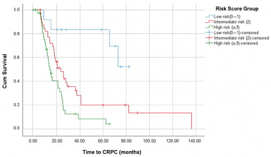
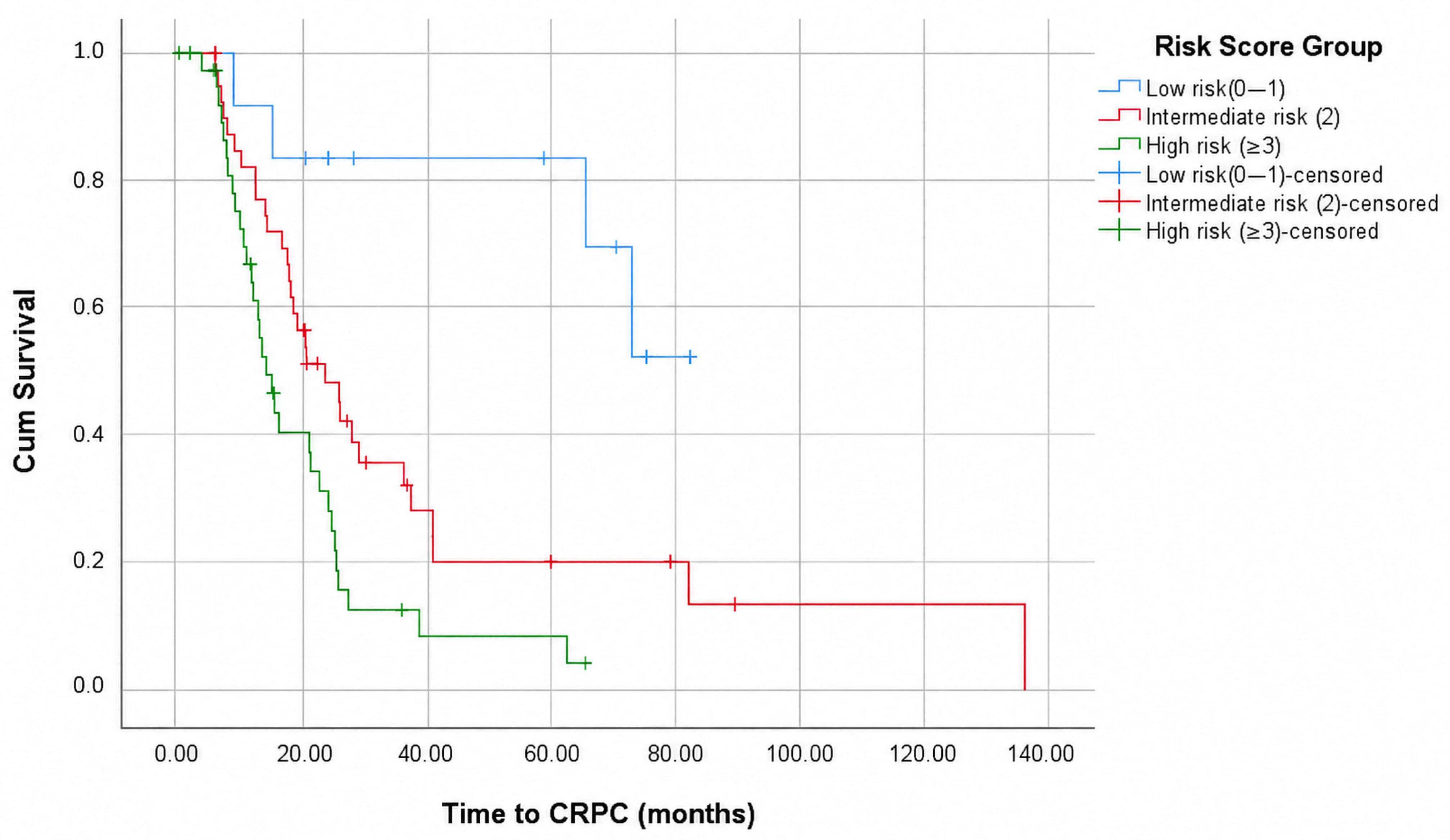
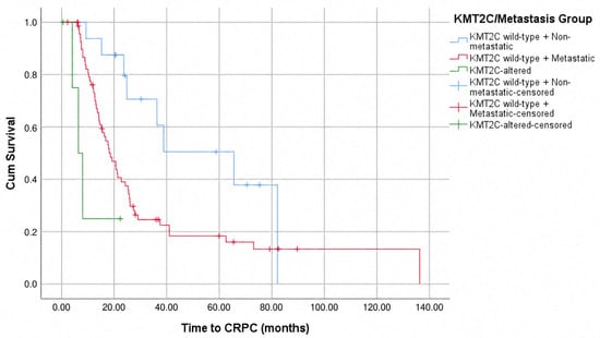
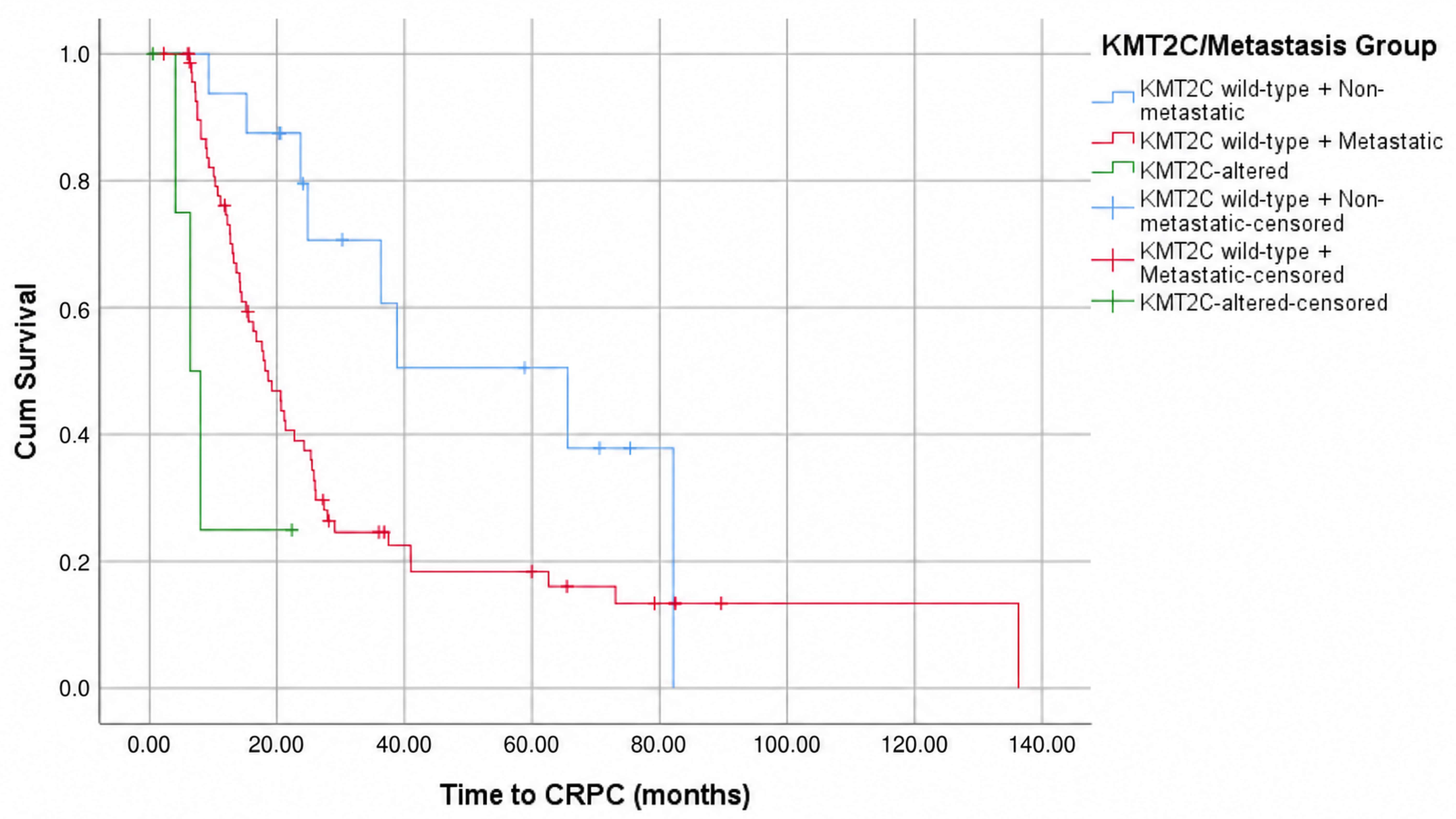
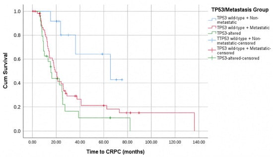
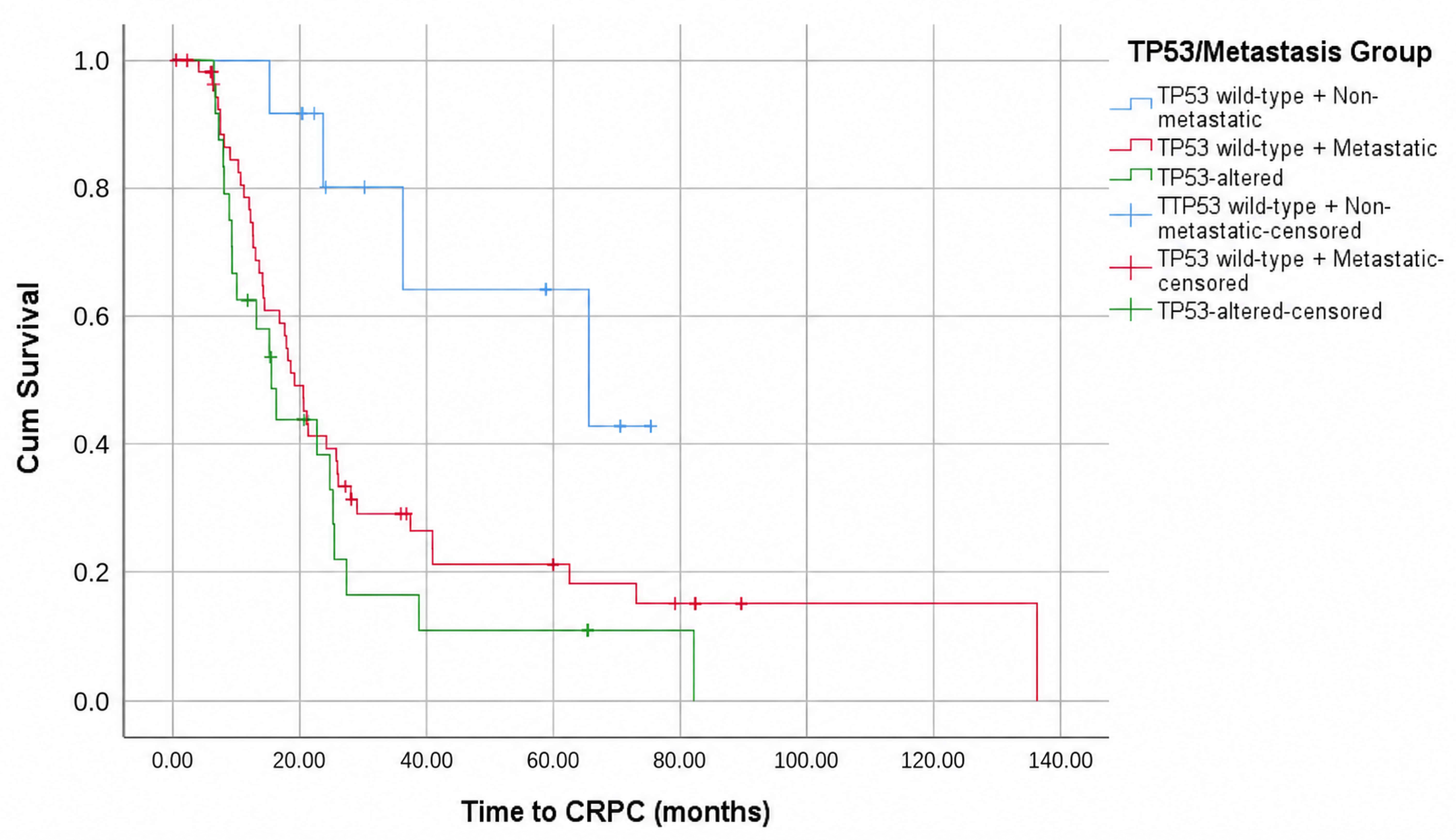
Among composite variables, high genomic burden (17.9 vs. 25.9 months; p = 0.011) and the high-risk genomic group (15.5 vs. 23.7 months; p = 0.031) were significantly associated with shorter time to CRPC (Table 4). The exploratory risk score stratified patients into low (median not reached), intermediate (23.7 months), and high-risk (14.3 months) groups (overall p < 0.001), with all pairwise comparisons significant (Figure 1).The KMT2C/metastasis group demonstrated a significant stepwise pattern: non-metastatic KMT2C wild-type (65.6 months), metastatic KMT2C wild-type (18.5 months), and KMT2C-altered (6.4 months; overall p = 0.001), with significant pairwise differences between the non-metastatic group and both other groups (p = 0.015 and p = 0.001) (Figure 2). The TP53/metastasis group showed a similar pattern (65.6 vs. 19.2 vs. 15.5 months; overall p = 0.003) (Figure 3). All results are summarized in Table 4.
Figure 1.
Kaplan-Meier curves for time to CRPC according to the exploratory composite clinical-genomic risk score. Low risk (n = 12), Intermediate risk (n = 40), High risk (n = 40). Overall log-rank p < 0.001. Tick marks represent censored observations.
Figure 2.
Kaplan-Meier curves for time to CRPC according to KMT2C alteration status and metastatic disease. KMT2C wild-type non-metastatic (n = 16), KMT2C wild-type metastatic (n = 71), KMT2C-altered (n = 5). Median: 65.6, 18.5, 6.4 months. Overall log-rank p = 0.001. Tick marks represent censored observations.
Figure 3.
Kaplan-Meier curves for time to CRPC according to TP53 alteration status and metastatic disease. TP53 wild-type non-metastatic (n = 12), TP53 wild-type metastatic (n = 56), TP53-altered (n = 24). Median: 65.6, 19.2, 15.5 months. Overall log-rank p = 0.003. Tick marks represent censored observations.
3.5. Time to CRPC: Cox Regression Analyses
3.5.1. Univariable Analyses
LATITUDE high-risk classification (HR = 2.198, p = 0.002), high-volume disease (HR = 3.063, p = 0.001), de novo metastatic disease (HR = 2.642, p = 0.010), high ISUP grade (HR = 2.338, p = 0.020), KMT2C alteration (HR = 3.589, p = 0.033), TP53 alteration (HR = 1.720, p = 0.045), high genomic burden (HR = 1.916, p = 0.012), high-risk genomic group (HR = 1.745, p = 0.034), KMT2C-altered vs. non-metastatic (HR = 7.446, p = 0.003), TP53-altered vs. non-metastatic (HR = 4.774, p = 0.004), and risk score high vs. low (HR = 7.504, p < 0.001) were significantly associated with time to CRPC. AR signaling pathway alteration was not significantly associated with time to CRPC (HR = 1.324, 95% CI: 0.762–2.302, p = 0.319) (Supplementary Figure S1). Full results are in Table 5.
Table 5.
Univariable Cox Proportional Hazards Regression for Time to CRPC (n = 92).
3.5.2. Multivariable Analyses
Five multivariable models were constructed. Model 2 (metastatic status, ISUP grade, KMT2C, TP53, genomic burden) demonstrated the best fit (−2LL = 464.300), with de novo metastatic disease (HR = 3.055, p = 0.006), KMT2C alteration (HR = 6.804, p = 0.003), and high genomic burden (HR = 1.917, p = 0.032) as independent predictors. Model 4, the most parsimonious, identified KMT2C (HR = 6.726, p = 0.003) and ISUP grade 4–5 (HR = 2.844, p = 0.007) as sole independent predictors. Model 3 confirmed the independent prognostic value of the exploratory risk score (high vs. low: HR = 7.904, p = 0.001). Results are summarized in Table 6.
Table 6.
Multivariable Cox Proportional Hazards Regression for Time to CRPC.
3.6. Overall Survival Analyses
3.6.1. Overall Survival from ADT Initiation (OS-ADT)
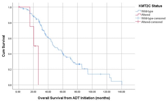
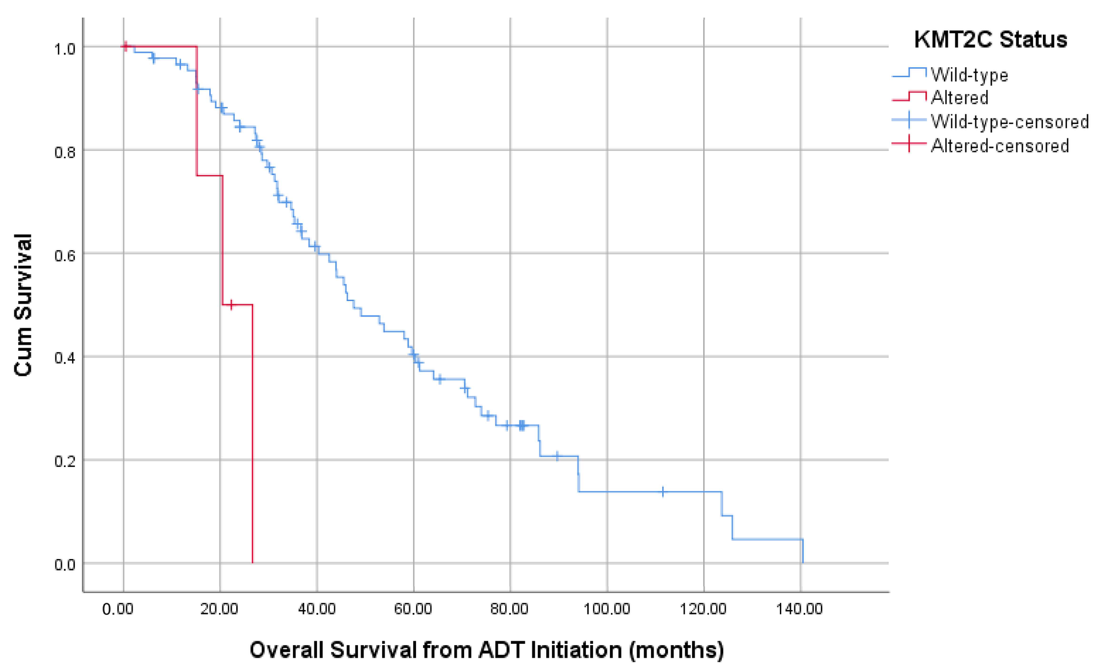
KMT2C alteration (20.5 vs. 38.4 months; p = 0.005) (Figure 4), ISUP grade 4–5 (35.3 vs. 88.5 months; p = 0.021), LATITUDE high risk (35.1 vs. 44.0 months; p = 0.009), TP53 alteration (34.7 vs. 44.0 months; p = 0.049), the high-risk genomic group (p = 0.043), and the risk score (p = 0.006) were significantly associated with shorter OS-ADT. MMR deficiency showed a borderline association (20.7 vs. 44.0 months; p = 0.060). AR signaling pathway alteration was not significantly associated with OS-ADT (median 52.9 vs. 43.9 months; HR = 1.491, p = 0.186) (Supplementary Figure S2). In multivariable analysis, KMT2C (HR = 4.730, p = 0.019), TP53 (HR = 1.810, p = 0.038), and ISUP grade 4–5 (HR = 2.364, p = 0.050) were independently associated with shorter OS-ADT.
Figure 4.
Kaplan-Meier curves for OS from ADT initiation according to KMT2C alteration status. KMT2C wild-type (n = 87) vs. KMT2C-altered (n = 5). Median OS: 38.4 vs. 20.5 months. Log-rank p = 0.005. Tick marks represent censored observations.
3.6.2. Overall Survival from Diagnosis (OS-Diagnosis)
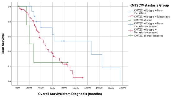
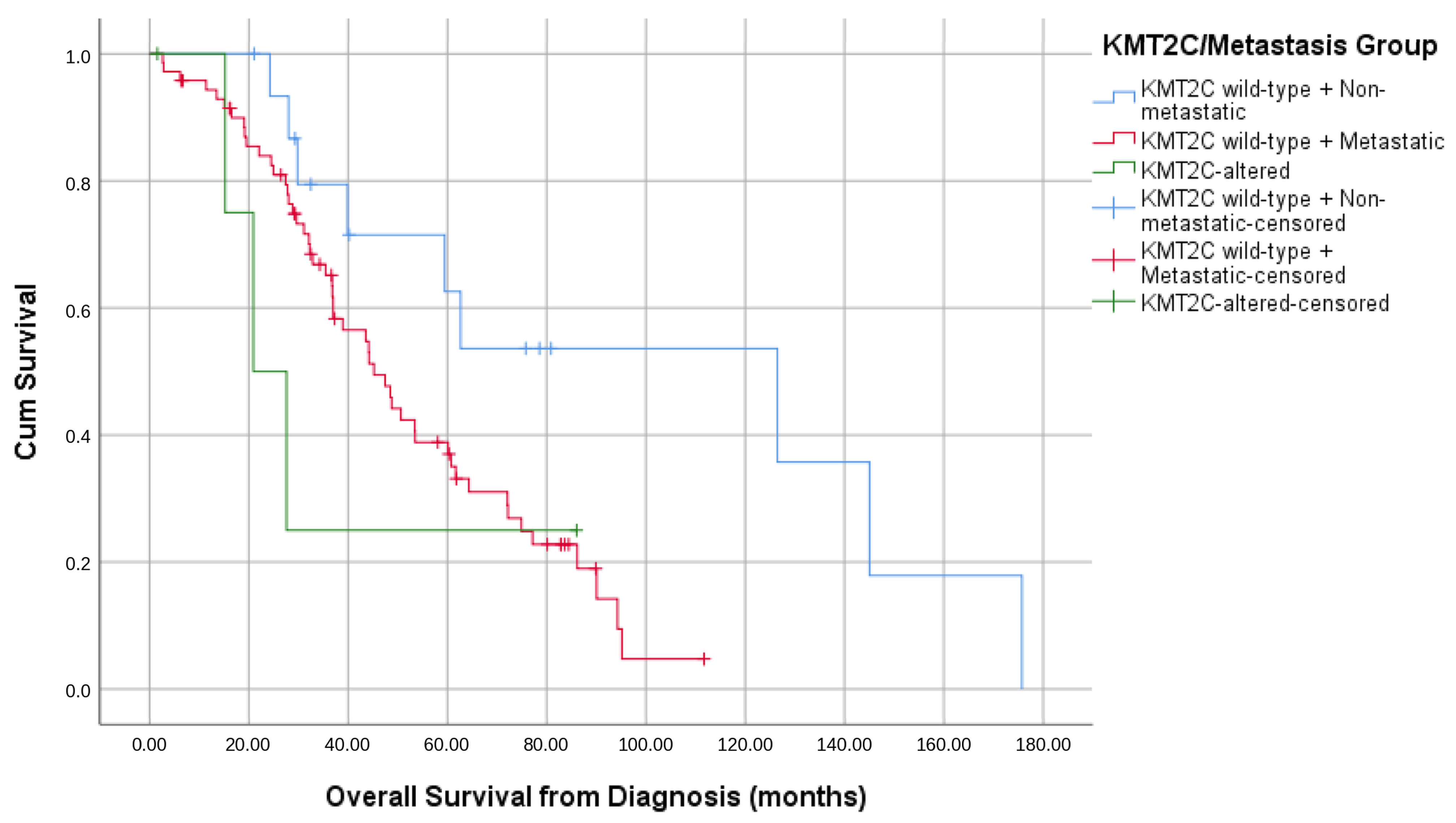
ISUP grade 4–5 (43.0 vs. 94.0 months; p = 0.002), de novo metastatic disease (44.0 vs. 126.0 months; p = 0.006), the KMT2C/metastasis group (p = 0.044) (Figure 5), and the risk score (p = 0.008) were significantly associated with shorter OS-diagnosis. KMT2C alteration, TP53, and the high-risk genomic group did not reach significance in OS-diagnosis analyses, suggesting their prognostic impact is predominantly confined to the post-ADT phase. AR signaling pathway alteration was not significantly associated with OS-diagnosis (median 50.7 vs. 44.2 months; HR = 1.268, p = 0.425) (Supplementary Figure S3). In multivariable analysis, ISUP grade 4–5 (HR = 2.504, p = 0.029) and KMT2C-altered vs. non-metastatic (HR = 5.101, p = 0.030) were independent predictors. Results are in Table 7.
Figure 5.
Kaplan-Meier curves for OS from diagnosis according to KMT2C alteration status and metastatic disease. KMT2C wild-type non-metastatic (n = 16), KMT2C wild-type metastatic (n = 71), KMT2C-altered (n = 5). Median OS: 126.0, 45.0, 24.0 months. Overall log-rank p = 0.048. Tick marks represent censored observations.
Table 7.
Overall Survival Analyses: Kaplan-Meier and Cox Regression Results.
4. Discussion
This study evaluated whether NGS panel findings obtained during the hormone-sensitive phase of prostate cancer were associated with time to CRPC and overall survival. In our real-world cohort of 92 patients, alterations in KMT2C and TP53, higher genomic burden, and metastatic disease at diagnosis were independently associated with earlier castration resistance. In contrast, classical HRR deficiency was not significantly associated with this endpoint. These results suggest that genomic findings from the hormone-sensitive stage may provide prognostic information beyond routine clinical factors, and could help guide treatment intensification before castration resistance develops.
4.1. KMT2C: An Epigenetic Predictor of Early Castration Resistance
KMT2C alteration was the strongest genomic predictor in this study. Patients with KMT2C alterations developed CRPC at a median of 6.4 months from ADT initiation, compared to 21.3 months in wild-type patients, and this remained independent of metastatic status and ISUP grade in multivariable analysis (HR = 6.804, p = 0.003). KMT2C was also the strongest independent predictor of OS from ADT initiation (HR = 4.730, p = 0.019), with a median OS of 20.5 versus 38.4 months.
These findings are supported by Zhu et al., who reported significantly shorter castration resistance-free survival (9.9 vs. 22.0 months, p = 0.015) and OS (71.9 vs. 137.4 months, p = 0.012) in KMT2C-mutated patients, with KMT2C confirmed as an independent predictor of OS (HR = 3.815) [16]. The agreement between two independent cohorts using different biopsy approaches—liquid biopsy versus tissue-based NGS—and from geographically distinct populations strengthens the clinical relevance of this finding. Our cohort showed an even shorter median time to CRPC, which may reflect differences in disease stage at NGS analysis, the earlier disease phase studied, or population-specific characteristics.
The mechanistic basis for this aggressive behavior has been clarified by Guo et al. [17]. Under ADT, KMT2C is normally recruited to enhancers of AR-regulated genes to maintain the luminal adenocarcinoma lineage. When KMT2C is lost, ASPP2 expression decreases, triggering ΔNp63-dependent transdifferentiation into double-negative prostate cancer (DNPC)—a phenotype lacking both AR and neuroendocrine markers. This DNPC state sustains tumor growth through fatty acid synthesis, bypassing androgen dependence entirely. This mechanism directly explains the rapid CRPC development seen in KMT2C-altered patients in our cohort.
To our knowledge, this is the first study to report the prognostic significance of tissue-based KMT2C alteration during the hormone-sensitive phase in a Turkish patient cohort, contributing real-world evidence from a population underrepresented in the genomic literature. KMT2C testing at the time of initial NGS may identify patients at high risk for early castration resistance who could benefit from treatment intensification or earlier inclusion in biomarker-driven trials.
4.2. The HRR Paradox: Why Did HRR Deficiency Fail to Predict CRPC?
One of the unexpected findings of this study was the lack of prognostic significance for HRR deficiency, despite its central role in the genomic landscape of advanced prostate cancer. Neither the restricted HRR definition nor the broader DDR deficiency variable was associated with time to CRPC or overall survival in any analysis.
A key explanation lies in the unusually high ATR alteration frequency in our cohort (35.9%), which drove HRR deficiency prevalence to 53.3%—well above the 19–28% reported in landmark mCRPC studies [10,13]. ATR is a DNA damage-sensing kinase that operates upstream of classical HRR effectors; while it is technically grouped within broader DDR classifications, its biological role differs substantially from core HRR genes such as BRCA1/2 and ATM. In the PROfound trial, ATR-altered patients were placed in Cohort B alongside 12 other non-BRCA/ATM genes, where olaparib showed markedly attenuated benefit compared to Cohort A [13]. The enrichment of ATR alterations in our cohort therefore introduced substantial heterogeneity into the HRR group, likely diluting any true prognostic signal. Consistent with this interpretation, separate analysis of HRR_core—comprising only BRCA1, BRCA2, and ATM—also failed to reach significance, though the small number of patients in this subgroup limits definitive conclusions.
Beyond the compositional issue, there is a broader biological argument. HRR deficiency may confer therapeutic vulnerability—particularly to PARP inhibitors—without necessarily accelerating the pace of castration resistance during the hormone-sensitive phase. Annala et al. showed that BRCA2 and ATM defects predict resistance to first-line abiraterone and enzalutamide in mCRPC [11], but the castration-sensitive phase represents a biologically distinct context where HRR status may carry less prognostic weight. Real-world data support this: Hommerding et al. reported HRR mutation prevalence of 20.8% in a mixed primary and mCRPC cohort, with BRCA2 frequency lower than in landmark mCRPC series, reflecting the stage-dependent enrichment of these alterations [18].
The unexpected direction of BRCA2 in our Kaplan-Meier analysis—where BRCA2-altered patients showed numerically longer time to CRPC—may also reflect the inability to distinguish germline from somatic alterations in our tissue-based panel. Germline BRCA2 carriers may exhibit a distinct clinical trajectory compared to those with somatic BRCA2 loss, and the absence of this distinction represents a recognized limitation of retrospective tissue NGS studies.
Taken together, the failure of HRR deficiency to predict time to CRPC in our cohort does not contradict its established role in advanced disease, but rather highlights that its prognostic relevance may be phase-dependent and that panel composition—particularly ATR inclusion—substantially influences HRR prevalence estimates across studies.
4.3. TP53 and Genomic Burden: The Case for Cumulative Genomic Damage
Beyond KMT2C, both TP53 alteration and high genomic burden independently predicted a shorter time to CRPC in the multivariable analysis. This finding suggests that castration resistance may reflect the combined effect of alterations across several pathways, rather than the impact of a single gene alone.
TP53 alteration was present in 26.1% of patients and was associated with shorter time to CRPC (15.5 vs. 23.7 months, p = 0.043) and shorter OS from ADT initiation (34.7 vs. 44.0 months, p = 0.049). In multivariable analysis incorporating metastatic status, ISUP grade, KMT2C, and genomic burden, TP53 lost independent significance for time to CRPC—suggesting that its prognostic effect is partly shared with other variables in the model. However, it retained independent significance for OS-ADT (HR = 1.810, p = 0.038), indicating that TP53 contributes prognostic information beyond CRPC development alone. This is consistent with findings from Annala et al., who demonstrated that TP53 defects independently predict primary resistance to abiraterone and enzalutamide in treatment-naive mCRPC [11], and with the mechanistic work of Mu et al. showing that compound TP53 and RB1 loss drives lineage plasticity through SOX2 upregulation [12]. Although concurrent TP53 and RB1 loss was rare in our cohort (n = 2), the independent prognostic impact of TP53 alone suggests that even partial disruption of p53-mediated tumor suppression is clinically meaningful during the hormone-sensitive phase.
High genomic burden—defined as alterations in two or more dysregulated pathways—was an independent predictor of shorter time to CRPC in our best-fitting multivariable model (HR = 1.917, p = 0.032). This finding aligns with the broader principle that genomic complexity, rather than any single alteration, reflects the biological aggressiveness of the disease. Patients with high genomic burden had a median time to CRPC of 17.9 months versus 25.9 months in the low burden group, a difference that remained significant after adjusting for metastatic status, ISUP grade, and KMT2C.
The exploratory composite risk score, which combinedKMT2C mutation, TP53 mutation, de novo metastatic disease, high ISUP grade, and high genomic burden, provided the clearest separation of risk in this analysis. Patients in the high-risk group had a median time to CRPC of 14.3 months, whereas the median was not reached in the low-risk group (HR = 7.904, p = 0.001). Because this score was developed post hoc, it should be tested prospectively before it is used in clinical practice. Still, these findings suggest that combining genomic and clinical features may offer a practical way to stratify patients at diagnosis.
4.4. MMR Deficiency: A Temporal Dissociation with Clinical Implications
MMR deficiency was identified in 16.3% of patients in this cohort—a rate higher than the 2–5% typically reported in mCRPC series, likely reflecting differences in panel composition, disease phase, and patient selection. Notably, MMR deficiency showed a strikingly different pattern from other genomic variables: it did not predict time to CRPC (p = 0.843) but was associated with shorter OS from ADT initiation, with a borderline significant difference in median OS of 20.7 versus 44.0 months (p = 0.060).
This temporal dissociation—where MMR deficiency influences survival but not the pace of castration resistance—is biologically plausible. MMR-deficient tumors accumulate somatic mutations at a higher rate, generating a hypermutated phenotype that may not inherently accelerate androgen escape but does progressively reshape the tumor microenvironment and immune landscape over time. The clinical effect of hypermutation may therefore become clearer after CRPC develops, when tumor heterogeneity is greater and immune escape mechanisms are more established.
The practical implication of this finding is significant. Boiarsky et al. demonstrated that MMR-deficient mCRPC patients treated with pembrolizumab experienced dramatically improved outcomes compared to MMR-proficient patients, with a median radiographic progression-free survival of 54 versus 3.7 months and a median OS not reached versus 17 months [19]. Four patients achieved durable remissions exceeding 2.5 years. These results, combined with the AUA/SUO 2026 guideline recommendation to offer pembrolizumab in dMMR/MSI-H mCRPC [20], establish a clear treatment pathway for this subset.
If MMR deficiency does not shorten time to CRPC, these patients may look similar to MMR-proficient patients during the hormone-sensitive phase. However, their prognosis after CRPC appears worse, and they may be candidates for immunotherapy once progression occurs. Identifying MMR-deficient patients before CRPC could therefore help clinicians plan ahead, including the early assessment of pembrolizumab eligibility at progression and consideration of clinical trials testing immunotherapy combinations with ADT in the hormone-sensitive setting.
4.5. AR Signaling Pathway in the Context of Co-Occurring Genomic Alterations
AR signaling pathway alterations were found in 30.4% of patients in our cohort, which is in line with previously reported rates in advanced prostate cancer [10]. In our analyses, AR pathway alterations were not independently associated with time to CRPC or overall survival. Still, their presence may be relevant when they occur together with other genomic changes. For example, when AR pathway alterations coexist with TP53 loss, SOX2-mediated transcriptional changes may support androgen-independent growth and promote lineage plasticity [12]. In MMR-deficient tumors, concurrent AR pathway alterations may also influence the tumor microenvironment and could be relevant to immunotherapy response, including pembrolizumab benefit [19,20]. In HRR-deficient tumors, AR amplification and splice variant expression have been described as possible mechanisms of resistance to PARP inhibitors [13]. For this reason, AR pathway status may be more informative when assessed together with other alterations rather than on its own. We did not perform formal subgroup analyses according to AR status within the TP53, HRR, or MMR-deficient subgroups because the sample size was too small for reliable comparisons. These possible interactions should be examined in larger prospective studies.
4.6. Limitations and Future Perspectives
This study has several limitations. The retrospective single-center design and small sample size limit statistical power, particularly for genomic subgroups with few positive cases such as KMT2C (n = 5). The inability to distinguish germline from somatic alterations may partly explain unexpected findings such as the non-significant direction of BRCA2, and paired germline testing would strengthen future analyses. Treatment heterogeneity across the cohort—including variation in ARPI use and chemotherapy—may also act as a confounding factor despite multivariable adjustment. The composite risk score and high-risk genomic group were constructed posthoc and require independent validation before clinical application.External validation of the present findings, particularly the prognostic role of KMT2C alteration, in larger publicly available cohorts such as the TCGA prostate adenocarcinoma dataset or SU2C-funded prostate cancer cohorts represents an important priority for future research.
Despite these limitations, the findings support several priorities for future research. The link between KMT2C loss and DNPC transdifferentiation [17] suggests that fatty acid synthesis inhibition may be a rational therapeutic target in KMT2C-altered patients. For MMR-deficient patients, whether earlier pembrolizumab deployment—in the hormone-sensitive phase—can prevent the poor post-CRPC outcomes observed here warrants prospective evaluation. More broadly, our data support routine NGS testing at ADT initiation not only for PARP inhibitor and immunotherapy eligibility in established mCRPC, but as a tool for proactive risk stratification before castration resistance develops.
5. Conclusions
This study demonstrates that NGS panel analysis during the hormone-sensitive phase of prostate cancer provides clinically meaningful prognostic information beyond established clinical parameters. KMT2C alteration emerged as the strongest independent genomic predictor of both early castration resistance and overall survival, a finding now supported by mechanistic evidence and corroborated by an independent cohort. TP53 alteration and high genomic burden were additional independent predictors, highlighting the prognostic value of cumulative genomic dysregulation. Conversely, HRR deficiency did not predict time to CRPC, likely due to the compositional influence of high ATR alteration frequency in this cohort. MMR deficiency demonstrated a clinically important temporal dissociation—not accelerating CRPC development but significantly shortening post-ADT survival—underscoring the value of early genomic identification for timely pembrolizumab planning. Taken together, these findings support the routine integration of NGS testing at ADT initiation as both a prognostic tool and a platform for biomarker-guided treatment decisions in prostate cancer.
Supplementary Materials
The following supporting information can be downloaded at https://www.mdpi.com/article/10.3390/curroncol33070416/s1, Figure S1: Kaplan–Meier curves for time to castration-resistant prostate cancer (CRPC) according to AR signaling pathway alteration status; Figure S2: Kaplan–Meier curves for overall survival from ADT initiation according to AR signaling pathway alteration status; Figure S3: Kaplan–Meier curves for overall survival from prostate cancer diagnosis according to AR signaling pathway alteration status.; Table S1: Variant-Level Details of KMT2C Alterations Identified in the Study Cohort (n= 5 patients).
Author Contributions
Conceptualization, M.T. and M.Ç.B.; methodology, M.T.; formal analysis, M.T.; investigation, M.T. and M.Ç.B.; resources, M.Ç.B.; data curation, M.T. and M.Ç.B.; writing—original draft preparation, M.T.; writing—review and editing, M.T. and M.Ç.B.; visualization, M.T.; project administration, M.T. All authors have read and agreed to the published version of the manuscript.
Funding
This research received no external funding.
Institutional Review Board Statement
The study was conducted in accordance with the Declaration of Helsinki, and approved by the Non-Interventional Clinical Research Ethics Committee of Aydın Adnan Menderes University Faculty of Medicine (protocol code 2026/178; date of approval: 7 May 2026).
Informed Consent Statement
Patient consent was waived due to the retrospective nature of the study.
Data Availability Statement
The data presented in this study are available on request from the corresponding author due to privacy and ethical restrictions.
Acknowledgments
During the preparation of this manuscript, the authors used Claude Sonnet 4.6 (Anthropic, San Francisco, CA, USA; claude.ai, accessed May–June 2026) for academic language improvement and English translation assistance. The authors have reviewed and edited all AI-assisted content and take full responsibility for the accuracy and integrity of this publication.
Conflicts of Interest
The authors declare no conflicts of interest.
Abbreviations
The following abbreviations are used in this manuscript:
| ADT | Androgen deprivation therapy |
| AR | Androgen receptor |
| ARPI | Androgen receptor pathway inhibitor |
| ATR | Ataxia telangiectasia and Rad3-related protein |
| CRPC | Castration-resistant prostate cancer |
| DDR | DNA damage response |
| DNPC | Double-negative prostate cancer |
| FFPE | Formalin-fixed paraffin-embedded |
| HRR | Homologous recombination repair |
| HR | Hazard ratio |
| IQR | Interquartile range |
| ISUP | International Society of Urological Pathology |
| KM | Kaplan-Meier |
| mCRPC | Metastatic castration-resistant prostate cancer |
| mHSPC | Metastatic hormone-sensitive prostate cancer |
| MMR | Mismatch repair |
| NGS | Next-generation sequencing |
| OS | Overall survival |
| OS-ADT | Overall survival from ADT initiation |
| PCa | Prostate cancer |
| PCWG3 | Prostate Cancer Working Group 3 |
| PSA | Prostate-specific antigen |
| SD | Standard deviation |
| UMI | Unique molecular identifier |
References
- Bray, F.; Laversanne, M.; Sung, H.; Ferlay, J.; Siegel, R.L.; Soerjomataram, I.; Jemal, A. Global cancer statistics 2022: GLOBOCAN estimates of incidence and mortality worldwide for 36 cancers in 185 countries. CA Cancer J. Clin. 2024, 74, 229–263. [Google Scholar] [CrossRef] [Scilit] [PubMed]
- Turan, G.; Turan, M.; Basbinar, Y.; Ellidokuz, H. Prostate cancer in Türkiye: Trend analysis of incidence and mortality rates. Semin. Oncol. 2025, 52, 152391. [Google Scholar] [CrossRef] [Scilit] [PubMed]
- Bonde, T.M.; Westerberg, M.; Aly, M.; Eklund, M.; Adolfsson, J.; Bill-Axelson, A.; Garmo, H.; Stattin, P.; Robinson, D. Time to castration-resistant prostate cancer and prostate cancer death according to PSA response in men with non-metastatic prostate cancer treated with gonadotropin-releasing hormone agonists. Scand. J. Urol. 2022, 56, 169–175. [Google Scholar] [CrossRef] [Scilit] [PubMed]
- Davies, A.; Conteduca, V.; Zoubeidi, A.; Beltran, H. Biological, evolution of castration-resistant prostate cancer. Eur. Urol. Focus 2019, 5, 147–154. [Google Scholar] [CrossRef] [Scilit] [PubMed]
- Shao, Y.; Liu, Z.; Ma, J.; Feng, Y.; Yu, J.; Huang, H.; Liu, Y.; Niu, Y.; Wang, Y. Molecular mechanisms and classification of castration-resistant prostate cancer: Insights into androgen receptor, cancer stem cells, and neuroendocrine features. Curr. Urol. 2026, 20, 1–14. [Google Scholar] [CrossRef] [Scilit] [PubMed]
- Pan, C.; He, Y.; Wang, H.; Yu, Y.; Li, L.; Huang, L.; Lyu, M.; Ge, W.; Yang, B.; Sun, Y.; et al. Identifying patients with rapid progression from hormone-sensitive to castration-resistant prostate cancer: A retrospective study. Mol. Cell Proteom. 2023, 22, 100613. [Google Scholar] [CrossRef] [Scilit] [PubMed]
- Kim, M.; Lee, J.; Jeong, C.W.; Ku, J.H.; Kim, H.H.; Kwak, C. Prostate-specific antigen kinetic profiles during androgen deprivation therapy as prognostic factors in castration-resistant prostate cancer. Urol. Oncol. 2015, 33, 203.e1–203.e9. [Google Scholar] [CrossRef] [Scilit] [PubMed]
- Mangolini, A.; Rocca, C.; Bassi, C.; Ippolito, C.; Negrini, M.; Dell’Atti, L.; Lanza, G.; Gafà, R.; Bianchi, N.; Pinton, P.; et al. Detection of disease-causing mutations in prostate cancer by NGS sequencing. Cell Biol. Int. 2022, 46, 1047–1061. [Google Scholar] [CrossRef] [Scilit] [PubMed]
- Karantanos, T.; Corn, P.G.; Thompson, T.C. Prostate cancer progression after androgen deprivation therapy: Mechanisms of castrate-resistance and novel therapeutic approaches. Oncogene 2013, 32, 5501–5511. [Google Scholar] [CrossRef] [Scilit] [PubMed]
- Abida, W.; Cyrta, J.; Heller, G.; Prandi, D.; Armenia, J.; Coleman, I.; Cieslik, M.; Benelli, M.; Robinson, D.; Van Allen, E.M.; et al. Genomic correlates of clinical outcome in advanced prostate cancer. Proc. Natl. Acad. Sci. USA 2019, 116, 11428–11436. [Google Scholar] [CrossRef] [Scilit] [PubMed]
- Annala, M.; Vandekerkhove, G.; Khalaf, D.; Taavitsainen, S.; Beja, K.; Warner, E.W.; Sunderland, K.; Kollmannsberger, C.; Eigl, B.J.; Finch, D.; et al. Circulating tumor DNA genomics correlate with resistance to abiraterone and enzalutamide in prostate cancer. Cancer Discov. 2018, 8, 444–457. [Google Scholar] [CrossRef] [Scilit] [PubMed]
- Mu, P.; Zhang, Z.; Benelli, M.; Karthaus, W.R.; Hoover, E.; Chen, C.C.; Wongvipat, J.; Ku, S.Y.; Gao, D.; Cao, Z.; et al. SOX2 promotes lineage plasticity and antiandrogen resistance in TP53- and RB1-deficient prostate cancer. Science 2017, 355, 84–88. [Google Scholar] [CrossRef] [Scilit] [PubMed]
- de Bono, J.; Mateo, J.; Fizazi, K.; Saad, F.; Shore, N.; Sandhu, S.; Chi, K.N.; Sartor, O.; Agarwal, N.; Olmos, D.; et al. Olaparib for metastatic castration-resistant prostate cancer. N. Engl. J. Med. 2020, 382, 2091–2102. [Google Scholar] [CrossRef] [Scilit] [PubMed]
- Almeeri, M.N.E.; Awies, M.; Constantinou, C. Prostate cancer, pathophysiology and recent developments in management: A narrative review. Curr. Oncol. Rep. 2024, 26, 1511–1519. [Google Scholar] [CrossRef] [Scilit] [PubMed]
- Scher, H.I.; Morris, M.J.; Stadler, W.M.; Higano, C.; Basch, E.; Fizazi, K.; Antonarakis, E.S.; Beer, T.M.; Carducci, M.A.; Chi, K.N.; et al. Trial design and objectives for castration-resistant prostate cancer: Updated recommendations from the Prostate Cancer Clinical Trials Working Group 3. J. Clin. Oncol. 2016, 34, 1402–1418. [Google Scholar] [CrossRef] [Scilit] [PubMed]
- Zhu, S.; Xu, N.; Liang, J.; Zhao, F.; Wang, Z.; Ni, Y.; Dai, J.; Zhao, J.; Zhang, X.; Chen, J.; et al. Mutations in epigenetic regulator KMT2C detected by liquid biopsy are associated with worse survival in prostate cancer patients. Oncol. Res. 2023, 31, 605–614. [Google Scholar] [CrossRef] [Scilit] [PubMed]
- Guo, J.; Li, N.; Liu, Q.; Hao, Z.; Zhu, G.; Wang, X.; Wang, H.; Pan, Q.; Xu, B.; Han, Y.; et al. KMT2C deficiency drives transdifferentiation of double-negative prostate cancer and confers resistance to AR-targeted therapy. Cancer Cell 2025, 43, 1261–1278. [Google Scholar] [CrossRef] [Scilit] [PubMed]
- Hommerding, M.; Hommerding, O.; Bernhardt, M.; Kreft, T.; Sanders, C.; Tischler, V.; Basitta, P.; Pelusi, N.; Wulf, A.L.; Ohlmann, C.H.; et al. Real-world data on the prevalence of BRCA1/2 and HRR gene mutations in patients with primary and metastatic castration resistant prostate cancer. World J. Urol. 2024, 42, 491. [Google Scholar] [CrossRef] [Scilit] [PubMed]
- Boiarsky, D.; Gulhan, D.C.; Savignano, H.; Lakshminarayanan, G.; McClure, H.M.; Silver, R.; Hirsch, M.S.; Sholl, L.M.; Choudhury, A.D.; Ananda, G.; et al. A panel-based mutational signature of mismatch repair deficiency is associated with durable response to pembrolizumab in metastatic castration-resistant prostate cancer. Clin. Genitourin. Cancer 2024, 22, 558–568. [Google Scholar] [CrossRef] [Scilit] [PubMed]
- American Urological Association; Society of Urologic Oncology. Advanced Prostate Cancer: AUA/SUO Guideline 2026. Available online: https://www.auanet.org/guidelines-and-quality/guidelines/advanced-prostate-cancer (accessed on 2 June 2026).

Disclaimer/Publisher’s Note: The statements, opinions and data contained in all publications are solely those of the individual author(s) and contributor(s) and not of MDPI and/or the editor(s). MDPI and/or the editor(s) disclaim responsibility for any injury to people or property resulting from any ideas, methods, instructions or products referred to in the content. |
© 2026 by the authors. Licensee MDPI, Basel, Switzerland. This article is an open access article distributed under the terms and conditions of the Creative Commons Attribution (CC BY) license.