


Simple Summary
Lynch syndrome is the most common hereditary condition associated with an increased risk of endometrial cancer, yet the biological mechanisms underlying cancer development in these women are not fully understood. Recent research suggests that the microbiota, the community of microorganisms living in the human body, may influence cancer risk through its effects on inflammation, immune function, hormone metabolism, and cellular health. This review summarises current evidence on the relationship between the microbiota and Lynch syndrome-associated endometrial cancer. Available studies indicate that women with Lynch syndrome may harbour microbial imbalances, characterized by an increase in potentially detrimental bacteria and a reduction in beneficial microorganisms that support intestinal and immune health. Although the evidence is limited, these findings suggest that microbiota profiling could contribute to future strategies for cancer prevention, risk assessment, and personalized treatment in this high-risk population.
Abstract
Lynch syndrome is the most common hereditary syndrome predisposing to endometrial cancer, but the mechanisms contributing to tumor development remain incompletely understood. This review aimed to summarise current evidence on the composition and potential pathogenic role of the microbiota in Lynch syndrome-associated endometrial cancer and to explore its clinical implications. A systematic literature search of PubMed/MEDLINE and Embase was conducted between October and November 2025 using predefined terms related to microbiota, carcinogenesis, endometrial cancer, and Lynch syndrome. Available evidence suggests that alterations in the microbiota may contribute to carcinogenesis through chronic inflammation, immune modulation, metabolic changes, and bacterial genotoxicity. Studies in Lynch syndrome carriers with endometrial or colorectal cancer report an increased abundance of potentially pro-inflammatory Gram-negative bacteria, including Bacteroides fragilis and Fusobacterium nucleatum, together with a reduction in short-chain fatty acid-producing bacteria such as Faecalibacterium, Blautia, and Butyrivibrio. Microbial metabolites, including butyrate, may have protective effects, whereas bacterial β-glucuronidase activity may increase estrogen bioavailability and contribute to a tumor-promoting environment. Emerging evidence also suggests that the microbiota may influence treatment response and toxicity. Overall, current evidence supports a potential role for the microbiota in Lynch syndrome-associated endometrial carcinogenesis, although data remain limited. Further studies are needed to determine whether microbiota profiling could contribute to risk stratification, prevention, and personalized therapeutic strategies.
1. Introduction
The human microbiota plays a crucial role in maintaining health and modulating disease processes. Recent advances in molecular sequencing, especially next-generation sequencing (NGS) of the 16S rRNA gene, has uncovered complex microbial communities in body sites, including the endometrial cavity [1]. The uterine microbiota, previously believed to be sterile, is now recognized as a distinct ecosystem with implications for reproductive health and cancer development [2].
Endometrial cancer (EC) is the most common gynecological malignancy in developed countries, and growing evidence suggests that dysbiosis of the reproductive tract microbiome may contribute to its pathophysiology [3,4].
Lynch syndrome (LS) is a common hereditary genetic condition that significantly increases the risk of developing various types of cancers, particularly colorectal and endometrial cancers. This condition is characterized by a germline mutation in DNA mismatch repair (MMR) genes normally responsible for DNA repair processes. Depending on the mutation, LS confers an EC risk ranging from 13% to 57%. The risk is 34–54% for MLH1, 21–57% for MSH2/EPCAM, 16–49% for MSH6, and 13–26% for PMS2 [5,6]. LS is also associated with a markedly increased lifetime risk of colorectal cancer, up to 61%, along with variable but significant risks for ovarian (up to 38%), gastric (up to 9%), urinary tract (up to 28%), pancreatic (up to 6.2%), prostate (up to 23.8%), and brain cancer (up to 7.7%) [6]. While the genetic basis of LS is well established, the variable penetrance of EC among mutation carriers suggests the presence of additional factors. In line with other studies on this topic, such as those related to the relationship between microbiota and the penetrance of other genetic mutations like BRCA for breast and ovarian cancers [7], it is plausible that also in this setting, the microbiota plays a determining role and may influence cancer risk.
Recent research has started to investigate the composition of the gut and reproductive tract microbiota in LS patients, the potential mechanisms by which these microorganisms might influence carcinogenesis, and the clinical implications for risk assessment and prevention [8,9,10,11,12,13]. Although the field is still developing, several bacterial taxa and mechanistic pathways have been suggested as contributors to endometrial carcinogenesis in LS, including inflammation, metabolic reprogramming, and immune modulation.
This review summarises the latest research on the composition, regulation, and pathogenic implications of the endometrial microbiota. It emphasises the roles of the microbiota in carcinogenesis, metabolic regulation, and therapeutic responsiveness, paying particular attention to the mechanisms involved in the penetrance of EC within the LS.
2. Materials and Methods
A structured literature search was conducted in PubMed/MEDLINE and Embase between October and November 2025 to identify literature relevant to microbiota in carcinogenesis, with particular focus on endometrial cancer (EC), and Lynch syndrome (LS).
The search included the following combinations of terms: “Microbiota AND cancer”, “Microbiota AND carcinogenesis”, “Microbiota AND endometrial cancer”, “Microbiota AND cancer AND Lynch Syndrome”. Titles and abstracts were screened for relevance, and selected original research articles were independently reviewed by three authors (SA, VEB, and MC). Study selection and synthesis considered relevance to the review question, study design, directness of evidence for LS-associated EC, methodological limitations, publication recency, and journal quality. Disagreements were discussed among the authors to reach a consensus.
The evidence was synthesized narratively, distinguishing findings directly obtained in LS-associated EC from evidence derived from other cancer types and preclinical models. This article is a structured narrative review.
3. Discussion
3.1. Composition and Regulation of the Endometrial Microbiota
Although historically considered sterile, the endometrial cavity hosts a low-biomass microbial community. Colonisation mainly occurs through two routes: an ascending pathway from the lower genital tract via cervical mucus permeability, and hematogenous translocation from distant sites such as the gut and oral cavity [3,4]. Over the past decade, an additional factor appears to influence the establishment of the endometrial microbiota, namely the widespread use of assisted reproductive technologies (ART). Microbial contamination associated with these procedures, which has been reported with increasing frequency, is likely to contribute to the formation and/or modulation of the endometrial microbial environment. Studies show that microbial concordance between vaginal and endometrial samples reaches only about 75%, supporting the endometrium as a partially distinct niche [4].
A longitudinal metagenomic analysis uncovered a gradient of microbial diversity along the female reproductive tract, showing a higher bacterial load and lower diversity in the vagina dominated by Lactobacillus species, with a reduced bacterial abundance in the upper reproductive tract [1].
However, endometrial microbiota studies are limited by methodological inconsistencies, including the risk of sample contamination from the cervix or vagina during collection, and small, heterogeneous patient cohorts [14]. These issues highlight the need for standardised protocols but the idea of the presence of an endometrial microbiota is now widely accepted and studied thanks to improved techniques of sequencing [15].
Sex hormones (closely dependent by the microbial composition such as determined by the β-glucuronidase (β-GUS) activity from gut microbiota) significantly modulate the endometrial microbiota composition. Use of gonadotropin-releasing hormone (GnRH) analogues has been associated with changes in microbial composition [16]. Postmenopausal women generally show lower Lactobacillus abundance [17], while phases of the menstrual cycle impact microbial diversity, with reduced diversity during ovulation and the luteal phase [18].
The hormonal factor also affects the microbial environment of the endometrium, since endometrial epithelial cells secrete antimicrobial molecules such as defensins, secretory leukocyte protease inhibitor (SLPI) and lactoferrin. The production of these molecules is modulated by estrogen levels. Estrogen enhances endometrial antimicrobial defenses, while decreasing vaginal antimicrobial secretion, establishing a hormonal balance between reproductive tolerance and immune protection [19].
3.2. Microbiota Alterations in Endometrial Cancer
Several studies have documented significant microbiota shifts in endometrial pathology. Microbiota diversity indices further distinguish benign, hyperplastic, and malignant endometrial conditions [20]. α-diversity and β-diversity are significantly increased in EC compared with benign cohorts, with hyperplasia showing an intermediate pattern [14]. EC patients exhibit increased microbial diversity and reduced Lactobacillus levels compared with healthy women [4]. EC-associated taxa include Anaerococcus, Porphyromonas, Prevotella, Fusobacterium, Bacteroides, and Peptoniphilus [4]. Atopobium vaginae and Porphyromonas somerae have even been proposed as upper tract oncogenic biomarkers [14]. Moreover, elevated vaginal pH (>4.5) due to Lactobacillus depletion correlates with EC risk [14]. These shifts indicate a structural reorganization of microbial communities during tumorigenesis.
The intratumoral microbiome has also been linked to molecular cancer characteristics, including microsatellite instability (MSI). Eleven microbial genera, bacterial and viral, (Acetivibrio, Alphapapillomavirus, Apibacte, Brackiella, Bracovirus, Epulopiscium, Legionella, Marinitoga, Paeniclostridium, Polyomavirus, Wolbanchia) have been found to be significantly associated with MSI-positive EC, suggesting potential microbial modulation of genomic instability [21] and the presence of a “microbiome axis”, an innovative concept that underscores the systemic interconnection of microbial communities across body sites [3,22]. Dysbiosis in one compartment, particularly the gut, can influence others through immune and metabolic signaling. In fact, there is considerable evidence suggesting that patients with EC exhibit reduced gut microbial diversity compared to healthy controls, with depletion of Firmicutes, Clostridia, and Ruminococcaceae, and enrichment of Proteobacteria, Enterobacteriaceae, and Shigella [22]. These shifts promote pro-inflammatory environments and an immunosuppressive state conducive to tumor development.
Recent metagenomic studies highlight shared microbial signatures between vaginal and rectal sites in EC patients. In EC patients, the vaginal and rectal microbial compositions differ significantly from those observed in women with benign gynecological conditions or endometrial hyperplasia [23]. Specifically, a decrease in Lactobacillus and Limosilactobacillus is observed in the vaginal microbiota, while Prevotella and Peptoniphilus are reduced in the rectal microbiota. Conversely, there is an increase in Anaerococcus, Porphyromonas, Prevotella, and Peptoniphilus in the vagina, and Buttiaxella in the rectum of EC patients [23]. Some bacterial species, such as Prevotella timonensis and Peptoniphilus A, are shared between the vagina and rectum in women with EC, suggesting a possible microbial interaction or cross-talk between the two sites. Interestingly, a higher abundance of vaginal Lactobacillus is associated with less sharing of bacteria from the rectum, confirming the potential protective role of Lactobacillus in maintaining vaginal microbial balance and preventing the colonisation of potentially harmful bacteria. Metabolic analyses further reveal dysfunctions in amino acid, complex carbohydrate, and hormone metabolism in EC patients, suggesting that microbial alterations are not only compositional but also functional, and may contribute to the pathogenesis and progression of the disease [23].
3.3. Links Between Microbiota and Carcinogenesis
Hanahan (2022) highlights that the polymorphic variability of microbiomes across individuals and tissues can profoundly influence cancer initiation, progression, and response to therapy, acting as an enabling characteristic of carcinogenesis. Through mechanisms involving genomic instability, inflammation, and immune modulation, distinct microbiota can either promote or inhibit the acquisition of cancer hallmarks, thereby shaping tumor behaviour and therapeutic outcomes [24].
One of the principal hallmarks of cancer is inflammation. Microbial metabolites and structural components activate inflammatory pathways central to carcinogenesis. Increased lipopolysaccharide (LPS) levels from gut dysbiosis act on tight junctions and stimulate increased gut permeability, linking toll-like receptor 4 (TLR4) to a massive NF-κB activation, and promoting cytokine release (IL-6, TNF-α, IL-1β) and chronic inflammation [3,25].
In addition to the chronic inflammatory mechanism generated by the gut microbiota from a distance, the local endometrial microbiota can also exert influence. The mechanisms are related to Atopobium vaginae and Porphyromonas somerae infection of EC cells, which has been associated with elevated IL expression [26], suggesting direct local inflammatory involvement, and this has been confirmed in EC patients where overexpression of Micrococcus and IL-6 was found [27].
A key risk factor in EC is hyperestrogenism. Bacterial β-GUS, encoded by gut commensals such as Bacteroides fragilis and Escherichia coli, hydrolyzes conjugated estrogens, increasing their bioavailability [28,29,30]. The set of gut microbial genes capable of metabolizing estrogens, known as the estrobolome, plays a pivotal role in regulating systemic estrogen levels. Dysbiosis enhances β-GUS expression, which in turn, deconjugates hormones, augmenting circulating estrogen that then stimulates the target site and functions as a driver of endometrioid EC, as is widely recognized in the literature [31,32]. Elevated β-GUS activity and inflammatory cytokines (IL-6, IL-11, TGF-β) have been correlated with EC-associated microbial shifts and fungal overgrowth of Penicillium species [33].
The estrogens connection is also related to the metabolic state. Obesity, a major EC risk factor, alters microbial and hormonal homeostasis. Adipose tissue with its aromatase activity increases systemic estrogen production in postmenopausal women, stimulating endometrial proliferation [34]. Gut dysbiosis associated with obesity, characterized by altered Firmicutes/Bacteroidetes ratios and increased Akkermansia, further promotes metabolic inflammation [35].
In addition to having an impact on all mechanisms related to the onset of cancer, the microbiota also influences progression and metastasis. Specific bacterial taxa, such as Ruminococcus sp. N15.MGS-57, correlate with elevated plasma levels of unsaturated fatty acids (C16:1, C20:2), which accumulate in tumor tissues and activate mTOR signaling, enhancing EC invasiveness and metastasis [36].
In addition to promoting tumor growth, the microbiota can also play a protective role in certain situations. Short-chain fatty acids (SCFA), particularly butyrate, exert anti-inflammatory and anti-proliferative effects [37]. Butyrate-producing genera such as Faecalibacterium and Butyrivibrio promote regulatory T-cell expansion and immune tolerance. Conversely, secondary bile acids may induce DNA damage and pro-tumorigenic NF-κB signaling [37].
3.4. Microbiota and Endometrial Cancer Penetrance in Lynch Syndrome
Studies on the penetrance of EC in patients with LS are few and have small sample sizes. However, in line with different cancer studies, emerging data suggest that women with LS who develop EC display a distinct microbiota profile compared to healthy controls and sporadic EC cases. In a pilot study, LS patients with gynecological cancers (including EC) showed significant enrichment of gut bacteria such as Faecalibacterium prausnitzii, Parabacteroides distasonis, Ruminococcus bromii, Bacteroides plebeius, Bacteroides fragilis, and Bacteroides uniformis [8]. These taxa are known for their roles in modulating inflammation and influencing immune responses. Notably, the same microbiota pattern was observed in LS patients with colorectal and gynecological cancers, suggesting a possible LS-specific microbial signature rather than a cancer-type-specific one [8].
In an observational study comparing the fecal microbiome of LS patients with cancer (LS-C) to those without cancer (LS-NC), LS-C patients showed higher proportions of Bacteroidetes and Verrucomicrobia, and lower Firmicutes, compared to LS-NC. At the genus level, LS-C patients showed increased Akkermansia and Bacteroides, and decreased Pseudobutyrivibrio. Although these differences were not statistically significant due to the small sample size, the results suggest that the fecal microbiome in LS patients with extraintestinal cancers, including EC, may differ from those without cancer [9].
In the vaginal and cervical microbiota, women with EC (not limited to LS) exhibited increased prevalence of Dialister pneumosintes and Mobiluncus curtisii, while protective species like Lactobacillus iners and Bifidobacterium were reduced [11]. These findings indicate that both gut and reproductive tract dysbiosis may contribute to carcinogenesis in LS [9].
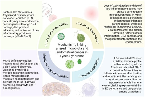
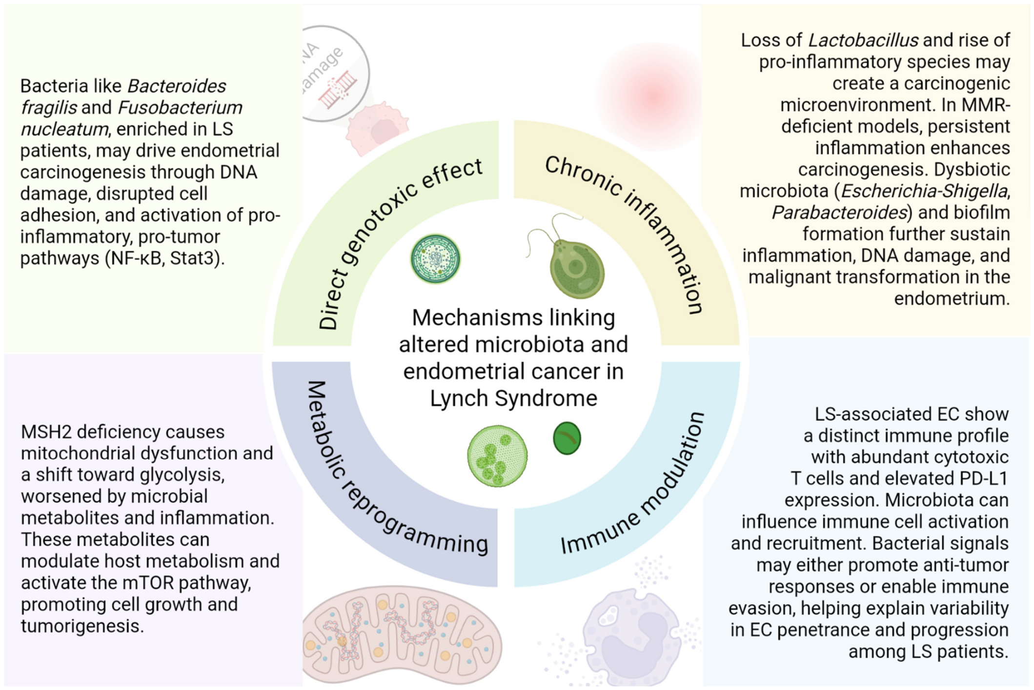
Several mechanisms have been proposed to explain how altered microbiota may influence endometrial carcinogenesis in LS (Table 1, Figure 1). In LS patients, the interplay between a dysbiotic microbiota and host factors can drive endometrial carcinogenesis through persistent inflammation, immune system modulation, metabolic shifts (notably via the mTOR pathway), and direct DNA damage from bacterial toxins. These mechanisms may help explain why only some LS carriers develop EC, highlighting the microbiota as a potential target for risk stratification and prevention.
Table 1.
Microorganisms and hypothesized carcinogenic mechanisms in Lynch syndrome-associated endometrial cancer. Lynch syndrome (LS), colorectal cancer (CRC), endometrial cancer (EC), short-chain fatty acid (SCFA).
Figure 1.
Hypothesized carcinogenic mechanisms in Lynch syndrome-associated endometrial cancer. This figure was created with BioRender.com.
- -
- Direct genotoxic effect: Certain bacteria such as Bacteroides fragilis, which has been found to be more abundant in LS patients with EC, can produce toxins that can directly damage host DNA causing a direct genotoxic effect, contributing to carcinogenesis [8].
Bacteroides fragilis is generally regarded as a commensal component of the human microbiota and is not intrinsically pathogenic. However, only a distinct phylogenetic subset of strains is able to produce the enterotoxin characteristic of enterotoxigenic Bacteroides fragilis (ETBF). This toxin promotes DNA damage, alters cell–cell adhesion, and triggers pro-carcinogenic inflammatory cascades via pathways such as NF-κB and Stat3. Although this mechanism is well documented in colorectal carcinogenesis, particularly at the earliest stages of neoplastic transformation, its relevance to endometrial tissue is currently speculative and it is now being investigated.
Fusobacterium nucleatum, which has been found to be enriched in subjects with LS and CRC, has been associated with an immunosuppressive microenvironment and increased cellular proliferation in colorectal models. However, most direct evidence derives from CRC studies and preclinical systems, and it has not been demonstrated in LS-associated EC. The detection of these bacteria and their toxins in the gut, and possibly in the reproductive tract, raises the hypothesis that analogous genotoxic and pro-inflammatory effects might contribute to endometrial carcinogenesis [38,39].
- -
- Chronic inflammation and microbial dysbiosis: Chronic inflammation is a well-recognized driver of carcinogenesis in LS. In the reproductive tract, the loss of Lactobacillus dominance and increased presence of pro-inflammatory species like Mobiluncus curtisii may promote a microenvironment conducive to carcinogenesis through persistent inflammation and altered pH [8,11]. In MMR-deficient mouse models (as seen in LS), persistent intestinal inflammation has been associated with increased tumor development. Inflammation may alter the local microbiota, and taxa such as Escherichia-Shigella, Parabacteroides and Atopobium vaginae have been reported in association with pro-inflammatory states. By analogy, it has been hypothesized that chronic inflammatory infiltrates, possibly triggered or sustained by dysbiotic microbiota, could create a microenvironment favouring DNA damage and proliferation in the endometrium of LS carriers. At present, this hypothesis lacks direct experimental validation in LS-associated EC [40,41].
- -
- Metabolic reprogramming and host–microbiota interactions: Another well-established hallmark of cancer is the metabolic reprogramming of tumor cells, classically described as the Warburg effect, which involves profound alterations in mitochondrial function and a preferential reliance on glycolytic metabolism [24]. Recent evidence from mouse models of LS-associated EC demonstrates that MSH2 deficiency leads to mitochondrial dysfunction, reduced mitochondrial content, and a metabolic shift toward glycolysis, which may be exacerbated by microbial metabolites and inflammation [42]. Microbial metabolites can influence host cell metabolism and signaling pathways, including the mTOR pathway, which is known to regulate cell growth and proliferation, potentially promoting tumorigenesis in the endometrial tissue of LS patients [43]. These metabolic and immune alterations, together with the accumulation of MMR-deficient endometrial glands, create a permissive environment for malignant transformation in LS carriers [44,45].
- -
- Immune modulation and tumor microenvironment: LS-associated endometrial cancers often display a distinct immune landscape compared to sporadic cases. Tumors in LS patients are typically characterized by high infiltration of cytotoxic T cells and increased expression of immune checkpoint molecules such as PD-L1. The microbiota can modulate this immune environment by influencing the recruitment and activation of immune cells. For example, certain bacterial products can stimulate pro-tumorigenic immune responses, while others may promote immune evasion by the tumor [41]. This interplay between the microbiota and the host immune system may help explain the variable penetrance and progression of EC in LS, as immune surveillance is both shaped by and responsive to microbial signals [41].
Current evidence is preliminary and based on small cohorts, with most studies highlighting associations rather than causality. Larger, longitudinal studies and mechanistic experiments are needed to clarify the role of the microbiota in modulating EC risk and penetrance in LS.
Potential clinical implications include the use of microbiota profiles as biomarkers for risk stratification and the exploration of microbiota-targeted interventions for prevention or adjunctive therapy in LS-associated EC [8,9,10,12,13].
3.5. Microbiota and Therapeutic Response
Emerging evidence from the PRIMMO trial has highlighted the role of the gut microbiota in shaping treatment outcomes and toxicity profiles among patients with cervical and endometrial cancer receiving pembrolizumab. Distinct microbial signatures were found to correlate with both therapeutic efficacy and immune-related adverse events, particularly gastrointestinal (GI) toxicity. Patients who developed severe GI toxicity exhibited an enrichment of Roseburia and Prevotella, whereas those without severe GI toxicity showed higher abundance of Barnesiella and members of the Barnesiellaceae family. Moreover, baseline fecal samples revealed that responders to pembrolizumab were enriched in taxa capable of producing SCFAs. In particular, the presence of the genus Blautia, known butyrate producers, was associated with improved clinical efficacy. Other butyrate-producing genera enriched in responders included Anaerostipes, Dorea, Faecalibacterium, and Roseburia. Conversely, enrichment of Enterobacteriaceae and Clostridium species was associated with poor therapeutic efficacy. Hierarchical clustering analysis identified two distinct microbial community clusters with markedly different survival outcomes, with a more than fourfold difference in mortality risk (HR 4.4, p < 0.001) [46].
Microbial butyrate has also been shown to sensitize EC tumors to MPA during fertility-sparing therapy. In MPA-responsive patients, tumor tissues were enriched with butyrate-producing bacteria (Butyrivibrio, Clostridium, Faecalibacterium), suggesting a role for microbial metabolites in promoting ferroptosis and progesterone receptor expression [47].
Probiotics such as Lactobacillus spp. produce soluble proteins (p75, p40) that protect intestinal epithelium from radiation-induced inflammation and apoptosis, reducing therapy-related mucosal injury [48,49].
Experimental organoid models demonstrate an anti-proliferative effect of L. crispatus-conditioned medium on EC cells, supporting a local protective role for Lactobacillus species [4].
Table 2 summarises the main studies reporting microbiota findings relevant to endometrial cancer and Lynch syndrome.
Table 2.
Main studies reporting microbiota findings relevant to endometrial cancer and Lynch syndrome. LS, Lynch syndrome; EC, endometrial cancer; CRC, colorectal cancer; LS-C/LS-NC, LS with/without cancer; MMR, mismatch repair; MSI, microsatellite instability; MPA, medroxyprogesterone acetate; SCFA, short-chain fatty acids; β-GUS, β-glucuronidase; GI, gastrointestinal; NS, not specified in the article; ↑ increased; ↓ decreased.
Limitations of the study: Overall, the current evidence is limited by small sample sizes, predominantly observational and cross-sectional designs, and considerable methodological heterogeneity, and therefore supports associations rather than causal relationships. Mechanistic insights are largely extrapolated from CRC and preclinical models, and prospective, adequately powered studies in LS-associated EC are needed.
4. Conclusions
The link between microbiota composition and EC in LS represents an emerging field with significant translational potential. Dysbiosis may amplify the effects of MMR deficiency through inflammation, metabolic changes, and immune modulation, acting as a co-factor in carcinogenesis. Conversely, commensal and SCFA-producing bacteria appear to exert protective effects by maintaining epithelial homeostasis and immune balance.
Distinct microbial signatures have been reported in LS carriers who develop EC compared with unaffected individuals, suggesting that the microbiome may partly explain interindividual variability in cancer risk. The gut microbiota also influences the efficacy and toxicity of EC treatment.
Despite these promising insights, current studies are small, heterogeneous, and mainly correlative, underscoring the need for more robust evidence.
Author Contributions
Conceptualization: S.A., and M.C.; Investigation: S.A., and M.C.; Supervision: M.C., G.P., A.F., and V.E.B.; Validation: M.C., G.P., A.F., and V.E.B.; Visualization: S.A., and M.C.; Writing—original draft: S.A.; Writing—review and editing: S.A., M.C., M.D., M.G., L.P.; M.P., B.C., A.F., and V.E.B. All authors have read and agreed to the published version of the manuscript.
Funding
This research received no external funding.
Data Availability Statement
No new data were created or analyzed in this study. Data sharing is not applicable to this article.
Acknowledgments
This work was supported by the Italian Ministry of Health, Ricerca Corrente 2026. AI-assisted tools (Grammarly, Deepl and ChatGPT 5.5) were used exclusively for language editing. No AI tools were used for the generation of scientific content, data analysis, or interpretation. The authors take full responsibility for the accuracy and integrity of the manuscript.
Conflicts of Interest
The authors declare no conflicts of interest.
Abbreviations
The following abbreviations are used in this manuscript:
| NGS | Next-generation sequencing |
| EC | Endometrial cancer |
| LS | Lynch syndrome |
| MMR | Mismatch Repair |
| ART | Assisted reproductive technologies |
| β-GUS | β-glucuronidase |
| GnRH | Gonadotropin-releasing hormone |
| SLPI | Secretory leukocyte protease inhibitor |
| MSI | Microsatellite instability |
| LPS | Lipopolysaccharide |
| TLR-4 | Toll-like receptor 4 |
| SCFA | Short-chain fatty acids |
| LS-C | LS patients with cancer |
| LS-NC | LS patients without cancer |
| ETBF | Enterotoxigenic Bacteroides fragilis |
| CRC | Colorectal cancer |
| GI | Gastrointestinal |
| MPA | Medroxyprogesterone acetate |
References
- Chen, C.; Song, X.; Wei, W.; Zhong, H.; Dai, J.; Lan, Z.; Li, F.; Yu, X.; Feng, Q.; Wang, Z.; et al. The microbiota continuum along the female reproductive tract and its relation to uterine-related diseases. Nat. Commun. 2017, 8, 875. [Google Scholar] [CrossRef] [Scilit] [PubMed]
- Moreno, I.; Codoñer, F.M.; Vilella, F.; Valbuena, D.; Martinez-Blanch, J.F.; Jimenez-Almazán, J.; Alonso, R.; Alamá, P.; Remohí, J.; Pellicer, A.; et al. Evidence that the endometrial microbiota has an effect on implantation success or failure. Am. J. Obstet. Gynecol. 2016, 215, 684–703. [Google Scholar] [CrossRef] [Scilit] [PubMed]
- Łaniewski, P.; Ilhan, Z.E.; Herbst-Kralovetz, M.M. The microbiome and gynaecological cancer development, prevention and therapy. Nat. Rev. Urol. 2020, 17, 232–250. [Google Scholar] [CrossRef] [Scilit] [PubMed]
- Semertzidou, A.; Whelan, E.; Smith, A.; Ng, S.; Roberts, L.; Brosens, J.J.; Marchesi, J.R.; Bennett, P.R.; MacIntyre, D.A.; Kyrgiou, M. Microbial signatures and continuum in endometrial cancer and benign patients. Microbiome 2024, 12, 118. [Google Scholar] [CrossRef] [Scilit] [PubMed]
- Dominguez-Valentin, M.; Sampson, J.R.; Seppälä, T.T.; Ten Broeke, S.W.; Plazzer, J.P.; Nakken, S.; Engel, C.; Aretz, S.; Jenkins, M.A.; Sunde, L.; et al. Cancer risks by gene, age, and gender in 6350 carriers of pathogenic mismatch repair variants: Findings from the Prospective Lynch Syndrome Database. Genet. Med. 2020, 22, 15–25, Erratum in Genet. Med. 2020, 22, 1569. https://doi.org/10.1038/s41436-020-0892-4. [Google Scholar] [CrossRef] [Scilit] [PubMed] [PubMed Central]
- Gupta, S.; Weiss, J.M.; Axell, L.; Burke, C.A.; Chen, L.M.; Chung, D.C.; Felder, S.; Foda, Z.; Grady, W.; Gustafson, S.; et al. NCCN Guidelines Version 1.2025: Genetic/Familial High-Risk Assessment: Colorectal, Endometrial, and Gastric. 2025. Available online: https://www.nccn.org/professionals/physician_gls/pdf/genetics_ceg.pdf (accessed on 28 October 2025).
- Actis, S.; Cazzaniga, M.; Bounous, V.E.; D’Alonzo, M.; Rosso, R.; Accomasso, F.; Minella, C.; Biglia, N. Emerging evidence on the role of breast microbiota on the development of breast cancer in high-risk patients. Carcinogenesis 2023, 44, 718–725. [Google Scholar] [CrossRef] [Scilit] [PubMed]
- Mori, G.; Orena, B.S.; Cultrera, I.; Barbieri, G.; Albertini, A.M.; Ranzani, G.N.; Carnevali, I.; Tibiletti, M.G.; Pasca, M.R. Gut Microbiota Analysis in Postoperative Lynch Syndrome Patients. Front. Microbiol. 2019, 10, 1746. [Google Scholar] [CrossRef] [Scilit] [PubMed]
- Gonzalez, A.; Kapila, N.; Melendez-Rosado, J.; Liang, H.; Castro-Pavia, F. An Evaluation of the Fecal Microbiome in Lynch Syndrome. J. Gastrointest. Cancer 2021, 52, 365–368. [Google Scholar] [CrossRef] [Scilit] [PubMed]
- Zheng, W.; Lin, X.; Chen, H.; Yang, Z.; Zhao, H.; Li, S.; Song, T.; Sun, Y. Gut microbiota and endometrial cancer: Research progress on the pathogenesis and application. Ann. Med. 2025, 57, 2451766. [Google Scholar] [CrossRef] [Scilit] [PubMed]
- Barczyński, B.; Fraszczak, K.; Grywalska, E.; Kotarski, J.; Korona-Głowniak, I. Vaginal and Cervical Microbiota Composition in Patients with Endometrial Cancer. Int. J. Mol. Sci. 2023, 24, 8266. [Google Scholar] [CrossRef] [Scilit] [PubMed]
- Stabile, G.; Doria, A.; Bruno, M.; D’Indinosante, M.; Gallotta, V.; Fanfani, F.; Scambia, G.; Restaino, S.; Vizzielli, G.; Carlucci, S.; et al. The Role of the Endometrial Microbiota in Endometrial Cancer: A Systematic Review of the Literature. J. Clin. Med. 2024, 13, 7135. [Google Scholar] [CrossRef] [Scilit] [PubMed]
- Prakash, A.; Nourianpour, M.; Senok, A.; Atiomo, W. Polycystic Ovary Syndrome and Endometrial Cancer: A Scoping Review of the Literature on Gut Microbiota. Cells 2022, 11, 3038. [Google Scholar] [CrossRef] [Scilit] [PubMed]
- Walther-António, M.R.; Chen, J.; Multinu, F.; Hokenstad, A.; Distad, T.J.; Cheek, E.H.; Keeney, G.L.; Creedon, D.J.; Nelson, H.; Mariani, A.; et al. Potential contribution of the uterine microbiome in the development of endometrial cancer. Genome Med. 2016, 8, 122. [Google Scholar] [CrossRef] [Scilit] [PubMed]
- Baker, J.M.; Chase, D.M.; Herbst-Kralovetz, M.M. Uterine Microbiota: Residents, Tourists, or Invaders? Front. Immunol. 2018, 9, 208. [Google Scholar] [CrossRef] [Scilit] [PubMed]
- Khan, K.N.; Fujishita, A.; Masumoto, H.; Muto, H.; Kitajima, M.; Masuzaki, H.; Kitawaki, J. Molecular detection of intrauterine microbial colonization in women with endometriosis. Eur. J. Obstet. Gynecol. Reprod. Biol. 2016, 199, 69–75. [Google Scholar] [CrossRef] [Scilit] [PubMed]
- Walsh, D.M.; Hokenstad, A.N.; Chen, J.; Sung, J.; Jenkins, G.D.; Chia, N.; Nelson, H.; Mariani, A.; Walther-Antonio, M.R.S. Postmenopause as a key factor in the composition of the Endometrial Cancer Microbiome (ECbiome). Sci. Rep. 2019, 9, 19213. [Google Scholar] [CrossRef] [Scilit] [PubMed]
- Vomstein, K.; Reider, S.; Böttcher, B.; Watschinger, C.; Kyvelidou, C.; Tilg, H.; Moschen, A.R.; Toth, B. Uterine microbiota plasticity during the menstrual cycle: Differences between healthy controls and patients with recurrent miscarriage or implantation failure. J. Reprod. Immunol. 2022, 151, 103611. [Google Scholar] [CrossRef] [Scilit] [PubMed]
- Wira, C.R.; Fahey, J.V.; Rodriguez-Garcia, M.; Shen, Z.; Patel, M.V. Regulation of mucosal immunity in the female reproductive tract: The role of sex hormones in immune protection against sexually transmitted pathogens. Am. J. Reprod. Immunol. 2014, 72, 236–258. [Google Scholar] [CrossRef] [Scilit] [PubMed]
- Fang, R.L.; Chen, L.X.; Shu, W.S.; Yao, S.Z.; Wang, S.W.; Chen, Y.Q. Barcoded sequencing reveals diverse intrauterine microbiomes in patients suffering with endometrial polyps. Am. J. Transl. Res. 2016, 8, 1581–1592. [Google Scholar] [PubMed]
- Byrd, D.A.; Fan, W.; Greathouse, K.L.; Wu, M.C.; Xie, H.; Wang, X. The intratumor microbiome is associated with microsatellite instability. J. Natl. Cancer Inst. 2023, 115, 989–993. [Google Scholar] [CrossRef] [Scilit] [PubMed]
- Li, Y.; Liu, G.; Gong, R.; Xi, Y. Gut Microbiome Dysbiosis in Patients with Endometrial Cancer vs. Healthy Controls Based on 16S rRNA Gene Sequencing. Curr. Microbiol. 2023, 80, 264. [Google Scholar] [CrossRef] [Scilit] [PubMed]
- Jimenez, N.R.; Herman, C.R.; Łaniewski, P.; Cope, E.; Lee, K.; Mahnert, N.D.; Chase, D.M.; Caporaso, J.G.; Herbst-Kralovetz, M.M. Navigating complexities of polymorphic microbiomes in endometrial cancer. npj Biofilms Microbiomes 2025, 11, 45. [Google Scholar] [CrossRef] [Scilit] [PubMed]
- Hanahan, D. Hallmarks of Cancer: New Dimensions. Cancer Discov. 2022, 12, 31–46. [Google Scholar] [CrossRef] [Scilit] [PubMed]
- Elkafas, H.; Walls, M.; Al-Hendy, A.; Ismail, N. Gut and genital tract microbiomes: Dysbiosis and link to gynecological disorders. Front. Cell. Infect. Microbiol. 2022, 12, 1059825. [Google Scholar] [CrossRef] [Scilit] [PubMed]
- Caselli, E.; Soffritti, I.; D’Accolti, M.; Piva, I.; Greco, P.; Bonaccorsi, G. Atopobium vaginae and Porphyromonas somerae Induce Proinflammatory Cytokines Expression in Endometrial Cells: A Possible Implication For Endometrial Cancer? Cancer Manag. Res. 2019, 11, 8571–8575. [Google Scholar] [CrossRef] [Scilit] [PubMed]
- Lu, W.; He, F.; Lin, Z.; Liu, S.; Tang, L.; Huang, Y.; Hu, Z. Dysbiosis of the endometrial microbiota and its association with inflammatory cytokines in endometrial cancer. Int. J. Cancer 2021, 148, 1708–1716. [Google Scholar] [CrossRef] [Scilit] [PubMed]
- Raftogianis, R.; Creveling, C.; Weinshilboum, R.; Weisz, J. Chapter 6: Estrogen Metabolism by Conjugation. JNCI Monogr. 2000, 27, 113–124. [Google Scholar] [CrossRef] [Scilit] [PubMed]
- Parida, S.; Sharma, D. The Microbiome–Estrogen Connection and Breast Cancer Risk. Cells 2019, 8, 1642. [Google Scholar] [CrossRef] [Scilit] [PubMed]
- McIntosh, F.M.; Maison, N.; Holtrop, G.; Young, P.; Stevens, V.J.; Ince, J.; Johnstone, A.M.; Lobley, G.E.; Flint, H.J.; Louis, P. Phylogenetic distribution of genes encoding β-glucuronidase activity in human colonic bacteria and the impact of diet on faecal glycosidase activities. Environ. Microbiol. 2012, 14, 1876–1887. [Google Scholar] [CrossRef] [Scilit] [PubMed]
- Yang, C.; Qin, L.H.; Li, L.; Wei, Q.Y.; Long, L.; Liao, J.Y. The causal relationship between the gut microbiota and endometrial cancer: A mendelian randomization study. BMC Cancer 2025, 25, 1947. [Google Scholar] [CrossRef] [Scilit] [PubMed]
- Pollet, R.M.; D’Agostino, E.H.; Walton, W.G.; Xu, Y.; Little, M.S.; Biernat, K.A.; Pellock, S.J.; Patterson, L.M.; Creekmore, B.C.; Isenberg, H.N.; et al. An Atlas of β-Glucuronidases in the Human Intestinal Microbiome. Structure 2017, 25, 967–977.e5. [Google Scholar] [CrossRef] [Scilit] [PubMed]
- Han, X.; Zheng, J.; Zhang, L.; Zhao, Z.; Cheng, G.; Zhang, W.; Qu, P. Endometrial microbial dysbiosis and metabolic alteration promote the development of endometrial cancer. Int. J. Gynecol. Obstet. 2024, 167, 810–822. [Google Scholar] [CrossRef] [Scilit] [PubMed]
- Westley, R.L.; May, F.E.B. A Twenty-First Century Cancer Epidemic Caused by Obesity: The Involvement of Insulin, Diabetes, and Insulin-Like Growth Factors. Int. J. Endocrinol. 2013, 2013, 632461. [Google Scholar] [CrossRef] [Scilit] [PubMed]
- Wu, J.; Wang, K.; Wang, X.; Pang, Y.; Jiang, C. The role of the gut microbiome and its metabolites in metabolic diseases. Protein Cell 2021, 12, 360–373. [Google Scholar] [CrossRef] [Scilit] [PubMed]
- Zhao, S.S.; Chen, L.; Yang, J.; Wu, Z.H.; Wang, X.Y.; Zhang, Q.; Liu, W.J.; Liu, H.X. Altered Gut Microbial Profile Accompanied by Abnormal Fatty Acid Metabolism Activity Exacerbates Endometrial Cancer Progression. Microbiol. Spectr. 2022, 10, e02467-22. [Google Scholar] [CrossRef] [Scilit] [PubMed]
- Zhang, B.; Mohd Sahardi, N.F.N.; Di, W.; Long, X.; Shafiee, M.N. The Gut–Endometrium Axis: Exploring the Role of Microbiome in the Pathogenesis and Treatment of Endometrial Cancer—A Narrative Review. Cancers 2025, 17, 1044. [Google Scholar] [CrossRef] [Scilit] [PubMed]
- Chung, L.; Orberg, E.T.; Geis, A.L.; Chan, J.L.; Fu, K.; DeStefano Shields, C.E.; Dejea, C.M.; Fathi, P.; Chen, J.; Finard, B.B.; et al. Bacteroides fragilis Toxin Coordinates a Pro-carcinogenic Inflammatory Cascade via Targeting of Colonic Epithelial Cells. Cell Host Microbe 2018, 23, 203–214.e5. [Google Scholar] [CrossRef] [Scilit] [PubMed]
- Lopez, L.R.; Bleich, R.M.; Arthur, J.C. Microbiota Effects on Carcinogenesis: Initiation, Promotion, and Progression. Annu. Rev. Med. 2021, 72, 243–261. [Google Scholar] [CrossRef] [Scilit] [PubMed]
- Lang, M.; Baumgartner, M.; Rożalska, A.; Frick, A.; Riva, A.; Jarek, M.; Berry, D.; Gasche, C. Crypt residing bacteria and proximal colonic carcinogenesis in a mouse model of Lynch syndrome. Int. J. Cancer 2020, 147, 2316–2326. [Google Scholar] [CrossRef] [Scilit] [PubMed]
- Gambini, D.; Ferrero, S.; Kuhn, E. Lynch Syndrome: From Carcinogenesis to Prevention Interventions. Cancers 2022, 14, 4102. [Google Scholar] [CrossRef] [Scilit] [PubMed]
- Yates, M.S. Abstract IA013: Unique metabolic changes in Lynch syndrome-associated endometrial cancer. Clin. Cancer Res. 2024, 30, IA013. [Google Scholar] [CrossRef] [Scilit]
- Jiang, I.; Yong, P.J.; Allaire, C.; Bedaiwy, M.A. Intricate Connections between the Microbiota and Endometriosis. Int. J. Mol. Sci. 2021, 22, 5644. [Google Scholar] [CrossRef] [Scilit] [PubMed]
- Hegazy, S.; Brand, R.E.; Dudley, B.; Karloski, E.; Lesnock, J.L.; Elishaev, E.; Pai, R.K. Mutation-specific Mismatch Repair–deficient Benign Endometrial Glands in Endometrial Biopsies and Curettings Are a Biomarker of Lynch Syndrome and Associate with Endometrial Carcinoma Development. Am. J. Surg. Pathol. 2023, 47, 835–843. [Google Scholar] [CrossRef] [Scilit] [PubMed]
- Wong, S.; Hui, P.; Buza, N. Frequent loss of mutation-specific mismatch repair protein expression in nonneoplastic endometrium of Lynch syndrome patients. Mod. Pathol. 2020, 33, 1172–1181. [Google Scholar] [CrossRef] [Scilit] [PubMed]
- De Jaeghere, E.A.; Hamerlinck, H.; Tuyaerts, S.; Lippens, L.; Van Nuffel, A.M.T.; Baiden-Amissah, R.; Vuylsteke, P.; Henry, S.; Trinh, X.B.; van Dam, P.A.; et al. Associations of the gut microbiome with outcomes in cervical and endometrial cancer patients treated with pembrolizumab: Insights from the phase II PRIMMO trial. Gynecol. Oncol. 2024, 191, 275–286. [Google Scholar] [CrossRef] [Scilit] [PubMed]
- Li, X.; Wang, Y.; Wu, Y.; Wang, J.; Qi, Y.; Bi, X.; Wang, J. Intratumor microbiome-derived butyrate enhances progesterone sensitivity by inducing ferroptosis in endometrial cancer. Pharmacol. Res. 2025, 219, 107046. [Google Scholar] [CrossRef] [Scilit] [PubMed]
- Barker, H.E.; Paget, J.T.E.; Khan, A.A.; Harrington, K.J. The tumour microenvironment after radiotherapy: Mechanisms of resistance and recurrence. Nat. Rev. Cancer 2015, 15, 409–425. [Google Scholar] [CrossRef] [Scilit] [PubMed]
- Bultman, S.J. The microbiome and its potential as a cancer preventive intervention. Semin. Oncol. 2016, 43, 97–106. [Google Scholar] [CrossRef] [Scilit] [PubMed]

Disclaimer/Publisher’s Note: The statements, opinions and data contained in all publications are solely those of the individual author(s) and contributor(s) and not of MDPI and/or the editor(s). MDPI and/or the editor(s) disclaim responsibility for any injury to people or property resulting from any ideas, methods, instructions or products referred to in the content. |
© 2026 by the authors. Licensee MDPI, Basel, Switzerland. This article is an open access article distributed under the terms and conditions of the Creative Commons Attribution (CC BY) license.