


Abstract
Cardiac sarcoidosis (CS) is an inflammatory cardiomyopathy with heterogeneous presentation and potentially life-threatening complications, including ventricular arrhythmia, atrioventricular block, and heart failure. Early diagnosis is challenging and a multidisciplinary approach is pivotal for optimal treatment and better outcome. We report the case of a 47-year-old man with newly diagnosed systemic sarcoidosis who rapidly developed heart failure and therapy-refractory ventricular arrhythmias despite combined immunosuppressive and antiarrhythmic therapy. Within eight months from the first diagnosis of CS, the patient required four hospitalizations, ICD implantation, ventricular tachycardia ablation, and ultimately orthotopic heart transplantation. Histological examination of the explanted heart revealed extensive myocardial fibrosis without residual granulomas or inflammation, consistent with end-stage, burned-out CS—highlighting the irreversibility of myocardial damage once the fibrotic phase has developed. This case illustrates the potentially devastating course of CS despite aggressive immunosuppressive therapy, emphasizing that the overall prognosis of sarcoidosis disease is largely influenced by cardiac manifestation. Indeed, complete metabolic remission based on a serial FDG-PET/CT scan did not prevent progressive functional deterioration—underscoring that irreversible myocardial fibrosis may progress beyond maximal medical therapy, including anti-inflammatory, immunosuppressive and neuro-humoral treatment. Therefore, prompt screening for CS is essential, and once cardiac involvement is suspected and respectively confirmed, the therapy window may become particularly narrow in the case of rapidly progressive CS.
1. Introduction
Sarcoidosis is a granulomatous disease with systemic or rarely unifocal manifestation of unknown etiology characterized by non-caseating epithelioid granulomas that may affect any organ. It predominantly affects adults aged 25–60 years, with an incidence of approximately 10 per 100,000 person-years in Western populations and an even higher prevalence in Northern Europeans and African Americans [1,2]. The average age at presentation of cardiac involvement is 50–55 years, with no major sex predominance in European cohorts [1]. The lungs and thoracic lymph nodes are affected in approximately 90% of cases, while cardiac involvement is clinically overt in only 5–10% of patients with systemic sarcoidosis, despite being detectable in 25–30% on post-mortem studies [1,2]. Cardiac sarcoidosis (CS) is particularly insidious: frequently underdiagnosed due to its heterogeneous presentation, it is associated with severe complications, including high-grade conduction disease, ventricular arrhythmias, heart failure, and sudden cardiac death [2,3]. Notably, cardiac disease can be the first manifestation of sarcoidosis: between 20% and 34% of patients with idiopathic high-grade atrioventricular block and a similar proportion of those with unexplained sustained ventricular tachycardia are ultimately found to have cardiac sarcoidosis as the underlying aetiology [2,3,4]. Early diagnosis and a multidisciplinary approach are crucial to improve outcomes, with 5-year survival reaching 90–96% in contemporary cohorts managed at specialist centres [3]. Cardiac MRI and FDG-PET/CT represent the gold standard for diagnostic evaluation and guidance of immunosuppressive therapy, respectively [1,2,4].
Despite significant advances in diagnosis and management of CS, the interplay between inflammation, scarring (fibrosis), and heart function is neither fully elucidated nor predictable [1,4]. Indeed, this case report aims to illustrate how rapidly CS may progress despite multimodality and immunosuppressive therapy, raising awareness of the narrow therapeutic window once CS is suspected.
In the following, we report the case of a 47-year-old man with systemic sarcoidosis who developed rapidly progressive CS with recurrent ventricular tachycardias and electrical storms, ultimately requiring orthotopic heart transplantation.
2. Case Presentation
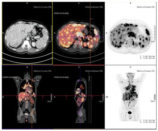
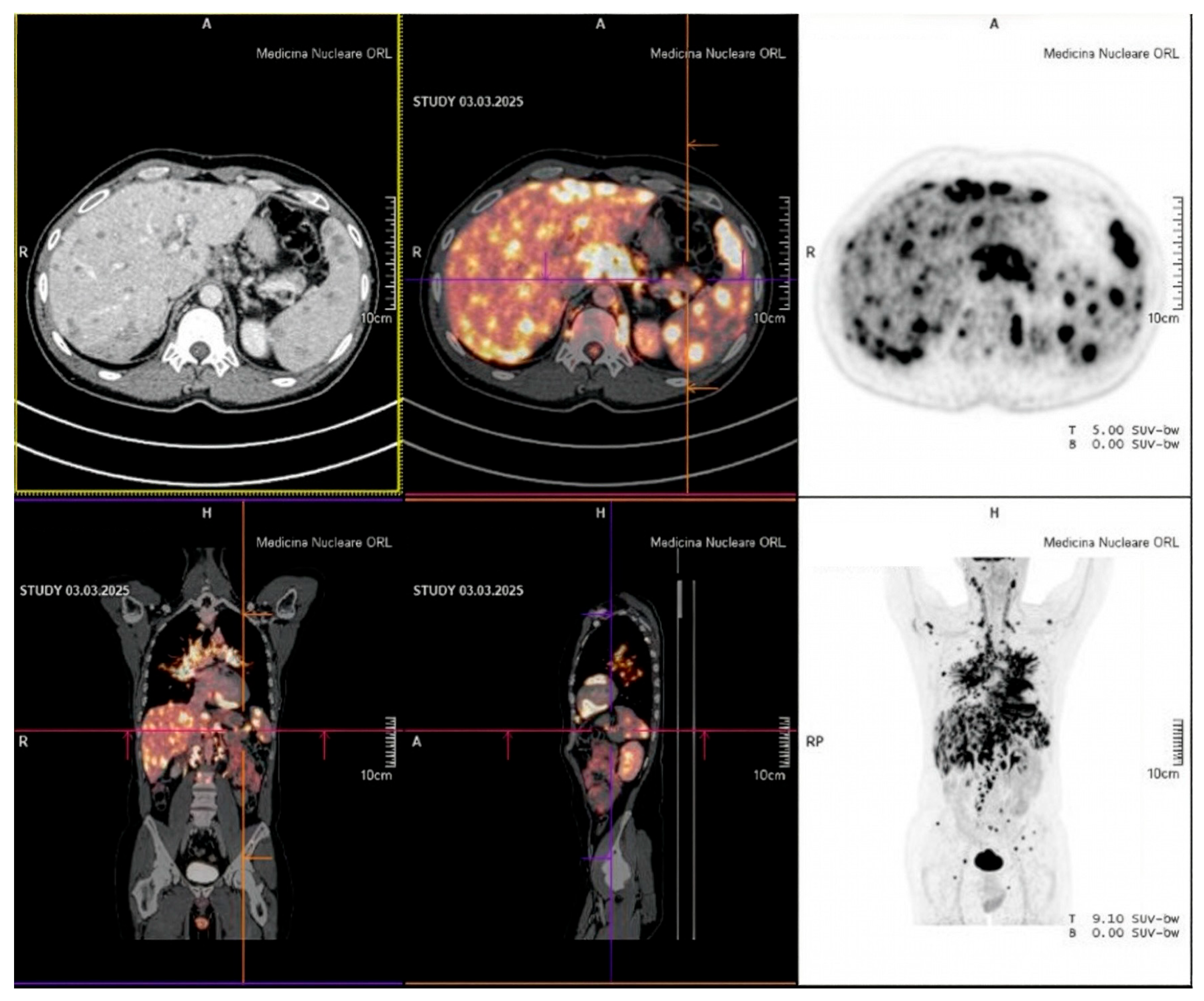
A 47-year-old man of south Italian origin, a computer engineer, with a positive history of bronchial asthma and a family history positive for coronary artery disease (grandfather, deceased at age 60) and heart failure (grandmother) but not for confirmed genetic cardiomyopathy or sudden cardiac death, presented with progressive dyspnoea on exertion (NYHA class II) since December 2024, dry cough, and recurrent short palpitations. He had been in good health until late 2024. In early 2025, a chest X-ray revealed bilateral hilar enlargement. The following thoraco-abdominal CT scan (January 2025) showed bilateral pulmonary nodules with reticular infiltrations, extensive bilateral mediastinal and hilar lymphadenopathy, and multiple nodular infiltrations in the liver and spleen (from 5 to 17 mm in size). Nevertheless, pulmonary function testing was unremarkable. Following his first pulmonological consultation (February 2025), the patient underwent transbronchial biopsy via EBUS which demonstrated non-caseating epithelioid granulomas with multinucleated giant cells and fibrosis. The subsequent whole-body FDG-PET/CT scan (March 2025) revealed markedly elevated metabolic activity at multiple sites—the lungs, pleura, supra- and infra-diaphragmatic lymph nodes, heart, liver, spleen, pancreas and bone (Figure 1). Therefore, the diagnosis of systemic sarcoidosis was established.
Figure 1.
Whole-body FDG-PET/CT scan (March 2025) showing markedly elevated metabolic activity at multiple sites—the heart, lungs, mediastinal and hilar lymph nodes, liver, spleen, pancreas, and bone—consistent with active systemic sarcoidosis with cardiac involvement at diagnosis. (Published with written informed consent of the patient).
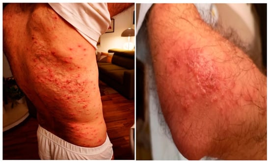
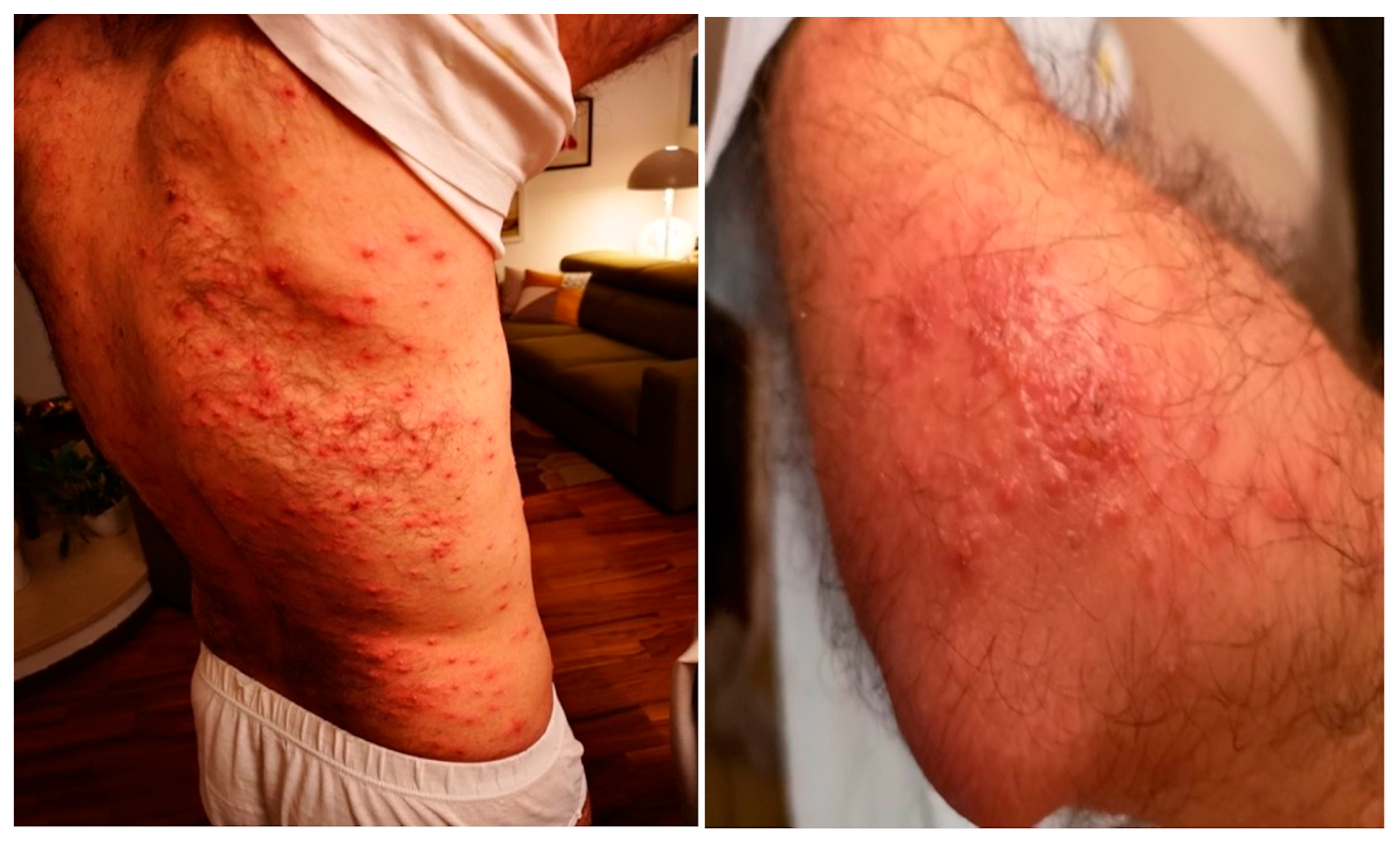
Immunosuppressive (IS) therapy was initiated with prednisolone 50 mg/d in addition to trimethoprim/sulfamethoxazole for Pneumocystis jirovecii prophylaxis. Of note, on retrospective review, the patient had experienced a maculo-papular exanthema in 2019, which was treated empirically with systemic corticosteroids without any skin biopsy—in hindsight, a missed first diagnosis of skin manifestation of systemic sarcoidosis (Figure 2).
Figure 2.
Maculo-papular exanthema mainly on the patient’s trunk consistent with cutaneous sarcoidosis. (Published with written informed consent of the patient).
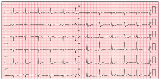
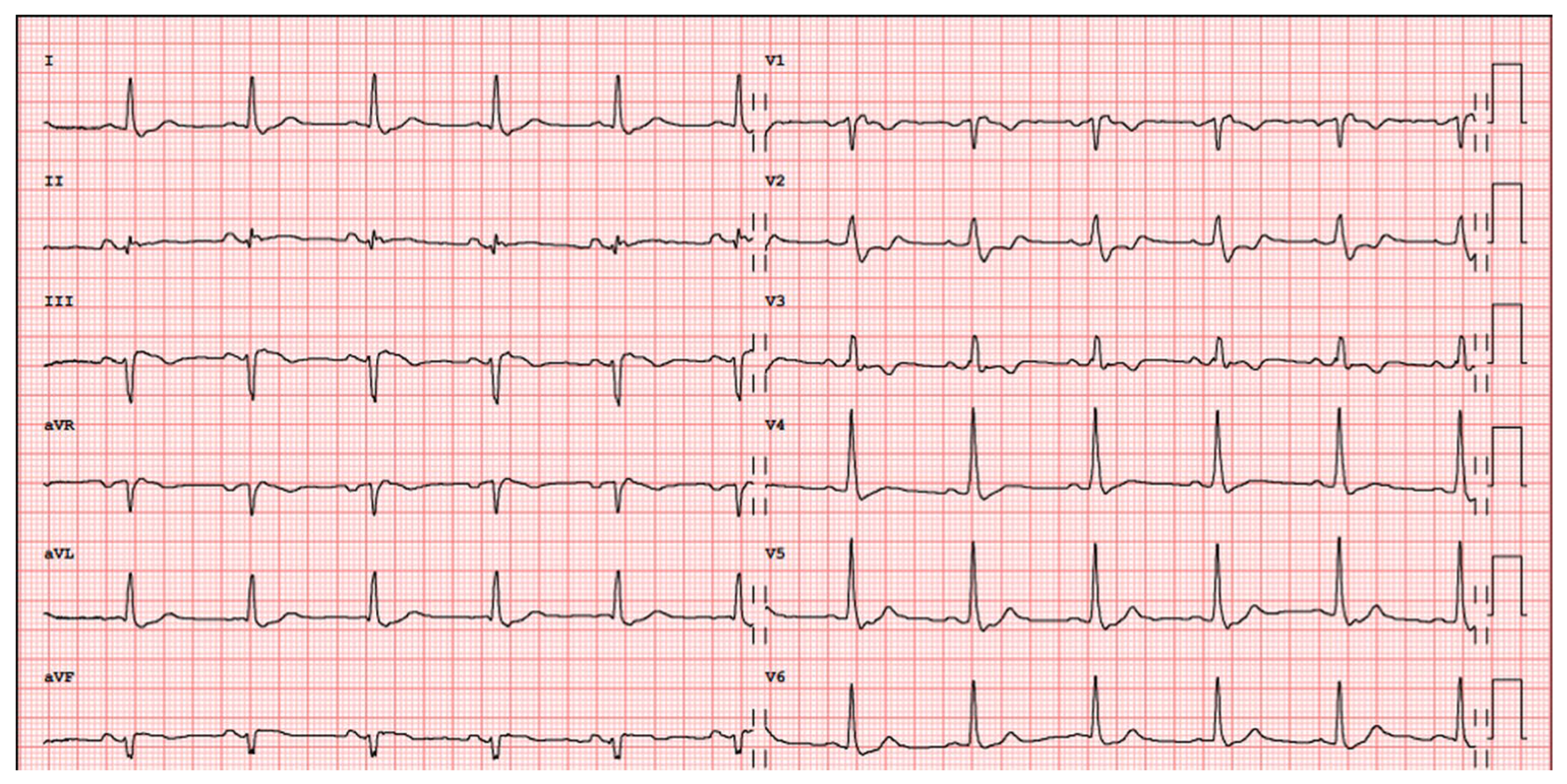
The patient was referred to the cardiologist in March 2025. Clinical examination revealed a normotensive patient with a subtle S3 gallop. Biomarkers showed mild elevation of levels of NT-proBNP (224 ng/L) and of the angiotensin-converting enzyme (ACE) (73 U/L), with normal high-sensitivity troponin-T (7 ng/L). The 12-lead ECG demonstrated right intraventricular conduction delay (Figure 3).
Figure 3.
Baseline 12-lead ECG (March 2025) showing normocardic sinus rhythm and right intraventricular conduction delay, but normal atrio-ventricular conduction (no AV block).
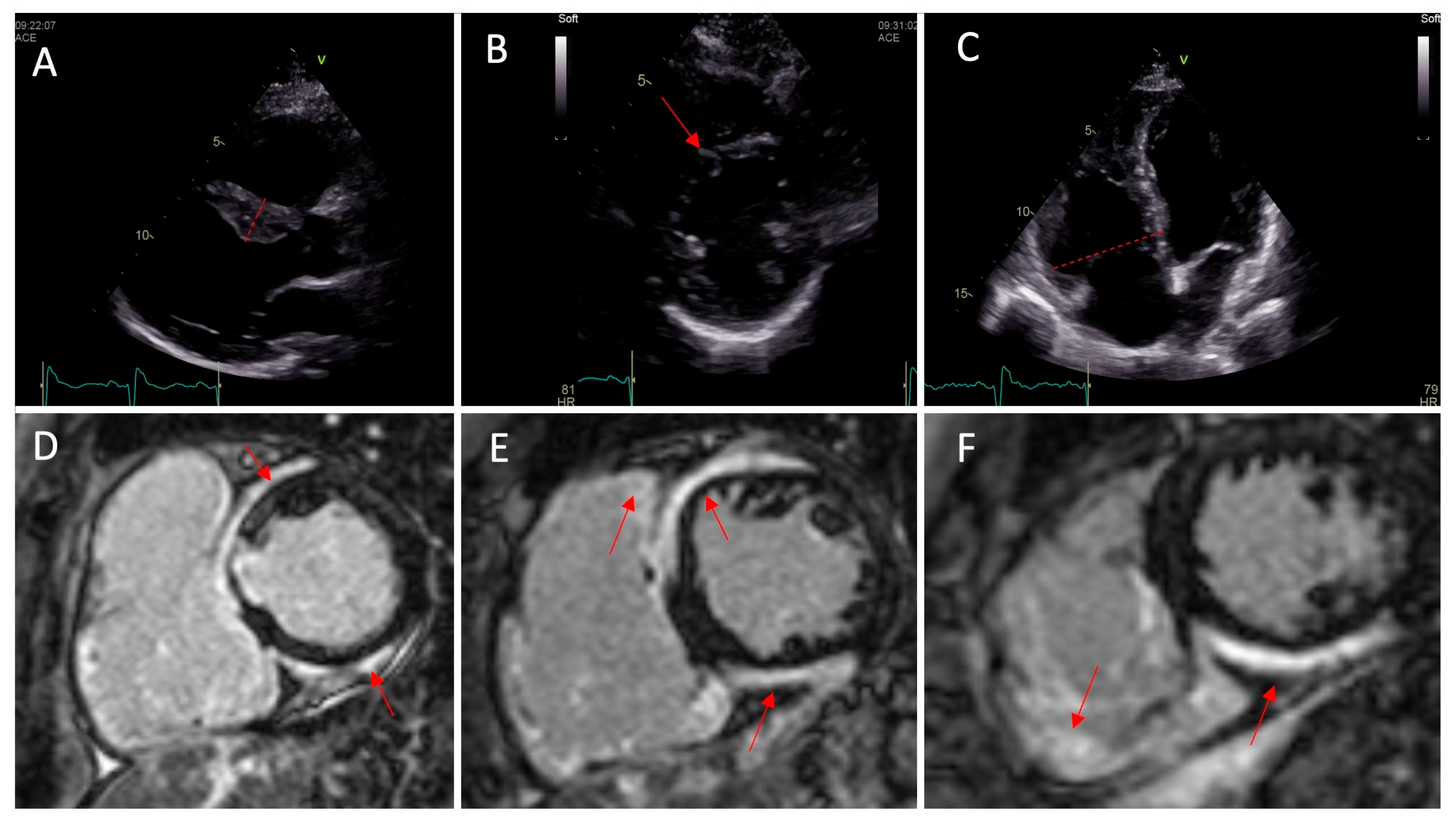
Transthoracic echocardiography (TTE) revealed a mild hypertrophy of the basal interventricular septum (12.6 mm); left ventricular (LV) ejection fraction (EF) was 51% (low-normal), with D-shape morphology and reduced global longitudinal strain (mean GLS) of −16.6%, suggesting heart failure with preserved EF (HFpEF). Right ventricular (RV) (radial) function was borderline impaired (FAC 30%, TAPSE 21 mm, S’ 12 cm/s).
The first 24 h Holter-ECG showed sinus rhythm with rare premature ventricular contractions (PVCs, burden = 0.3%), but with one polymorphic non-sustained ventricular tachycardia (NSVT) of six beats at 206 bpm of heart rate. A beta-blocker (Bisoprolol 2.5 mg/d) and a SGLT2 inhibitor were initiated. Only six days later, a second Holter-ECG was repeated showing alarming deterioration: PVCs burden increased to 44%, with over 26,000 monomorphic PVCs (left bundle branch block morphology) with more than 1000 NSVTs reaching up to 20 beats.
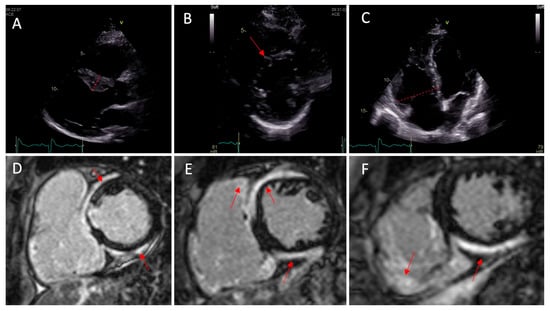
The patient was admitted urgently (first hospitalization). Coronary CT excluded obstructive coronary artery disease. Cardiac MRI (Figure 4) demonstrated T2 STIR positivity in multiple LV segments, consistent with active myocardial inflammation and diffuse gadolinium contrast media uptake in the LV and RV in late gadolinium enhancement (LGE) sequences, supporting the diagnosis of CS in the context of histologically confirmed systemic sarcoidosis. Following a multidisciplinary meeting (MDM), cardiac therapy was optimized (bisoprolol 5 mg/d, amiodarone 200 mg/d, spironolactone 12.5 mg/d, and dapagliflozin 10 mg/d) and a wearable cardioverter defibrillator (WCD) was prescribed. IS therapy was further escalated with intravenous pulse methylprednisolone (500 mg/d × 3 days) followed by oral prednisolone 50 mg/d with tapering; intravenous infliximab 5 mg/kg was given twice (March and June 2025), and mycophenolate mofetil 500 mg was given twice daily (added in May 2025).
Figure 4.
Baseline transthoracic echocardiography showing, in Panel (A), the thickening of the basal septum (12.6 mm, red lines) and left ventricle (LV) ejection fraction biplane of 51%. A flattening of the septum (D-shape) was also noted in Panel (B) as well as right ventricle (RV) dilatation with systolic dysfunction (FAC 30%, Tapse: 21 mm, S’: 12 cm/s) in Panel (C). Late gadolinium enhancement sequences in cardiac MRI highlighted the presence of an extensive contrast uptake in the septum and inferior wall at the basal level (Panel (D), red arrows), mid-ventricular level (Panel (E), red arrows) and apical level (Panel (F), red arrows). Gadolinium contrast uptake was also noticed in the RV (Panels (E,F), red arrows).
Almost three months later, the patient needed the second hospitalization (June 2025) for symptomatic ventricular tachycardia (VT). Repeated cardiac MRI showed LV EF ~50% and RV EF about 35%, with persistent T2 STIR positivity in the LV mid-distal segments and in RV, and stable gadolinium contrast media uptake in LGE sequences. However, FDG-PET/CT demonstrated complete metabolic resolution of cardiac and systemic sarcoid metabolic activity. A dual-chamber 3T MRI-compatible implantable cardioverter defibrillator (ICD) was implanted. Only six days later, the patient developed an electrical storm leading to 15 anti-tachycardia pacing (ATP) deliveries and one ICD shock, requiring hospitalization and emergency transfer to the University Hospital Inselspital of Berne (third hospitalization, June 2025). Intravenous lidocaine was administered for 2 days and the dual-chamber ICD was reprogrammed to DDD 90 bpm in order to prevent ventricular arrhythmia. Cardiac therapy was further optimized (bisoprolol 10 mg/d, amiodarone 200 mg/d, spironolactone 25 mg/d, and dapagliflozin 10 mg/d) alongside oral prednisolone unchanged at 10 mg and mycophenolate mofetil up-titrated to 750 mg twice daily; infliximab was definitively stopped because of its potential adverse effect (heart failure).
However, at the first outpatient follow-up visit in September 2025, a striking progression of heart failure from HFpEF to HF with reduced ejection fraction (HFrEF) was diagnosed. BP was 92/60 mmHg, NT-proBNP increased to 910 ng/L, and estimated GFR was reduced to 50 mL/min/1.73 m2. Indeed, current TTE revealed dramatic biventricular deterioration: LV end-diastolic diameter increased from 48 to 55 mm, LV EF decreased from 51% to 35%, and there was new functional mitral valve regurgitation. RV function decreased further as well: FAC from 30% to 26%, TAPSE from 21 to 14 mm, and S’ from 12 to 8 cm/s. ICD interrogation revealed a high burden of ventricular pacing (VP) of 41%, and the device was reprogrammed, activating the RYTHMIQ algorithm to minimize unnecessary RV pacing and risk of HF/tachy-cardiomyopathy. Indeed, the FDG-PET/CT follow-up scan confirmed persistent complete (cardiac and systemic) metabolic remission.
However, at the next ICD interrogation in early October 2025, slow but sustained ventricular tachycardia (SVT) was detected with QRS of 192 ms, HR of 112 bpm, and with superior axis morphology with QS from V1 to V5, consistent with mid-apical septal origin (focus). Cardiac MRI performed on the same day confirmed diffuse LV hypokinesis conditioning LV EF of 35%, extensive subepicardial fibrosis of the basal septum, inferior and basal lateral walls, and RV EF of only 25%. Catheter ablation of the ectopic focus was performed and was acutely successful. However, post-ablation ECG monitoring revealed recurrent ventricular arrhythmia leading to a new electrical storm. Once urgently transferred to Inselspital, the patient underwent right heart catheterization (RHC), confirming the absence of pulmonary hypertension (mean pulmonary artery pressure (mPAP) 18 mmHg, post-capillary Wedge pressure (PCWP) 5 mmHg). Given refractory life-threatening arrhythmia burden and advanced biventricular failure, the patient was listed as super-urgent for orthotopic heart transplantation, which was successfully performed a few weeks later in October 2025.
Histopathological examination of the explanted heart revealed extensive multi-segmental myocardial scarring/fibrosis with no residual non-caseating granulomas and no residual inflammation/myocarditis—confirming end-stage fibrotic CS with burned-out granulomatous inflammation.
Under triple IS therapy (oral prednisolone, mycophenolate mofetil and rapamune-sirolimus) after successful heart transplantation (HTx), the clinical follow-up has been uneventful so far, except for asymptomatic re-activation of pulmonary sarcoidosis, detected by a FDG-PET/CT scan follow-up (at week 30 post-HTx), which has been treated by increasing the dose of prednisolone from 5.0 mg to 20 mg per day, with success.
3. Discussion
The diagnosis of cardiac sarcoidosis (CS) represents one of the biggest challenges with regard to inflammatory cardiomyopathies. The histopathological hallmark is the presence of non-caseating granuloma formation driven by dysregulated CD4+ T-helper type 1 activation. In the heart, granulomas preferentially involve the basal interventricular septum, the AV nodal region, and the subepicardial layers [1]. If left untreated or refractory, this inflammatory process leads to irreversible myocardial fibrosis—as dramatically illustrated by the histopathological findings in the explanted heart of our patient. Importantly, cardiac disease may be the first manifestation of sarcoidosis.
The most common presentations (phenotypes) are high-degree atrioventricular block (AVB) with prevalence rates about 10–52%, ventricular arrhythmias presenting as an initial manifestation in up to 24% of patients with clinically manifest CS, and heart failure affecting approximately 14–33% at diagnosis [1,2,4]. Cutaneous manifestations such as erythema nodosum or maculo-papular exanthema may represent the first—and often overlooked—systemic manifestation [5]. As illustrated in our case, such early cutaneous signs may precede the cardiac diagnosis by several years, missing the opportunity to recognize and adequately treat this dangerous inflammatory systemic disease. Once systemic sarcoidosis is diagnosed, cardiac involvement should always be actively investigated with cardiac history (palpitations in younger age), 12-lead ECG, biomarkers (NT-proBNP, high-sensitivity troponin), and echocardiography. Holter-ECG monitoring should be added when arrhythmias are suspected, as many patients remain asymptomatic despite significant arrhythmic burden [1,2].
The diagnosis is most commonly achieved through a combination of extra-cardiac histological confirmation and compatible cardiac manifestations on multimodal imaging, reviewed in a multidisciplinary team (MDT) setting, following two main distinct diagnostic pathways recommended by international guidelines [1,2,4].
The first pathway applies to patients with known extra-cardiac sarcoidosis, in whom at least one of the following criteria is present: cardiac symptoms, rhythm abnormalities, structural echocardiographic changes, or elevated cardiac biomarkers—any of which should prompt CMR and FDG-PET/CT as the next diagnostic step. The second pathway applies to patients presenting with otherwise-unexplained cardiac manifestations such as advanced AVB, sustained VT, or LV impairment—in whom extra-cardiac sarcoidosis evaluation and advanced cardiac imaging should be performed to confirm or exclude the diagnosis, acknowledging that histological proof may be very difficult to obtain.
Our patient followed the first pathway: histologically confirmed systemic (pulmonary, extra-cardiac) sarcoidosis prompted cardiac evaluation, leading to the definitive diagnosis through CMR and FDG-PET/CT. Of note, symptoms such as palpitations and exertional dyspnoea in younger subjects have to be considered very carefully (red flags). Importantly, cardiac-specific biomarkers—including NT-proBNP, high-sensitivity troponin (hs Tn), and the angiotensin-converting enzyme (ACE)—represent very useful adjunctive tools in the diagnostic work-up [1]. In our patient, mildly elevated NT-proBNP and ACE levels further supported the suspicion of cardiac involvement, while hs Tn remained normal, suggesting, rather, the absence of active inflammation (fibrosis). Though standard 12-lead ECG has limited sensitivity for this disease (8–21%), in our patient we found right intraventricular conduction delay. Twenty-four-hour Holter-ECG monitoring, with higher sensitivity (50–67%) [1], proved the key arrhythmic alarm. Of particular note, the dramatic increase in ventricular arrhythmia in the second Holter-ECG turned out to be associated time-wise with the intravenous pulse methylprednisolone (500 mg per day) therapy (administered over three days). Echocardiography, despite its high specificity (95–98%), has limited sensitivity (25–27%) [1,2]; yet, it provided an important early clue through RV dilatation and dysfunction, and reduced GLS despite preserved LV EF—a pattern consistent with subclinical myocardial involvement [1].
Cardiac magnetic resonance (CMR) plays a central role in the diagnosis of cardiac sarcoidosis by providing comprehensive tissue characterization and assessment of myocardial involvement. The hallmark CMR finding is late gadolinium enhancement (LGE) in a non-ischemic pattern, typically involving the subepicardial or mid-myocardial layers and often affecting the basal septum and lateral wall. T2-weighted imaging and T2 mapping can identify myocardial edema, reflecting active inflammation. In addition, CMR allows for accurate evaluation of ventricular function, chamber volumes, and regional wall motion abnormalities. The presence and extent of LGE carry important prognostic implications and are associated with an increased risk of VT and adverse cardiac events. Of note, RV LGE is associated with advanced disease and a worse prognosis [1,2]. In our patient, serial CMR tracked disease evolution: from active inflammation, through a mixed inflammatory-fibrotic pattern, to predominantly extensive subepicardial fibrosis with declining ventricular function.
FDG-PET/CT has a sensitivity of 84–89% and a specificity of 78–83% in the diagnosis of CS. It complements CMR by detecting active myocardial inflammation, but also enables more accurate therapy monitoring and prognostic stratification according to the experts and the available literature [1,3,4]. In our patient, serial FDG-PET/CT confirmed complete metabolic remission, though cardiac function continued to deteriorate—suggesting longer-standing, ‘burned-out’ CS in which metabolically inactive fibrotic tissue has replaced active granulomas and inflammation. These findings emphasize that once CS is suspected, the therapeutic window may be very narrow since immunosuppression is most effective before irreversible fibrosis has developed [1]. Metabolic remission does not prevent functional decline. However, it is of major help to clinicians to keep from further up-titrating IS therapy with potentially life-threatening side effects (first of all, infections) [1,4]. Recent evidence further supports the integration of FDG-PET/CT into the diagnostic pathway, particularly when CMR is inconclusive, and puts FDG-PET/CT follow-up scans in front for guiding immunotherapy [6].
Three sets of diagnostic criteria are currently available (HRS = Heart Rhythm Society, JCS = Japanese Cardiology Society, and WASOG = World Association of Sarcoidosis and other granulomatous disorders) [1,2,4]. HRS and WASOG criteria are closely aligned, both mandating histological confirmation alongside compatible cardiac manifestations on multimodality imaging. The updated JCS algorithm uniquely allows for diagnosis of isolated CS in the absence of extra-cardiac involvement based on imaging findings alone, a clinically relevant distinction as isolated CS is associated with a higher rate of adverse cardiovascular events [7]. In the present case, the diagnosis was established without endomyocardial biopsy (EMB), fulfilling both HRS and JCS criteria through histologically confirmed systemic sarcoidosis, compatible arrhythmic and functional cardiac manifestations, T2 STIR-positive CMR, and a cardiac FDG-PET/CT pattern consistent with active CS.
Nonetheless, differential diagnosis (DD) remains relevant in CS, especially in the cases without endomyocardial histological confirmation. Indeed, it has been shown that CS may masquerade (like a chameleon) as genetic cardiomyopathy; in particular, as arrhythmogenic right ventricular dysplasia (ARVD) or arrhythmogenic biventricular cardiomyopathy (ACM). Moreover, CS and arrhythmogenic-cardiomyopathy-associated genetic variants may coexist [1,8], and sustained ventricular arrhythmias may also independently increase myocardial FDG uptake, irrespective of active granulomatous disease [9]. Due to this challenging overlap between CS and ACM, experts recommend genetic counselling/testing for the majority of CS cases that lack histological confirmation, given the implications for treatment and cascade family screening. In particular, genetic testing is recommended for the following cases: in the case of isolated, clinically apparent CS without extra-cardiac disease (potential carriers of pathogenic LMNA, TPM1, and MYBPC3); in the case of de novo unexplained AV block, ventricular arrhythmia or heart failure without a prior diagnosis of sarcoidosis; in the case of a positive family history suggestive of genetic cardiomyopathy; in the case of overlap features suggesting desmosomal disease–desmoplakin pathogenic variants (producing myocarditis-like presentations); and in the case of septal LGE with conduction disease in known ACM variant carriers (patients with known ACM-associated variants (like PKP2, DSG2, DSP, and TTN) who develop AV-block, PR prolongation, or septal involvement) [1,4]. Back to our clinical case, the absence of any sign of inflammation/myocarditis and of myocardial granulomas in the explanted heart may raise the question of whether CS is the only correct final diagnosis. The negative family history for confirmed genetic cardiomyopathy and/or SCD, the exact chronological order between the first cardiac symptoms (dyspnoea and palpitations) and the histologically confirmed (through EBUS-guided biopsy) diagnosis of systemic, multi-organ sarcoidosis based on 18-FDG-PET/CT with positive cardiac involvement, and the prior history of skin rash (see Figure 2) without histological diagnosis (no skin biopsy performed at that time) suggesting longer-standing sarcoidosis (conditioning cardiac fibrosis/scarring) left us in no doubt about the diagnosis of advanced, end-stage CS. Of course, underlying genetic susceptibility or additional genetic cardiomyopathy cannot be ruled out a priori without targeted genetic testing, and should always be considered in the DD of any unclear form of cardiomyopathy.
Regardless of these diagnostic considerations, CMR-derived LGE features carry important prognostic implications for arrhythmic risk stratification. LGE extent ≥ 7.4% of LV mass is independently associated with life-threatening ventricular arrhythmic events, while specific high-risk features—LGE on the right side of the interventricular septum, LGE in the RV free wall, and multifocal LGE—independently predict arrhythmic events even with preserved LVEF. A novel four-tier risk stratification algorithm integrating LVEF, LGE extent, and high-risk LGE features outperforms traditional HRS criteria and may help to guide ICD implantation decisions [10].
Management requires a parallel and integrated approach addressing both cardiac complications and the underlying inflammatory process. Rhythm control includes antiarrhythmic agents (amiodarone or sotalol as preferred options), device therapy, and catheter ablation when refractory. In our patient, ICD implantation was indicated given the combination of sustained VT, reduced LVEF, and extensive myocardial scarring on CMR, which are well-established risk factors for sudden cardiac death in CS [1,2,4]. The success rate of ablation is modest due to the extensive and often epicardial arrhythmogenic substrate [2,3], as confirmed in our case by the recurrence of an electrical storm shortly after an initially successful procedure. HF management follows current guideline-directed medical therapy (GDMT). IS therapy should be initiated when active myocardial inflammation is confirmed [1,2,4], using corticosteroids as first-line agents, with additional steroid-sparing immunosuppressants (methotrexate, mycophenolate mofetil, and azathioprine) and biologics (infliximab, adalimumab) for refractory cases—as employed in the present case. Rapidly progressive disease, as seen here, warrants upfront addition of a second immunomodulator or intravenous methylprednisolone pulses [3,4], though corticosteroids may paradoxically increase ventricular arrhythmia burden [11,12]. This potential paradox of pro-arrhythmic hazards of corticosteroid therapy comes from a few observational studies and deserves particular attention. Indeed, in a multicentre registry of 441 patients with CS, a constant hazard for life-threatening ventricular arrhythmias was observed following corticosteroid initiation, with 5- and 10-year estimated incidence rates of 19.2% and 31.6%, respectively [12]. In the single-centre prospective study of 20 patients with an ICD/pacemaker and clinically manifest, FDG-PET/CT positive CS, corticosteroid initiation led to a significant increase in PVCs burden (an up-to-three-fold increase) and of VTs [11]. The underlying mechanisms are not fully understood and rely on the particular interplay between the presence and burden of inflammation/myocarditis and scarring/fibrosis, both being well known arrhythmogenic substrates. If steroids can treat (by suppressing) inflammation/myocarditis (detected by FDG-PET/CT), they have no effect on scarring/fibrosis (detected by MRI) [1,11,12]. That is why, in our opinion/experience, especially high-dose corticosteroid therapy (initiation) in CS may be associated with a pro-arrhythmic risk, particularly if scarring/fibrosis is predominant. These findings highlight the importance of close arrhythmic monitoring during initiation and escalation of corticosteroid therapy in CS [1,11,12]. Careful tapering of corticosteroids to 5–10 mg/day under serial FDG-PET is recommended [1,4,13]. Notably, symptomatic cardiac sarcoidosis may relapse when corticosteroids are tapered in up to 75% of patients, underscoring the need for long-term maintenance strategies [4]. A key finding in this patient was the progressive HFpEF to HFrEF transition despite complete metabolic remission—a reminder that metabolic remission on FDG-PET/CT does not necessarily translate into functional recovery once extensive fibrosis has been established. Furthermore, ventricular arrhythmia-induced cardiomyopathy and increasing electro-mechanical dyssynchrony are the other potential mechanisms contributing to ventricular dysfunction and overt heart failure.
Last, heart transplantation represents a viable option for end-stage CS refractory to medical therapy, with published 5-year post-transplant survival rates of 79–83%, comparable to other non-ischaemic cardiomyopathies [1,3]. Of note, disease may recur in the allograft in ~5% of cases without graft loss [1]. A recent systematic review confirmed the rarity of post-transplant CS recurrence, with only 13 cases identified in the literature, predominantly managed with intensification of immunosuppressive therapy [14]. In our case, 30 weeks after successful heart transplantation (HTx), FDG-PET/CT follow-up revealed asymptomatic relapse of pulmonary sarcoidosis though ongoing triple IS therapy post-HTx (including oral prednisolone tapered to 5 mg, mycophenolate mofetil 360 mg twice and tacrolimus 2.5 mg twice per day). The patient was therefore put on 20 mg prednisolone per day, and the MDM recommended a much slower tapering of prednisolone as well as a long-term maintenance dose of at least 7.5 mg per day.
As nicely highlighted throughout this case, a true MDT approach—comprising cardiologists, pulmonologists, electrophysiologists, advanced imaging specialists, and transplant teams—proved decisive in guiding key management decisions [1,2,4].
4. Conclusions
This case illustrates the potentially devastating course of cardiac sarcoidosis (CS), even under aggressive immunosuppressive therapy. Complete metabolic remission on serial FDG-PET/CT did not prevent progressive functional deterioration—underscoring that irreversible myocardial fibrosis may progress beyond the anti-inflammatory treatment. Thus, the therapy time window in rapidly progressive disease may be very narrow. Early cardiac evaluation in all patients with confirmed systemic sarcoidosis is essential, as subclinical cardiac involvement may already be present at diagnosis. In selected cases, differential diagnosis may be very challenging since genetic (arrhythmogenic) cardiomyopathy may masquerade as CS. Importantly, complete metabolic remission on serial FDG-PET/CT should not be automatically equated with disease control, as irreversible myocardial fibrosis may progress silently beyond the reach of immunosuppressive therapy. Close rhythm monitoring during high-dose corticosteroid therapy is advisable [11,12]. The seamless collaboration of a dedicated MDT across the healthcare system proved to be essential in navigating complex management decisions and ensuring timely access to successful heart transplantation.
Author Contributions
A.R.: Conceptualization, investigation, writing—original draft preparation. A.G.P.: Investigation, writing—review and editing. M.M.: Investigation, writing—review and editing. G.M.: Conceptualization, investigation, supervision, writing—review and editing. All authors have read and agreed to the published version of the manuscript.
Funding
This research received no external funding.
Institutional Review Board Statement
Ethical review and approval were not required for this publication under the Swiss Federal Act on Research involving Human Beings (HRA), as this was a single case report arising from routine clinical care and not a method-driven research project requiring approval under the HRA (Art. 3(a) HRA & Art. 2 para. 2 HRA).
Informed Consent Statement
Written informed consent was obtained from the patient for publication of this case report, including permission for educational and scientific use of clinical data.
Data Availability Statement
The original contributions presented in this study are included in the article. Further inquiries can be directed to the corresponding author.
Acknowledgments
The authors would like to thank all the nurses and medical staff who took care of the patient, and, in particular, the patient and the family caregiver, for their consent and approval for publication of the following case report and review. During the preparation of this manuscript, the first author used Claude (Claude Sonnet 5, Anthropic, San Francisco, CA, USA) for language editing and translation assistance. The authors have reviewed and edited the output and take full responsibility for the content of this publication.
Conflicts of Interest
All authors declare no conflicts of interest in relation to the submitted work. Giorgio Moschovitis (GM) has received advisory-board or speaker’s fees from Astra Zeneca, Bayer, Boehringer Ingelheim, Daiichi Sankyo, Gebro Pharma, Novartis and Vifor, all outside of the submitted work. Anna Giulia Pavon (AGP) has received research grant support from the Swiss Heart Foundation and Swiss National Foundation, outside the submitted work.
References
- Sharma, R.; Kouranos, V.; Cooper, L.T.; Metra, M.; Ristic, A.; Heidecker, B.; Baksi, J.; Wicks, E.; Merino, J.L.; Klingel, K.; et al. Management of cardiac sarcoidosis. Eur. Heart J. 2024, 45, 2697–2726. [Google Scholar] [CrossRef] [Scilit] [PubMed]
- Birnie, D.H.; Nery, P.B.; Ha, A.C.; Beanlands, R.S. Cardiac Sarcoidosis. J. Am. Coll. Cardiol. 2016, 68, 411–421. [Google Scholar] [CrossRef] [Scilit] [PubMed]
- Lehtonen, J.; Uusitalo, V.; Pöyhönen, P.; Mäyränpää, M.I.; Kupari, M. Cardiac sarcoidosis: Phenotypes, diagnosis, treatment, and prognosis. Eur. Heart J. 2023, 44, 1495–1510. [Google Scholar] [CrossRef] [Scilit] [PubMed] [PubMed Central]
- Cheng, R.K.; Kittleson, M.M.; Beavers, C.J.; Birnie, D.H.; Blankstein, R.; Bravo, P.E.; Gilotra, N.A.; Judson, M.A.; Patton, K.K.; Rose-Bovino, L. American Heart Association Heart Failure and Transplantation Committee of the Council on Clinical Cardiology, and Council on Cardiovascular and Stroke Nursing. Diagnosis and Management of Cardiac Sarcoidosis: A Scientific Statement from the American Heart Association. Circulation 2024, 149, e1197–e1216, Erratum in Circulation 2024, 150, e197. https://doi.org/10.1161/CIR.0000000000001275. [Google Scholar] [CrossRef] [Scilit] [PubMed]
- Löfgren, S. Primary pulmonary sarcoidosis. Acta Medica Scand. 1953, 145, 424–431. [Google Scholar] [CrossRef] [Scilit]
- Baldini, M.; Paci, G.; Metzger, G.; Satilmis, G.; Suwalski, P.; Gruenewald, E.; Balzer, F.; Margonato, A.; Kelle, S.; Furth, C.; et al. FDG PET-CT uncovers cardiac sarcoidosis when CMR is inconclusive: The impact of multimodal imaging. Eur. J. Heart Fail. 2026, 28, xuag034.002. [Google Scholar] [CrossRef] [Scilit]
- Sato, K.; Kawamatsu, N.; Yamamoto, M.; Machino-Ohtsuka, T.; Ishizu, T.; Ieda, M. Utility of Updated Japanese Circulation Society Guidelines to Diagnose Isolated Cardiac Sarcoidosis. J. Am. Heart Assoc. 2022, 11, e025565. [Google Scholar] [CrossRef] [Scilit] [PubMed] [PubMed Central]
- Rossi, V.A.; Palazzini, M.; Ammirati, E.; Gasperetti, A.; Grubler, M.; Brunckhorst, C.; Manka, R.; Giannopoulos, A.; Tanner, F.C.; Medeiros-Domingo, A.; et al. Coexistence of cardiac sarcoidosis and arrhythmogenic cardiomyopathy-associated genetic variants: A multicentre case-control study. Heart 2025, 111, 480–486. [Google Scholar] [CrossRef] [Scilit] [PubMed]
- Kovacs, B.; Giannopoulos, A.A.; Bogun, F.; Pazhenkottil, A.P.; Bonetti, N.R.; Manka, R.; Medeiros-Domingo, A.; Gruner, C.; Schmidt, D.; Flammer, A.J.; et al. Sustained Ventricular Tachyarrhythmias are Associated With Increased 18F-Fluorodeoxyglucose Uptake Mimicking Cardiac Sarcoidosis. Circ. Cardiovasc. Imaging 2024, 17, e016316. [Google Scholar] [CrossRef] [Scilit] [PubMed]
- Azzu, A.; Antonopoulos, A.S.; Okafor, J.; Morosin, M.; Androulakis, E.; Hatipoglu, S.; Almogheer, B.; Ahmed, R.; Mohiaddin, R.; Alpendurada, F.; et al. Extent and Features of Late Gadolinium Enhancement Stratify Arrhythmic Risk in Patients with Biopsy-Proven Sarcoidosis. JACC Cardiovasc. Imaging 2025, 18, 768–780. [Google Scholar] [CrossRef] [Scilit] [PubMed]
- Medor, M.C.; Spence, S.; Nery, P.B.; Beanlands, R.; Promislow, S.; Juneau, D.; de Kemp, R.; Ha, A.C.; Rivard, L.; Gula, L.; et al. Treatment with corticosteroids is associated with an increase in ventricular arrhythmia burden in patients with clinically manifest cardiac sarcoidosis: Insights from implantable cardioverter-defibrillator diagnostics. J. Cardiovasc. Electrophysiol. 2020, 31, 2751–2758. [Google Scholar] [CrossRef] [Scilit] [PubMed]
- Fujimoto, Y.; Nabeta, T.; Kitai, T.; Naruse, Y.; Taniguchi, T.; Tanaka, H.; Morimoto, R.; Baba, Y.; Yoshioka, K.; Matsue, Y. Constant hazard for life-threatening ventricular arrhythmias following corticosteroid initiation in patients with cardiac sarcoidosis. Heart Rhythm 2025, 23, 1221–1223. [Google Scholar] [CrossRef] [Scilit] [PubMed]
- Stievenart, J.; Le Guenno, G.; Ruivard, M.; Rieu, V.; André, M.; Grobost, V. Cardiac sarcoidosis: Systematic review of the literature on corticosteroid and immunosuppressive therapies. Eur. Respir. J. 2022, 59, 2100449. [Google Scholar] [CrossRef] [Scilit] [PubMed]
- Khan, S.P.; Seplowe, M.; Vemulakonda, L.; Shakil, F.; Aggarwal-Gupta, C.; Lanier, G.; Levine, E.; Ohira, S.; Spielvogel, D.; Gass, A.; et al. Early recurrence of cardiac sarcoidosis after orthotopic heart transplantation. J. Heart Lung Transplant. 2024, 43, 100146. [Google Scholar] [CrossRef] [Scilit]
Disclaimer/Publisher’s Note: The statements, opinions and data contained in all publications are solely those of the individual author(s) and contributor(s) and not of MDPI and/or the editor(s). MDPI and/or the editor(s) disclaim responsibility for any injury to people or property resulting from any ideas, methods, instructions or products referred to in the content. |
© 2026 by the authors. Licensee MDPI, Basel, Switzerland. This article is an open access article distributed under the terms and conditions of the Creative Commons Attribution (CC BY) license.