


Summary
The history of development of adult interventional cardiology in the last quarter of the 20th century is reduced – somewhat arbitrarily – to the detailed description of a crucial period in life of three engaged personalities who have left, by their pioneering work, a lasting mark in percutaneous coronary intervention: 1. Andreas Grüntzig and the beginning of the balloon dilatation; 2. Ulrich Sigwart and his essential contribution to the application of intracoronary stenting; 3. Vitali Verin and the intravascular beta irradiation to decrease coronary restenosis. All three investigators have worked during a certain period with the author and have achieved these milestones in Switzerland. Some thoughts how clinically meaningful progress can be achieved also in the future conclude this description.
Zusammenfassung
Die Geschichte der interventionellen Kardiologie beim Erwachsenen im letzten Viertel des zu Ende gegangenen Jahrhunderts, wird – etwas willkürlich – auf die detaillierte Beschreibung eines Lebensabschnittes von drei engagierten Persönlichkeiten beschränkt, von denen jede ein bleibendes Zeichen in der Entwicklung der koronaren Interventionen hinterlassen hat: 1. Andreas Grintzig und der Beginn der Ballon-Dilatation; 2. Ulrich Sigwart und sein entscheidender Beitrag in der Anwendung intrakoronarer Stents; 3. Vitali Verin und die intravaskuläre Beta-Bestrahlung zur Verminderung der koronaren Restenose.
Alle drei Forscher haben während einer gewissen Periode mit dem Autor zusammengearbeitet und ihre bahnbrechenden Arbeiten in der Schweiz realisiert. Zum Schluss werden einige Gedanken zur Frage, wie klinisch relevante Fortschritte in Zukunft erzielt werden können, aufgerollt.
Introduction
The surgery of the heart has always aimed at the shortest way to the diseased organ by larger or smaller openings in the skin and through the tissues to the field of operation. On the contrary, the interventional treatment follows the natural ways to the heart (veins and arteries) by using instruments attached to catheters. The history of interventional cardiology begins therefore in 1964 by the extraction of a foreign body from the heart by I. Thomas, using catheter tweezers or, in 1967, by the atrioseptotomy by W.J. Raskind, respectively by the closure of a Ductus botalli by W. Porstmann in the same year. Because congenital heart disease concerns a relatively restricted number of patients, treated generally in pediatric centers, and the bulk of interventions today in cardiology of the adult concerns coronary artery disease, the birth of interventional cardiology is generally dated the 16th of September 1977, when Andreas Grüntzig achieved the first successful coronary balloon dilatation in a conscious patient [1].
In the following decade many cardiologists considered stenotic coronary arteries as a sort of calcified water conducting distribution system, instead of a living and highly differentiated system with a very delicate inner carpet providing many highly specific biological properties. Therefore, in wild phantasies every type of instrument used in the plumbing industry to reopen or widen tubes was widely used: cutters, drillers, grinders, milling cutters, and naturally the laser light which, through its precision, seemed very attractive. The problem with laser light is only that it always goes straight and the coronary arteries have a lot of curves ...
It is nearly useless to go through the list of all what has been called new devices because, except the stent, none of those have resisted the test of time. I think of the “directional angioplasty”, which was rarely directional, the rotational angioplasty, which occluded by embolisation more smaller downstream vessels than all other methods, the cutting balloon, the hot or cold balloon, the various lasers up to the excimer laser angioplasty, etc. None of these devices has shown a longer or better survival compared to simple balloon or stent application, rather the contrary was true. The midterm results of these devices were very unsatisfactory because they showed a very high rate of restenosis, clearly higher than the balloon alone. Many of these devices are complicated and they do not at all respect the endothelium with its many biological functions, even in its dysfunctional status, and often lead to bleeding in the media and even in the adventitia. One thing has gotten very clear: the more the artery is maltreated, the stronger is its reaction, what induces this high restenosis rate.
Here we are at the problem of restenosis which has often been called the Achille’s heel of interventional cardiology, a disease which actually was created by Andreas Grüntzig. Already in the original patients dilated in Zurich the restenosis rate was about 30%. Very many systemic pharmacologic attempts were made to decrease the restenosis rate, but none had more than a borderline success. The same was true with the local application of substances which, except if delivered by a stent, do not stay there for a sufficient time where one would like to have a high concentration.
The main hope is set today on gene therapy, because many scientists believe that the deletion of a single gene or a growth factor or induction of expression of a positively acting gene could inhibit the hypertrophic scar formation, including the remodeling of the artery. But we should not forget that woundhealing with a strong scar, and specially the intravascular woundhealing with a long lasting scar, is an essential goal of nature after a laceration of a vessel. And nature had millions of years to develop adequate mechanisms and certainly has found for such an important problem a network of eventually redundant mechanisms. Nature has probably involved multiple pathways with several factors, respectively genes. This is why the deletion of restenosis by selective gene therapy has proved difficult and very few clinical results are available.
The brachytherapy, instead, fights genetic material non selectively. Ionising radiation damages DNA, but many alterations are reversible, specifically those in which only one and not both strands of DNA are damaged. Up to the end of the last century brachytherapy was the only method which in the clinic was able to fight restenosis with and without stents successfully. It is essentially a massive, “non directed” gene therapy.
I would like to recall the history of interventional adult cardiology based on the three most successful methods, namely (1.) balloon dilatation; (2.) stenting and (3.) brachytherapy by beta-rays. This seems justified even more so as the researchers who have made these three essential methodological steps forward in the clinical arena, Andreas Grüntzig, Ulrich Sigwart and Vitali Verin were – at least during a certain period – my collaborators in the Cardiology Division at the University Hospital in Zurich or at the Cardiology Center of the University Hospital in Geneva; and I have carefully observed their personal evolution and their essential realizations. I agree that this leads to a very incomplete, one-sided history of interventional cardiology; on the other side I can give more authentic elements and personal observations as I recall it or some of my documents prove. And we can be proud that these three essential steps all occurred in Switzerland.
Andreas Grüntzig was born on June 25th, 1939. His father was geograph and officer in the German army during world war II. He was lost on the battlefield in Russia when Andreas was 5 years old. After having gone to primary school in Rochlitz/Sachsen and a period in Argentina, about which Andreas was never very specific, he went to the Thoma college in Leipzig – well known for its outstanding choir. He graduated oriented toward old languages and obtained his baccalaureate in 1957. According to the socialist regime in East Germany he should do bricklaying in builder’s yards. Instead, with his courageous mother and with his brother, he fled in 1957 to West Germany before the wall was erected. In 1958 he repeated the maturity test in Heidelberg. Then he studied medicine at the Heidelberg University from 1958 until 1964 and promoted to medical doctor under Prof. Gotthard Schettler (later my opponent in the World Heart Federation). For approbation as a physician Andreas Grüntzig was trained first in the Surgical Department of Mannheim, in gynecology in Hannover and, in 1965, in internal medicine in Bad Harzburg. In 1966 he worked in vascular surgery in Ludwigshafen and learned to know the Ratschow Clinic in Darmstadt.
From fall 1966 until fall 1969 Andreas Grüntzig was an assistant in the Institute for “Sozial- und Arbeitsmedizin” of the University of Heidelberg under Professor Schafer. After a visit in London in 1967, to attend an epidemiologic and statistics course of Geoffrey Rose, he studied the validity of the Rose-WHO questionnaire concerning angina pectoris and myocardial infarction by retesting it after a two weeks delay in German railway workers [2]. The reproducibility was only 85%, which did not satisfy Andreas.
Attracted by the book “Differentialdiagnose innerer Krankheiten” of Prof. Robert Hegglin, which had just appeared in its 11th edition, Grüntzig came into the Medical Policlinic in Zurich and started to work in the Division of Angiology, under the guidance of Prof. Alfred Bollinger. The Cardiology Division, under my guidance, was side to side with Angiology. Andreas Grüntzig, with his very good observational facilities, realized that the relaxation of the reflex of the Achille’s tendon was slowed after workloads leading to claudication. This observation prompted us to the first clinical measurement of negative dp/dt in the left ventricle in patients with 3-vessel disease, a parameter which decreased also significantly during ischemia [3]. Together with Grüntzig, Bollinger and Mahler we studied the venous backflow from the legs under various conditions [4]. We also published a paper with Andreas in which the validity of the Rose-WHO questionnaire was tested before coronary angiography against the definitive result of this invasive method in our patients [5]. 12% of patients with significant coronary artery disease had given negative answers in the questionnaire. At that time nobody spoke of silent ischemia; a few years later articles concerning this topic mushroomed.
Andreas Grüntzig discussed the procedure of Dotter and Judkins in angiology and saw its practical application by Zeitler and Schoop in the Aggertal Clinic in Engelskirchen. Obviously, the application of two or more concentrically arranged catheters of increasing diameter, with a big entry hole in the femoral artery and mainly longitudinal forces scratching the endothelium and the plaques leading to their embolisation, was not a very gentle procedure. Instead we discussed the utilization of a balloon which could be inserted in the empty state through a small entry hole into the artery and advanced into the stenosis and, by expansion, exerted only radial forces to dilate the stenosis, a very simple idea!
Andreas realized that he had to work as an assistant in radiology and he started, after a short period in the bed unit of the Medical Policlinic, on the 1st of August 1971 under Prof. Wellauer, who made strong obstruction that any X-ray contrast medium was used outside of his Department; only his direct collaborators were allowed to inject contrast medium. There was one single table and room for peripheral angiography and angiocardiography in the Institute of Radiology of the University Hospital in Zurich at that time. Here we witnessed not rarely that most of the contrast volume was jetted at the ceiling while the Schonander machine with its internal noise exposed the whole series of large films or that, on the contrary, the whole volume of contrast was injected into the circulation, producing enormous heat feelings in the patients, but the angiographic machine stayed silent and did not expose one single image. With such equipment and with a mediocre type of radioscopy Andreas Grüntzig made his experiences with the Dotter technique to dilate femoral arteries; the first case was assisted by Dr. Zeitler.
The realization of a balloon with adequate dimensions which did not assume simply the hourglass shape in the stenosis but was expanded in its whole length, rather like a sausage, was no simple enterprise. After many unsuccessful or partially successful attempts, together with Maria and her husband Walter Schlumpf, on the already well known kitchen table in Grüntzig’s apartment, it was possible, through the influence of Prof. Hopff, a material specialist of the Federal Technical University in Zurich, to create catheters from polyvinylchloride. Some catheters were individually manufactured according to the length and the diameter of the artery in the diagnostic angiography before the intervention [6]. The single lumen was occluded by a metallic tip-occluder so that the balloon could be filled up with diluted contrast medium once placed into the stenosis. After about 50 patients treated with the Dotter method Andreas used definitively these balloon catheters to dilate iliac and femoral stenoses and published his work in German journals [7,8].
The success with percutaneous opening of leg arteries leading to patients who were to be amputated being able to walk swiftly out of the door of the University Hospital of Zurich made great effect. It was obvious for me, as Head of Cardiology, that I had to bring Grüntzig into our division; after all it was the same disease which created the stenoses and occlusions in the coronary arteries.
On the 15th of October 1973 Andreas Grüntzig entered as an assistant into cardiology against the protest of one of my “Oberärzte”. I promoted him already on March 1st, 1974 to “Oberassistent” and, on the 1st August 1974, to “Oberarzt”. He stayed on this post without further promotion during his whole further time in Zurich. Hanspeter Krayenbuhl directed the Cardiology Division after I had been called to the Geneva University, starting there on the 1st of April 1976.
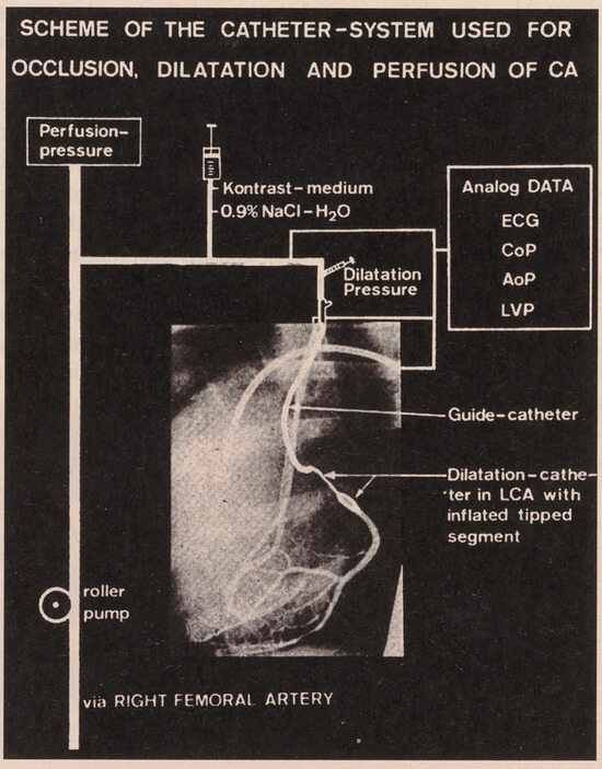
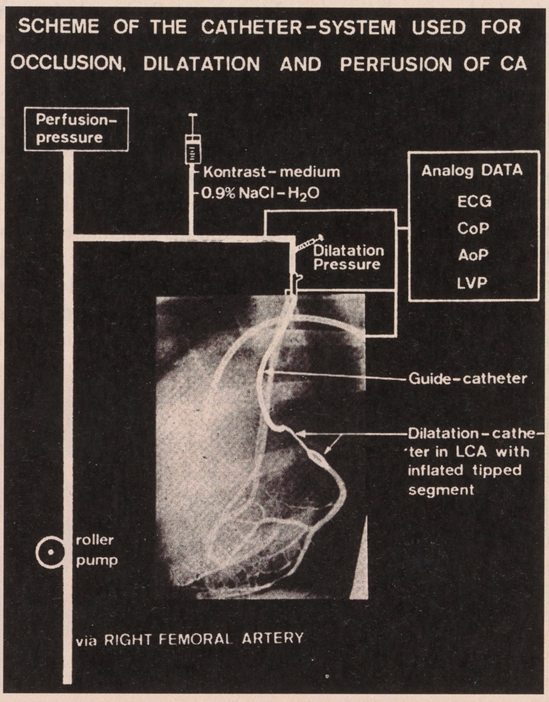
It was a great pleasure to teach Andreas right and left heart catheterization, transseptal puncture, coronary angiography, and many other aspects of our fascinating speciality; also that each step into a new area has to be planed most carefully. Overall the perspective to use catheters for something else than the eternal diagnosis was extremely attractive. The first step in this direction were anesthetized dogs in our animal facilities, in which Marko Turina had sewed acute coronary stenoses with 6-0 silk. They could be opened by balloon catheters inserted from the femoral artery. I was convinced, from these animal experiments, that it would be impossible to interrupt even for a short time the coronary circulation without perfusion of the peripheral coronary bed. This prompted the realization of the coronary double lumen balloon, made by Schneider AG, so that during the dilatation blood from the other femoral artery could be infused by a roller pump through the central lumen into the distal coronary artery (Figure 1). What was necessary and advantageous in animals with acute coronary stenosis proved however not to be necessary in man with chronic stenoses. We used the central lumen from all the beginning also for distal pressure measurement.
Figure 1.
Experimental setting of coronary artery dilatation in dogs with a coronary artery stenosis surgically created with 6–0 silk around a proximal branch of the left coronary artery. A week later, in the closed chest animal, a guide-catheter was positioned under fluoroscopy into the left main coronary stem. Through it the double lumen dilatation catheter was advanced to the stenosis, with its central lumen connected to a three-way stopcock through which the distal coronary pressure could be measured, while the perfusion with arterial blood from the other femoral artery was shortly stopped. During this X-ray exposure contrast medium was injected to visualize the circumflex coronary artery. Through the lateral lumen of the dilatation catheter the balloon with minimal compliance was inflated with a tuberculin syringe to 6 or 8 atmospheres, by which most silk threads bursted. However, sub-total stenoses could not be opened because, when we tried to push the dilatation catheter in the severe stenoses, the guide catheter left the coronary ostium. The ECG, the coronary artery (CoP), the aortic (AoP) and the left ventricular (LVP) pressures, as well as the perfusion pressure were registered on an Electronics for Medicine recorder. Interruption of the perfusion led quickly to ST-changes in the ECG, extrasystoles or ventricular fibrillation [9].
We presented the percutaneous opening of coronary stenoses in dogs in the spring meeting 1976 of the “Deutsche Gesellschaft für Kreislaufforschung” in Bad Nauheim to a public which was convinced that this technique could not be transferred to man with chronic coronary artery stenoses. The work was published in extenso in the “Archiv der Deutschen Gesellschaft für Kreislaufforschung” [9] more than one year earlier than the in USA often cited abstract of Grüntzig at the American Heart Association in fall 1977.
Andreas liked speed and efficiency. With 20 years he was fascinated, as his brother recalled, in touring with a car in Spain. Among the skiers in our Cardiology Division he wanted to be the speediest but Schurle Rhomberg, an assistant from the Austrian alps specially experienced and gifted, and as a happy nature as Andreas, often left him behind. Andreas also took flying lessons with a Piper and later a Cessna plane on the small airport of Pfaffikon, in the vicinity of Zurich. No doubt he was a “high flyer”. Flying became his second hobby, the first one was his extraordinary professional involvement. He told me once: “Flying proves to myself that I have no fear.”
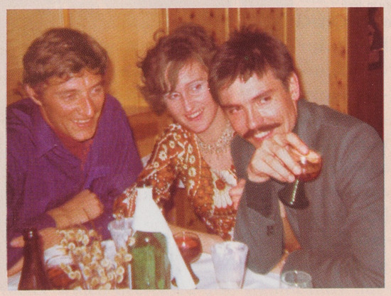
To realize a dream, one has to have first the facility for dreaming. We were sure to be on the right track. Imagination, elan and perseverance are omnipotent. It was a happy time for Andreas. His constant optimism, his persistent character and his pragmatic approach to each problem, and also his never ending energy had to reach the goal. We had also many happy hours out of the hospital. During the festivities, which were frequent in our cardiology team, the handsome Andreas with his charming and radiant smile was very much liked by our feminine personnel and the nurses (Figure 2).
Figure 2.
Andreas Grüntzig (right) in happy company after a day of skiing in the Austrian alps.
It lasted nearly two years from the animal experiments until the first successful coronary dilatation in a conscious patient. There was no ethical committee at that time but the ingenious heart surgeon Ake Senning backed up fully Andreas and was ready to help in emergency should something go wrong, but it was not simple to find the right patient. After having made with R. Myler, in San Francisco, some intraoperative balloon dilatations, and also after the group in Frankfurt under Prof. Kaltenbach was ready to dilate coronary arteries together with Andreas in their place, the pressure was high enough in Zurich that Grüntzig got the permission from his superiors, and naturally also the place of the first patient who had an optimal anatomy. It was a young entrepreneur, with his 38 years of the same age as Grüntzig, with a proximal LAD stenosis, and it was a plain success [1]. It was obviously not necessary to use the roller pump with arterial blood because the pressure in the periphery of the LAD rose considerably with the first deflation of the balloon and, after a second inflation, the angiographic result looked very good. This happy patient was until recently without any complaints. The third patient, who was dilated even at two sites, was also a plain success. The second and the fourth patient, both in Frankfurt, each with a stenosis of the main stem, had unfortunately not the same and long lasting result; the second had to be operated somewhat later than half a year after the first dilatation as expression of restenosis, and the fourth died after an anterior myocardial infarction 2<sup>1</sup>/<sub>2</sub> months later, but the main stem was patent. The seventh patient in Zurich was bypass-operated immediately after an unsuccessful attempt, so that the surgical standby arranged by Prof. Senning and his collaborators was used for the first time [10].
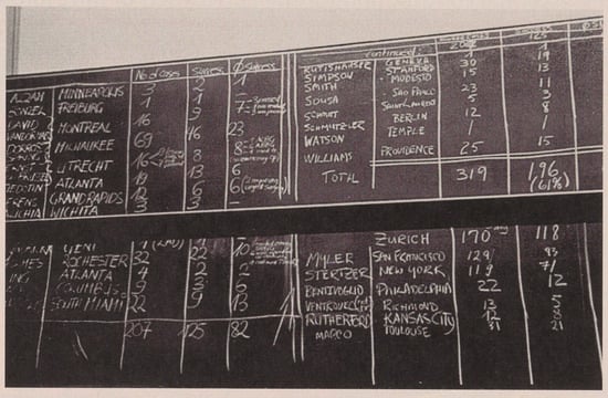
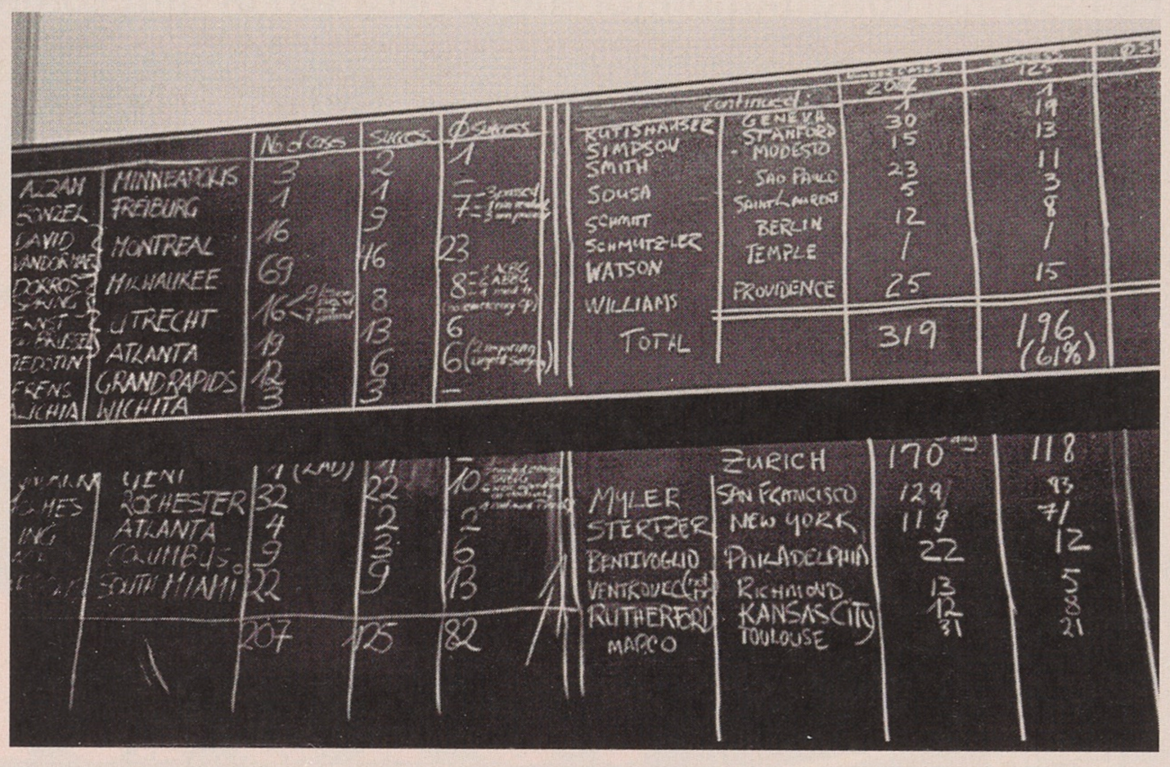
Since the interest in the method grew very rapidly Andreas started with the first courses, during which he transmitted with great courage the procedures from the catheterization room into the auditorium, also a new method at that time, which continues today with satellite-broadcast transmitting life procedures from both sides of the Atlantic. Grüntzig has invited me to all the four courses in Zurich: The first took place in August 1978, the second in August 1979, the third in January 1980, and the last in August 1980. I have conserved all my notes very carefully (Figure 3).
Figure 3.
Blackboard in an auditorium of the Zurich University Hospital during the last demonstration course when the world statistics of coronary balloon dilatation were completely summarized on the wall by Grüntzig.
In September 1980 Andreas Grüntzig followed a call of Willis Hurst and Spencer King to the Emory University in Atlanta, where he had much better support than in Zurich. When Andreas died on the 27th of October 1985 in the cockpit of his new plane, in a severe thunderstorm, Hurst asked me to say something about Andreas’ earlier life in Europe at the funeral on the 1st of November 1985 in Atlanta and on the 12th November at the American Heart Association; I certainly followed his wish [11].
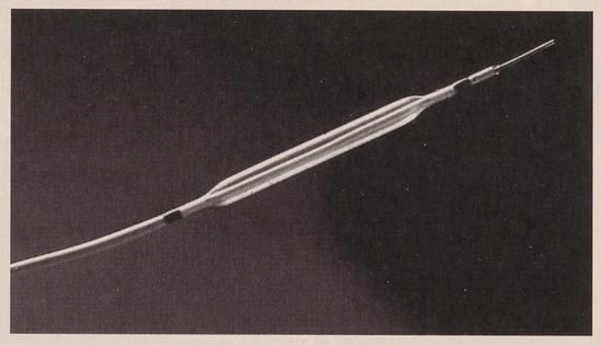
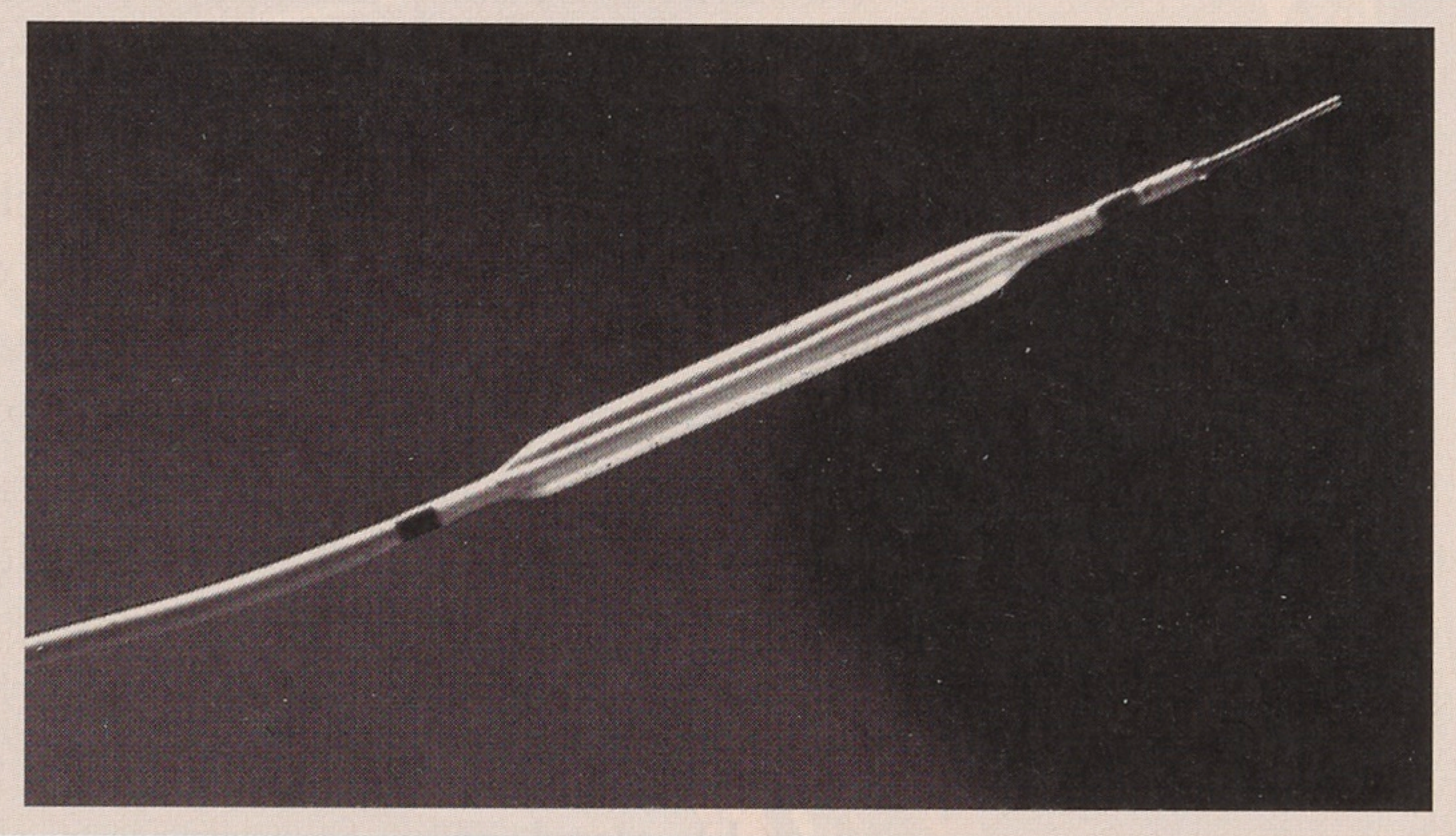
Considerable progress in interventional cardiology on top of Grüntzig’s double lumen catheter (Figure 4) brought obviously the steerable guide wire of Simpson in the central lumen [12], because it allowed to reach even the most distal coronary segment. The other great simplification is the procedure of Bonzel [13], which allows very quick introduction and exchange of the balloon catheters, avoiding partly radioscopy, and has quickly suppressed in Europe the long wire technique introduced by Kaltenbach [14].
Figure 4.
Non steerable double lumen coronary balloon catheter DG 20-30 (i.e., 20 mm long, 3 mm Ø). With this instrument 170 patients were attempted in Zurich and 133 were successfully dilated by Grüntzig before he left to Atlanta. (Courtesy of Schneider [Europe] AG, Bulach).
Further very important contributions came from Patrick Serruys, Michel Bertrand and Bernhard Meier. Bernhard, who followed Grüntzig from Zurich to Atlanta and, after his training, guided many years our catheter laboratories in Geneva [15] before he was elected Chief of Cardiology at the University Hospital in Bern, had the great 3 S principle: “simple, safe and successful intervention”. Recently, however, pushed by the patient Prof. Meier deviated from accepted indications. He dilated again and stented the proximal LAD of the very first patient of Grüntzig on September 4th, 2000 (23 years after the first intervention), even the patient had a normal exercise test and a normal fractional regional flow reserve! On December 4th, 2000 Meier had to fight already an instant restenosis. As long as the restenosis problem is not solved, plaque sealing has no place.
The legacy of Andreas Grüntzig in Zurich were 170 patients. In 133 there was a direct and obvious success. A new stenosis on the same spot in the coronary arteries occurred within 6 months in 36 patients and, in 6 patients, later. The problem of restenosis will occupy us in the next two chapters.
Even Jacques Puel has the merit to have implanted the first coronary stent worldwide on the 28th of March 1986 in Toulouse, the contribution of Ulrich Sigwart as a pioneer in this field is at least as great. He persisted, in spite of many sceptical voices, setbacks and pitfalls, and proceeded experimentally and in the clinic to improve and simplify the procedures to insert and impact the stent into the wall; together with the relevant branches of the industry he improved the design of these metallic devices. The most important steps were, however, to inhibit more selectively the function of the platelets. The importance of platelet adhesion and agglomeration on the surface of the stents was only gradually discovered. The inhibition of the final common pathway of these platelet functions by glycoprotein IIb/IIIa inhibitors allows now that stents can be safely inserted, even in unstable lesions. The ease of manipulation and the angiographically nearly always perfect appearance of the stented vessel led in my mind in the late nineties even to an excess of stent implantations, the so-called “stentomania”.
Ulrich Sigwart was born on the 9th of March 1941 in Wuppertal, Germany, as son of a physician. He also lost his father very early. He studied medicine at the Universities of Freiburg i.Br. and Basle and realized his “Staatesxamen” at the University of Münster in Essen, with the mark “very good”. In Basle he had met his charming future wife Christine. In 1967 he promoted to Dr. med. with “magna cum laude” at the University of Freiburg i.Br. After the obligatory “Famulatur” he longed for the new world, where he worked at very famous places: in Framingham with Prof. Tedechi, in Boston with Prof. Zimmermann, and at the Baylor College in Houston with Prof. Henry McIntosh.
In the years 1971 and 1972 he came in our stimulating team in Zurich. At that time Grüntzig worked in radiology with the concentric Dotter catheters for opening iliac and femoral arteries. Sigwart was very skilled and showed great dexterity in the cath lab. Hardly any assistant was faster in the left atrium during transseptal access, which we often used at that time to introduce tipmanometers into the left atrium and the left ventricle or retrograde into the ascending aorta to measure high fidelity gradients over the mitral and aortic valves without having a catheter passing through these valves. We measured those pressures also during pacing, handgrip and bicycle ergometry. His interest in the domain of coronary artery disease gained a good ground in the study of the transfer function of the coronary vascular bed and the measurement of coronary blood flow, when we injected instantaneously Cardiogreen in the left coronary artery and sampled with the aid of a fiberoptic catheter the dye-concentration in the coronary sinus again under various conditions which lead to ischemia, a condition which changed the transfer function considerably [16,17].
From 1972 to 1979 Ulrich Sigwart was “Oberarzt” at the Gollwitzer-Meier Institute in Bad Oeynhausen under Prof. Gleichmann. Angiographic studies under ergometry was one of his main fields. There he could fully exploit his skill; he worked with great pleasure in the catheterization laboratory. In 1978 he presented his habilitation in order to get the “venia legendi” at the University of Düsseldorf with his book “Die automatische Erfassung von Herzkatheterdaten”, which I could appreciate as an external reviewer.
Then Ulrich came back to Switzerland and got the post of “medécin-associé” in Lausanne at the Medical-Surgical Department (the first modern structure in Switzerland) under Prof. Rivier and Prof. Sadeghi. In 1982 he was even commissary chief. In those years Sigwart studied mainly the regional and global left ventricular function before, during and after angioplasty and the effect of collaterals. Already at that time he showed great interest in the obstructive cardiomyopathy for which he later introduced an optimal interventional therapy. From Geneva, the neighbour of Lausanne, I had again good contact with Sigwart, specially when we prepared the first international congress on “Silent ischemia”, for which Sigwart made an essential contribution [18].
At the succession of the cardiology chair in Lausanne, our commission presented a primo and ex-aequo situation. The medical faculty and the government opted for Professor Lukas Kappenberger as future chief, Sigwart becoming “Professeur associé” at the Cardiology Division. This gave Ulrich again more freedom and time to work experimentally and in the catheterization laboratory.
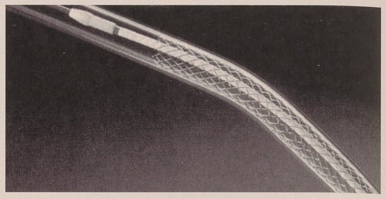
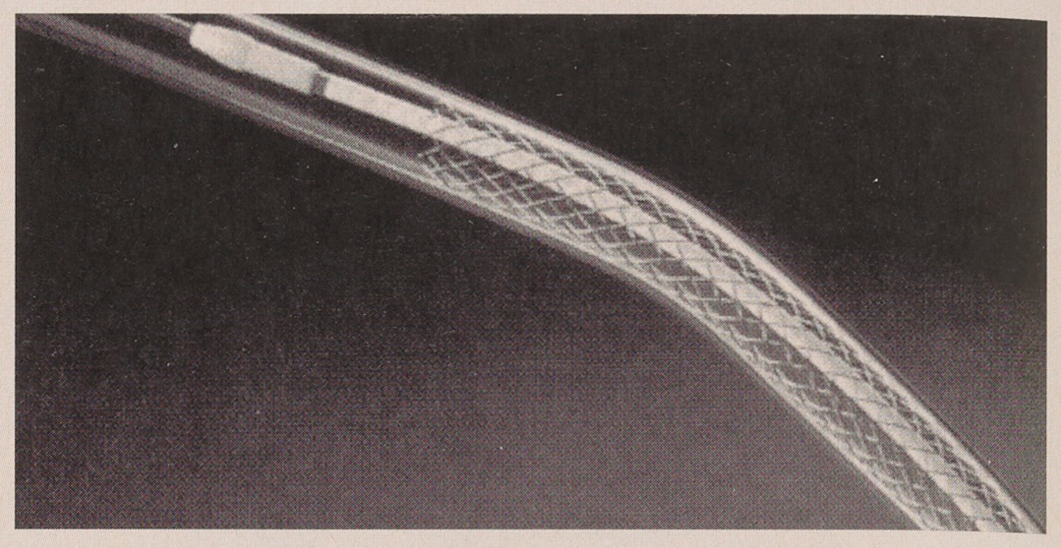
The industrial development of the first stent, the Wallstent, by the Medinvent Company, a few kilometers outside of Lausanne, was essential. Sigwart had a close interchange with this company. This self-deploying stent of Medinvent was delivered in the vessel by a special application system (Figure 5). Even today in the coronaries superseded by other types of stents, this type was the first stent which, after experimental application in dogs by Sigwart in Lausanne and in sheeps by Puel in Toulouse, was used in man. In March 1986 the ethical committee at the University Hospital of Lausanne consented to the proposal of Sigwart to implant this stent in case of vessel occlusion after angioplasty, in restenosis, and in bypass grafts in the periphery and in the heart.
Figure 5.
Wallstent of the firm Medinvent (near Lausanne) with its innovative delivery system self-expanded in a plastic tube. (Courtesy of Medinvent SA/Schneider [Europe] AG).
On June 12th, 1986, during an interventional course at the University of Lausanne in which I participated, a 51-year-old woman, after a balloon dilatation of the LAD and the right coronary artery in the morning, closed her LAD during the mid-day pause. Ulrich Sigwart opened it with a Wallstent in the presence of the heart surgeons in the catheterization laboratory. The same patient got about one month later a second stent because of a restenosis in the right coronary artery. This essential new step in interventional cardiology, shown in the afternoon by Sigwart to the astonished participants of the course, was very convincing. Sceptics argued, however, that metal spirals of copper were very well suited and actually used to create intravascular thromboses. But Sigwart’s procedure had clearly documented that an acute closure of a balloon dilated vessel could be treated by stenting, circumventing an urgent surgical intervention which was always a procedure with a relatively high mortality. This has developed to the actual situation in which only very exceptionally a surgical standby is still exercised during an interventional procedure. On the contrary, today the interventionalist is sometimes called by the surgeon to dilate or stent a bypass which is postoperatively closing up. Today it is well documented that the probability of an urgent surgical intervention is well below 0,5%, so that a surgical team in the same center is useful but not any more a prerequisite for coronary dilatations [19].
Already in the earliest time of stenting Sigwart observed that the self-expanding Medinvent stent in stenosed vessels not always took a cylindrical form over its whole length. Therefore, he reasoned to open the stent, first in these areas and later over its whole length by supplementary balloon inflations (the so-called “Swiss kiss”). By this procedure a better apposition resp. impaction of the stent in the arterial wall was achieved. IVUS investigations proved later that the full apposition of all stent struts at the intima was favorable to endothelial overgrowth of the stent and decreased the tendency to thrombosis [20]. Antonio Colombo has finally very much propagated the high pressure implantation of stents. By this procedure the trauma in the arterial wall layers may be very much increased and has, as a consequence, also a stronger reaction of the vessel wall. By the way a kiss never should be traumatic! In daily practise an individual compromise has to be looked for. To achieve an optimal “impacting” of all the stent struts with a minimal trauma of the media and adventitia is the best solution we have to aim at.
The second stent of the first patient in Lausanne was implanted by Sigwart under the new and very important point of view to circumvent or decrease the restenosis. The first essential paper of Sigwart and collaborators in the New England Journal of Medicine in 1987 [21] combines in its title the two goals: “Intravascular stents to prevent occlusion and restenosis after transluminal angioplasty”. The many unsuccessful previous attempts to fight the restenosis by drugs given orally called for non-pharmacologic methods. The stent, by its potential to open the vessel wider, could accept a certain proliferation towards the lumen without compromising the blood flow (“bigger is better”). The two randomized studies STRESS [22] and BENESTENT [23] confirmed the superiority of stents in focal lesions (<15 mm) and vessel diameters ≥3 mm and, in the BENESTENT II study [24], the heparin-coated stent showed its advantage in lesions in the LAD, and specially in unstable patients. The very attractive angiographic aspect immediately after stent implantation has guided to use the stent always more widely and under other conditions. In the daily work in the catheterization laboratory stenting of long lesions and utilization of stents in small vessels may, however, lead to an increased percentage of chronic in-stent restenosis, for which an efficient therapy was lacking until a few years ago.
During the development and application of different stents thrombotic phenomena played a crucial role. Initially Sigwart and we all faced the problem of subacute thrombosis, which was treated with strong anticoagulation and aspirine plus dipyridamol over longer periods after stenting. But late thrombosis sometimes with catastrophic outcome damped the initial enthusiasm around 1990. Only after experimental results showed that the platelets were the main players in the in-stent thrombosis, the combination of aspirine and ticlopidine and, more recently, the combination of aspirine and clopidogrel led to new excellent results. In unstable angina the intravenous monoclonal antibodies of IIb/IIIa-receptors showed further great advantages. These drugs would be wider used if their high price would not limit their application.
Elective stents have also shown to be advantageous after opening chronic occlusions of vessels of ≥ 2,75 mm and in acute myocardial infarction, mostly in vessels of ≥ 3 mm of diameter with the platelet passivation indicated above. Today stents are used preferentially if a suboptimal result after balloon angioplasty is present, but primary stenting is more and more widely applied. Even more side branches are lost there is now a tendency for stentomania – specially in young cardiologists – because primary stenting with present models is so easy and the angiographic result of the day looks so well.
In his Grüntzig Lecture [25] at the European Congress of Cardiology in 1996 Sigwart expressed, based on the experience in the past, an enthusiastic vision of endovascular stents of different configuration and specially impregnation and foresaw wide application of stenting in the next decade in interventional cardiology and angiology.
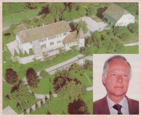
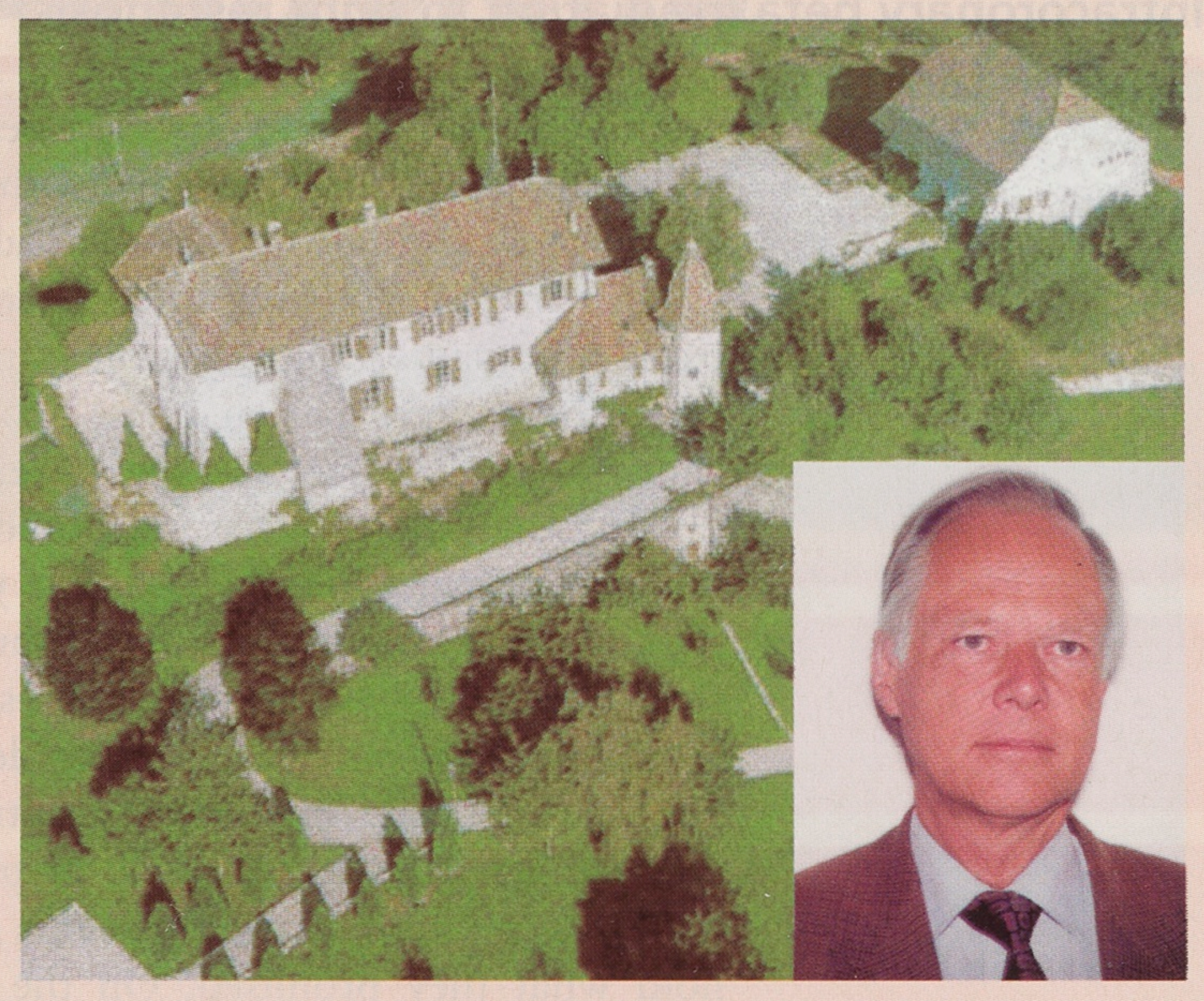
In 1989 Sigwart left Lausanne, heading for London where he became Director of the Department of Invasive Cardiology at the Royal Brompton Hospital and consultant in various clinics where he has, among others, to deal with Royal families. He became – even being Swiss – also a perfect Englishman in as much as the word “an Englishman’s house is his castle” turned into reality for him and his family (Figure 6). After getting a honorary doctorate at the University of Lausanne on the 30th of October 1999, he invited us all to his marvellous Château Bioley-Magnoux in the great canton de Vaud. It is a great satisfaction that Ulrich Sigwart will soon take over the chair of Cardiology at the University of Geneva.
Figure 6.
Ulrich Sigwart and his château Bioley-Magnoux in the beautiful Vaud landscape.
The restenosis was up to now the essential limit why balloon dilatation with and without stents and even less with other devices could not be applied in patients with three-vessel disease, except one wants that the patient as in dentistry has another intervention soon. Most studies show a restenosis rate of the treated lesion of 20-40% in 6 months. Assuming about 1 Million angioplasties in the USA in the year 1999 the costs of restenosis amount to a sum of more than 1 Billion US $. This underscores the enormous interest in efficient means to decrease or abolish restenosis. An effective genetic therapy was not available and also local pharmacological applications without stents have not hold what they promised. On the contrary, brachytherapy was up to recently the only method that has shown a clearcut decrease in restenosis in patients with and without stents. As in coronary angioplasty the basic experience in treatment of restenosis by ionizing radiation was made in the periphery. The first study in 30 patients with gamma irradiation in patients with repeated femoral occlusion was made in 1989-1990 in Frankfurt. The follow-up, however, was far from complete.
Vitali Verin was born as a son of an engineer on the 20th September 1962 and went to school in Tashkent (USSR). Then he studied medicine in Moscow and got his diploma in July 1985 at the Second Medical Institute Moscow “with honour”. In June 1986 he got a second diploma after formation in surgery. Then he worked at the First Moscow Hospital as an interventional radiologist during 5 years; among other important work he closed the Ductus botalli in 35 patients.
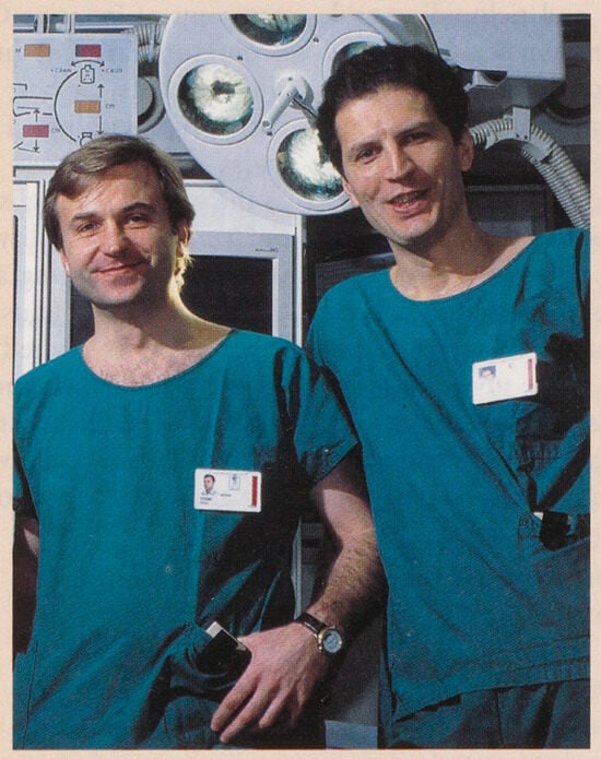
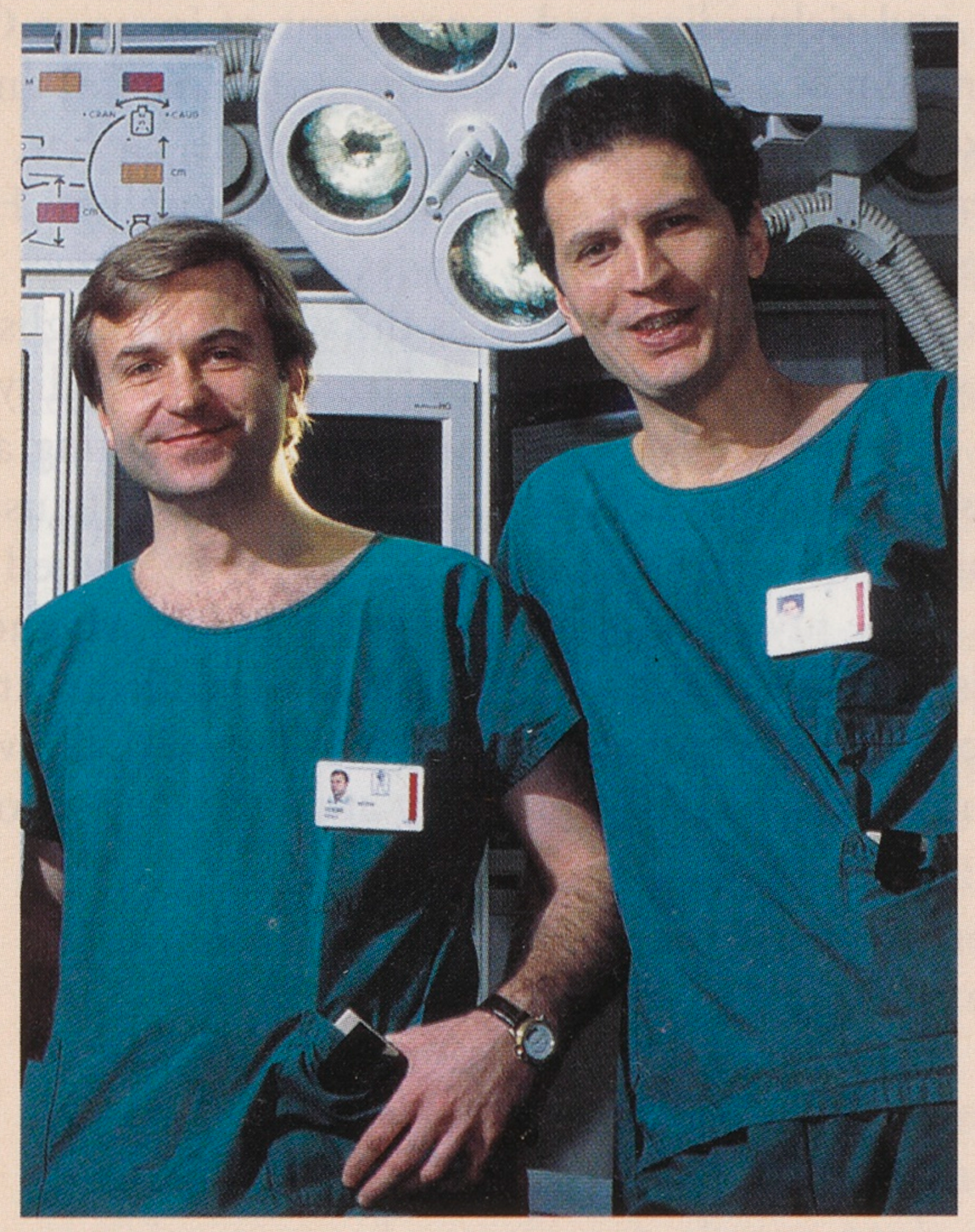
In April 1991 he came as an assistant in interventional cardiology at our Cardiology Center in Geneva and showed from all the beginning not only interest in the daily work doing PCI but in different research projects. A happy accident was that another collaborator of Russian origin, Youri Popowski, was working at the Radiocologic Division of our University Hospital (Figure 7). Verin and Popowski proposed to tackle the restenosis problem with radioactive radiation, since also other exaggerated wound healing as the keloid can be treated by this method. I agreed immediately. We were at this moment not aware of the work in Frankfurt. Naturally such a method had to be studied first experimentally. I sent Vitali Verin to Profs. Baumgartner and Hoff in Basle who experimented with hypercholestorelomic rabbits. The diet with 2% cholesterol had to be alternated by normal chow each two weeks because, otherwise, the animals would die before getting sufficient atheroma.
Figure 7.
Vitali Verin (left) and Youri Popowski in a Siemens biplane cath-lab at the University Hospital in Geneva.
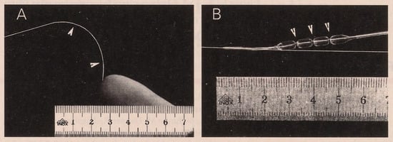
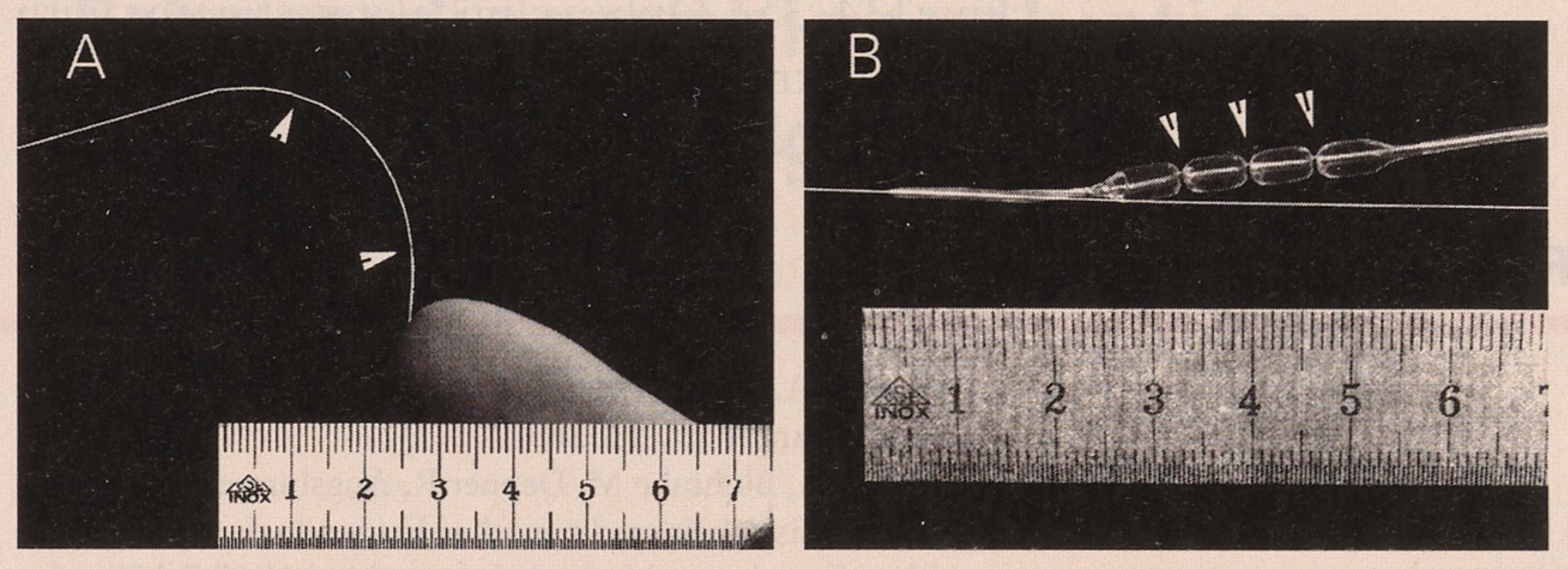
Multiple contacts with Russian physicists led to the choice of Yttrium-90, one of the most potent beta emitters (2,28 MeV) in the form of a flexible spiral, which was scaled with a cover of titan (Figure 8A). Together with the firm Schneider Europe AG in Bulach a centering system was developed, so that the 90Y-spiral stays in the center of the vessel, in which the restenosis is to be prevented. It consists of a segmented balloon which is introduced into the vessel to be irradiated by the method of Bonzel [13]. Four interconnected compartments are mounted on a double lumen shaft (Figure 8B) of which the central blind ending lumen contains the 90Y-spiral [26].
Figure 8.
A. Flexible Yttrium-90 coil coated with the titanium and two markers (arrows) delineating the radioactive source. B. Centering balloon with four interconnected chambers and three waists (arrows) allowing to center the Yttrium-90 coil relative to the arterial lumen [31].
Careful measurements of the dose by Youri Popowski [27] preceded the animal experiments; they confirmed that beta irradiation can be easily shielded by lucite (plexiglass) and that the dose in animal tissue falls about at its half by every mm of tissue. This property of 90Y limits an unnecessary irradiation of distant tissue and allows simple manipulation and shielding in the catheterization laboratory, with intravascular irradiation time of only a few minutes.
I promoted Vitali Verin to “chef de clinique scientifique” by January 1st, 1995. On June 21st 1995 Urban, Verin and coll. inserted for the first time the mentioned beta source with the goal of decreasing restenosis into the right coronary artery of a 68-year-old man in our Cardiology Center at the University Hospital of Geneva. When Condado and colleagues [28,29] presented at the American College of Cardiology, in March 1995, the first results after angioplasty by non-centered gamma radiation, and when Teirstein was about to start his randomized study with gamma radiation to decrease the in-stent restenosis [30], we expressed our scepticism on wider clinical application of this approach in the light of the difficulties of radiation protection and handling in the catheterization laboratory. In our feasibility study in 15 patients [31] the dose of 18 Gy at the balloon surface (the same which was successful in rabbits and was allowed by our ethical committee) was too low.
Verin and Popowski have also studied the biological effect of beta irradiation on the scar formation regarding apoptosis and other tissue alterations [32], what explains that not only proliferation is hampered but that a negative remodeling of the irradiated vessel may occur. In the meantime Verin, Popowski et al. have realized the first multicenter dose finding study with intracoronary beta irradiation in 181 patients [33]. In this landmark study with a dose of 18 Gy 1 mm inside the tissue (= 1 mm from the surface of the balloon) only in 4% of balloon dilated lesions a restenosis was present. These excellent results – no other study with intravascular radiation (SCRIPPS, GAMMA, BERT, BETAWRIST, etc.) to fight restenosis comes near to it – show clearly that centered beta irradiation with the right dose fights the Achille’s heel of coronary dilatation created by Grüntzig very efficiently. This success over the restenosis is very much the merit of the arduous work of Vitali Verin and Youri Popowski. Most recently they experiment with the isotope 144Cerium-Praeseodymium, a beta source with a half-life of 285 days and even a higher energy than Yttrium-90.
Final remarks
Andreas Grüntzig – Ulrich Sigwart – Vitali Verin, it is not only an alphabetic and temporal sequence of successful investigators in interventional cardiology in Switzerland in the last quarter of the gene century. Each one stands on the shoulders of his predecessor with the ideas for the realization of a better solution, every 9th year after his predecessor, of an important practical problem for the patients.
Success depends mainly on the firm conviction that borders which earlier theories and conventions have set can be transgressed. Optimism, hard work and confidence that a farflung goal can be reached are necessary. Each step must be approached with steadiness and persistence despite unfortunate setbacks. Nobody can attain today an important goal alone. Good leaders choose their colleagues wisely, help them to optimize their potential, create good conditions for their work that let them reach out for the success.
In the next 25 years much greater progress will be realized. After all the balloon, the stent and brachytherapy are crutches which improve symptoms but very rarely lead to cure of the disease. In the future the main thrust of research should be directed at the question how the basic process of atheromatosis, including vascular aging and myocardial dysfunction, with its devastating consequence, namely heart failure, can be avoided or at least considerably delayed. Relevant results of basic research should be weighted early whether they allow safe translation into the clinical arena. This is best achieved by close mental and local collaboration of scientists working in the basic sciences and in the hospital or ideally – as has been the case with the three mentioned investigators – scientists able to work in both domains. Tissue and animal experiments should be planned and executed based on concepts which can be ultimately applied in man, so that there is a continuity from the molecular and cellular level to in vitro and in vivo experiments and the future the clinical investigator will be the key person leading such research.
References
- Grüntzig, A. Transluminal dilatation of coronary artery stenosis. Lancet 1978, 1, 2631. [Google Scholar]
- Grüntzig, A.; Blohmke, M.; Depner, R.; Augsburger, W. Prufung der Zuverlassigkeit medizinischer Fragen in der epidemiologischen Forschung. Methods Inf. Med. 1968, 7, 159–65. [Google Scholar] [CrossRef] [Scilit] [PubMed]
- Rutishauser, W.; Amende, I.; Schoenbeck, M. Relaxation velocity of contractile elements in ischemic coronary heart disease. Circulation 1971, 44, II–220. [Google Scholar]
- Bollinger, A.; Rutishauser, W.; Mahler, F.; Grüntzig, A. Zur Dynamik des Ruckstroms aus der Vena femoralis. Z. Kreislaufforsch. 1970, 59, 963–71. [Google Scholar] [PubMed]
- Grüntzig, A.; Schonbeck, M.; Winzeler; Lichthen, P.; Rutishauser, W. Die Treffsicherheit einer Fragebogendiagnose bei coronarer Herzkrankheit mit der selektiven Coronarangiographie als Referenztest. Verh. Dtsch. Ges. Inn. Med. 1972, 78, 1017–20. [Google Scholar] [CrossRef] [Scilit] [PubMed]
- Grüntzig, A.; Hopff, H. Perkutane Rekanalisation chronischer arterieller Verschlusse mit einem neuen Dilatationskatheter. Modifikation der Dotter-Technik. Dtsch. Med. Wochenschr. 1974, 99, 2502–10. [Google Scholar] [CrossRef] [Scilit] [PubMed]
- Grüntzig, A. Die perkutane Rekanalisation chronischer arterieller Verschlusse mit einem doppellumigen Dilatationskatheter. Fortschritte Roentgenstr 1976, 12, 48–80. [Google Scholar]
- Grüntzig, A. Die perkutane transluminale Rekanalisation chronischer Arterienverschlusse mit einer neuen Dilatationstechnik; G. Witzstrock-Verlag: Baden-Baden, 1977. [Google Scholar]
- Grüntzig, A.; Riedhammer, H.; Turina, M.; Rutishauser, W. Eine neue Methode zur perkutanen Dilatation von Koronarstenosen – tierexperimentelle Prufung. Verh. Dtsch. Ges. Kreislaufforsch. 1976, 42, 282–5. [Google Scholar] [CrossRef] [Scilit] [PubMed]
- Grüntzig, A.R.; Senning, A.; Siegenthaler, W.E. Nonoperative dilatation of coronary-artery stenosis: percutaneous transluminal coronary angioplasty. New Engl. J. Med. 1979, 301, 61–8. [Google Scholar] [CrossRef] [Scilit] [PubMed]
- Rutishauser, W. Memorial tribute to Andreas R. Grüntzig given at the American Heart Association annual scientific sessions in Washington, D.C., November 12th, 1985. Cardiology 1986, 73, 192–3. [Google Scholar]
- Simpson, J.B.; Baim, D.S.; Robert, E.W.; Harrison, D.C. A new catheter system for coronary angioplasty. Am. J. Cardiol. 1982, 49, 1216–22. [Google Scholar] [CrossRef] [Scilit] [PubMed]
- Bonzel, T.; Wollschlager, H.; Kasper, W.; Meinertz, T.; Just, H. The sliding rail system (monorail): description of a new technique for intravascular instrumentation and its application to coronary angioplasty. Z. Kardiol. 1987, 76 (Suppl. 6), 119–22. [Google Scholar] [PubMed]
- Kaltenbach, M. The long wire technique – a new technique for steerable balloon catheter dilation of coronary artery stenoses. Eur. Heart J. 1984, 5, 1004–9. [Google Scholar] [CrossRef] [Scilit] [PubMed]
- Meier, B. Long-term results of coronary angioplasty. Annu Rev. Med. 1991, 42, 47–59. [Google Scholar] [CrossRef] [Scilit] [PubMed]
- Sigwart, U.; Hirzel, H.; Burian, H.; Rutishauser, W. Transferfunktion des Koronargefassbettes. Verh. Dtsch. Ges. Inn. Med. 1972, 78, 1020–3. [Google Scholar] [CrossRef] [Scilit] [PubMed]
- Sigwart, U.; Rutishauser, W. A selective coronary indicator dilution technique with fiberoptic recording. Z. Kardiol. 1975, 64, 607–15. [Google Scholar] [PubMed]
- Sigwart, U.; Grbic, M.; Payot, M.; Goy, J.J.; Essinger, A.; Fischer, A. Ischemic events during coronary artery balloon obstruction. In Silent Myocardial Ischemia; Rutishauser, W., Roskamm, H., Eds.; Springer Verlag: Berlin Heidelberg, 1984; pp. 29–36. [Google Scholar]
- Meier, B. Surgical cover for angioplasty. Br. Heart J. 1994, 72, 506–8. [Google Scholar] [CrossRef] [Scilit]
- Colombo, A.; Hall, P.; Nakamura, S.; Almagor, Y.; Maiello, L.; Martini, G.; et al. Intracoronary stenting without anticoagulation accomplished with intravascular ultrasound guidance. Circulation 1995, 91, 1676–88. [Google Scholar] [CrossRef] [Scilit] [PubMed]
- Sigwart, U.; Puel, J.; Mirkovitch, V.; Joffre, F.; Kappenberger, L. Intravascular stents to prevent occlusion and restenosis after transluminal angioplasty. New Engl. J. Med. 1987, 316, 701–6. [Google Scholar] [CrossRef] [Scilit] [PubMed]
- Fischman, D.L.; Leon, M.B.; Baim, D.S.; et al. for the Stent Restenosis Study Investigators. A randomized comparison of coronary-stent placement and balloon angioplasty in the treatment of coronary artery disease. New Engl. J. Med. 1994, 331, 496–501. [Google Scholar] [CrossRef] [Scilit] [PubMed]
- Macaya, C.; Serruys, P.W.; Ruzygrok, P.; et al. for the Benestent Study Group. Continued benefit of coronary stenting versus balloon angioplasty. One-year clinical follow-up of Benestent study. J. Am. Coll. Cardiol. 1996, 27, 255–61. [Google Scholar] [CrossRef] [Scilit] [PubMed]
- Serruys, P.; van Hout, B.; Bonnier, H.; et al. for the Benestent Study Group. Randomized comparison of implantation of heparin-coated stents with balloon angioplasty in selected patients with coronary artery disease (Benestent II). Lancet 1998, 352, 673–81. [Google Scholar] [CrossRef] [Scilit] [PubMed]
- Sigwart, U. The 1996 Grüntzig Lecture. Stents: a mechanical solution for a biological problem? Eur. Heart J. 1997, 18, 1068–72. [Google Scholar] [CrossRef] [Scilit] [PubMed]
- Verin, V.; Popowski, Y.; Urban, P.; Belenger, J.; Redard, M.; Costa, M.; et al. Intraarterial beta-irradiation prevents neointimal hyperplasia in a hypercholesterolemic rabbit restenosis model. Circulation 1995, 92, 2284–90. [Google Scholar] [CrossRef] [Scilit] [PubMed]
- Popowski, Y.; Verin, V.; Papirov, I.; Nouet, P.; Rouzaud, M.; Grob, E.; et al. Intra-arterial 90-yttrium brachytherapy: preliminary dosimetric study using a specially modified angioplasty balloon. Int. J. Radiat. Oncol. Biol. Phys. 1995, 33, 713–7. [Google Scholar] [CrossRef] [Scilit] [PubMed]
- Condado, J.A.; Gurdiel, O.; Espinosa, R.; Gonzalez Brito, J.; Harrison, M.L.; Lyoner, L.R.; et al. Percutaneous transluminal coronary angioplasty and intracoronary radiation therapy: a possible new modality for the treatment of coronary restenosis. J. Am. Coll. Cardiol. 1995, Suppl., 188A. [Google Scholar]
- Condado, J.A.; Waksman, R.; Gurdiel, O.; Espinosa, R.; Gonzalez, J.; Burger, B.; et al. Long-term angiographic and clinical outcome after percutaneous transluminal coronary angioplasty and intracoronary radiation therapy in humans. Circulation 1997, 96, 727–32. [Google Scholar] [CrossRef] [Scilit] [PubMed]
- Teirstein, P.S.; Massullo, V.; Jani, S.; Popma, J.J.; Mintz, G.S.; Russo, R.J.; et al. Catheter-based radiotherapy to inhibit restenosis after coronary stenting. New Engl. J. Med. 1997, 336, 1697–1703. [Google Scholar] [CrossRef] [Scilit] [PubMed]
- Verin, V.; Urban, P.; Popowski, Y.; Schwager, M.; Nouet, P.; Dorsaz, P.A.; et al. Feasibility of intracoronary beta-irradiation to reduce restenosis after balloon angioplasty. A clinical pilot study. Circulation 1997, 95, 1138–44. [Google Scholar] [CrossRef] [Scilit] [PubMed]
- Verin, V.; Popowski, Y.; Bochaton-Piallat, M.L.; Belenger, J.; Urban, P.; Neuville, P.; et al. Intraarterial beta irradiation induces smooth muscle cell apoptosis and reduces medial cellularity in a hypercholesterolemic rabbit restenosis model. Int. J. Radiat. Oncol. Biol. Phys. 2000, 46, 661–70. [Google Scholar] [CrossRef] [Scilit] [PubMed]
- Verin, V.; Popowski, Y.; de Bruyne, B.; Baumgart, D.; Sauerwein, W.; Lins, M.; et al. Endoluminal beta-radiation therapy for the prevention of coronary restenosis after balloon angioplasty. N Engl. J. Med. 2001, 344, 243–9. [Google Scholar] [CrossRef] [Scilit] [PubMed]
- Burnett, J.C., Jr. Biomedical research at Mayo Clinic: A tradition of collaboration and a vision for year 2000 and beyond. Mayo Clin. Proc. 2000, 75, 337–9. [Google Scholar] [CrossRef] [Scilit] [PubMed]
- Chien, K.R. Mentors for the new millennium. Advice to a young cardiologist. Circulation 2000, 101, 1616–8. [Google Scholar] [PubMed]
© 2001 by the author. Attribution - Non-Commercial - NoDerivatives 4.0.